How experience amplifies escape: an inhibitory connection is released
A male-mouse study finds that prior threat experience recruits prefrontal input and endocannabinoids to weaken pontine-to-hypothalamic inhibition, strengthening escape responses.

Escape can be innate, yet its intensity changes with experience. After repeated exposure to a threatening odor, mice escape faster and make more attempts during subsequent rat or carbon-dioxide challenges. A study published on May 27, 2025 in Nature Communications links this enhancement to CCK cells in the hypothalamic dorsal premammillary nucleus (PMd). A central change involves weakened inhibition from the pontine ventral tegmental nucleus of Gudden (VTg). Prefrontal input and endocannabinoids help connect prior threat experience to current defensive action.
Enhanced escape after learning accompanies reduced VTg inhibitory-terminal activity and increased local endocannabinoid signaling. Reducing CB1-receptor signaling in VTg PV cells weakens this additional enhancement.
Contents · 7 sections
An odor experience changes subsequent escape
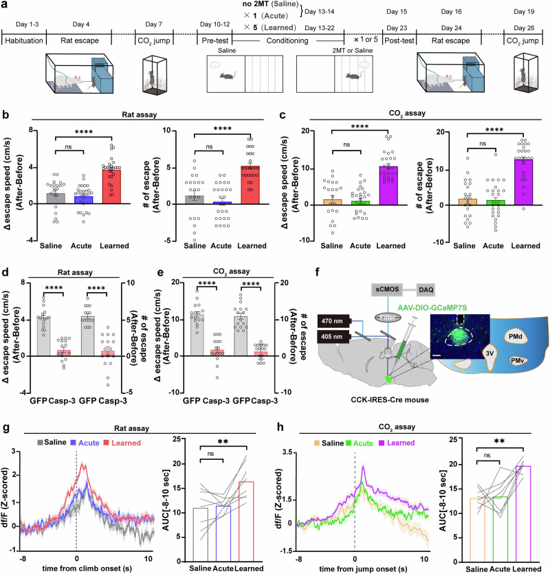
The study first exposes mice repeatedly to a threatening odor. They subsequently spend less time in the odor-associated compartment, demonstrating measurable place aversion. Single or fewer exposures do not produce comparably clear memory-related effects and escape enhancement.
The change extends to another setting. In the rat assay, mice climb away faster and make more escape attempts; during the carbon-dioxide challenge, jump speed and number also rise. The prior experience influences the intensity of action against subsequent, different threats rather than only the response to the original odor.
Source: Chai R et al. · Nature communications · CC BY-NC-ND
Original figure and English caption ↗
PMd receives multiple defense-associated inputs
PMd CCK cells are already active at escape onset, with a greater increase after repeated threat experience. Ablating these cells weakens baseline escape and interferes with the extra enhancement from experience, identifying them as an important component of this defensive action.
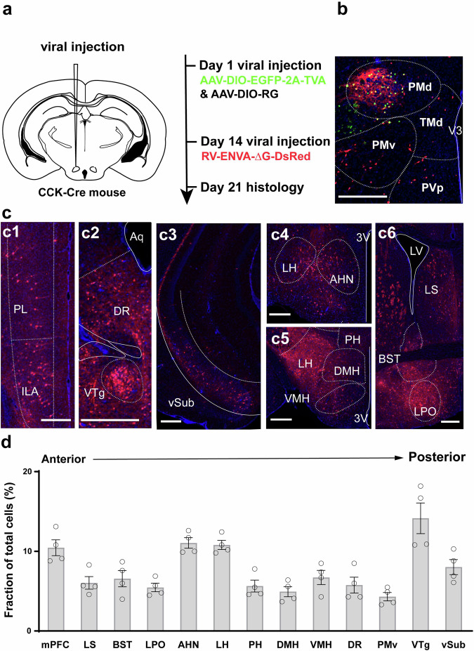
Upstream tracing identifies input from prefrontal cortex, ventral subiculum and several hypothalamic regions, with particularly dense input from VTg. The node thus receives experience-associated information alongside pontine modulation, providing two pathways to investigate.
Source: Chai R et al. · Nature communications · CC BY-NC-ND
Original figure and English caption ↗
VTg PV cells directly inhibit PMd
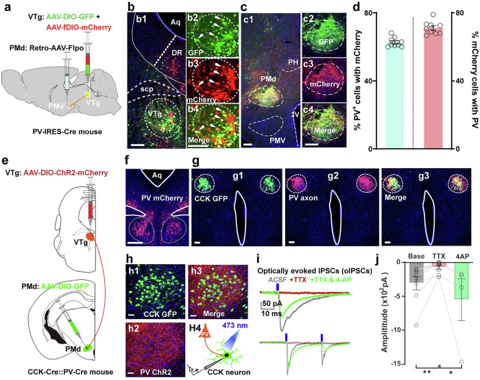
Further analysis shows that PMd-projecting VTg cells are predominantly PV cells, whose terminals densely surround PMd CCK cells. Slice-current recordings support a direct inhibitory connection: activating VTg terminals evokes inhibition in PMd cells.
Behavioral modulation follows the same direction. Enhancing this connection slows escape and reduces its frequency; suppressing it strengthens escape. The inhibitory input acts as a brake on action intensity.
Source: Chai R et al. · Nature communications · CC BY-NC-ND
Original figure and English caption ↗
The local brake weakens as experience strengthens escape
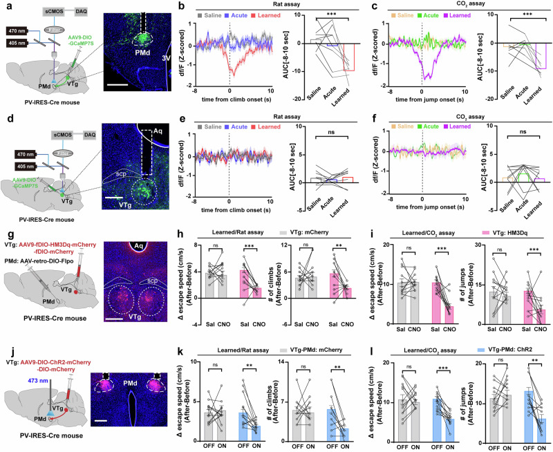
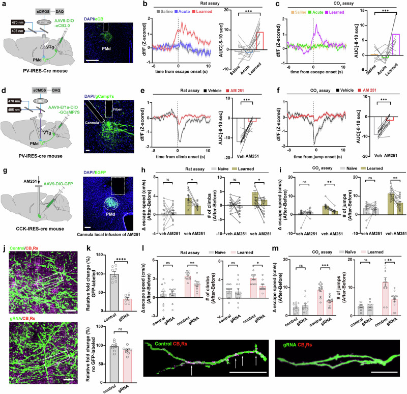
In mice with repeated threat experience, calcium signals in VTg terminals within PMd fall sharply at escape onset. Naive mice and those with a single exposure show no equivalent pattern. Enhancing the connection again weakens the escape enhancement after learning.
VTg somatic calcium signals show no corresponding decrease, and evoked spike counts remain broadly similar. The change points toward regulation near axon terminals rather than silencing of the entire VTg population. This leads to the next question: is a local signal suppressing terminal transmission?
Source: Chai R et al. · Nature communications · CC BY-NC-ND
Original figure and English caption ↗
Endocannabinoids release the inhibition
At escape onset after learning, the study detects increased endocannabinoid signaling near PMd. These endogenous lipids can reduce presynaptic transmitter release through CB1 receptors. At inhibitory terminals, reduced transmission makes downstream cells more readily active. This disinhibition involves two steps: a local lipid signal weakens the brake, making the output it previously restrained easier to recruit. The direction of an effect thus depends on the connection being modulated.
Blocking local CB1 signaling weakens the terminal decrease and the extra escape response. Reducing CB1 receptors in VTg PV cells likewise attenuates the learning-associated enhancement, while baseline escape and odor-related aversion remain broadly unchanged. Lipid signaling contributes to how experience modulates response intensity.
Source: Chai R et al. · Nature communications · CC BY-NC-ND
Original figure and English caption ↗
Experience-associated modulation reaches local synapses
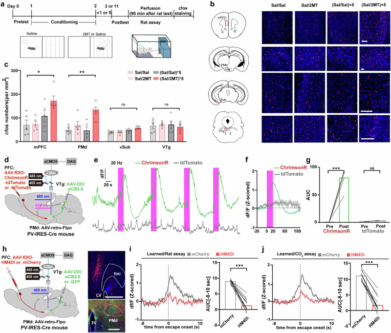
After repeated threat experience, activity-associated markers increase in prefrontal cortex and PMd, without corresponding changes in ventral subiculum or VTg. Following the prefrontal-to-PMd input reveals that activating it increases local endocannabinoid signaling.
Suppressing this input weakens the endocannabinoid rise during escape after learning. Rather than requiring direct silencing of VTg somata, prefrontal input can recruit signaling near a different synapse to alter pontine inhibition of the hypothalamus.
Source: Chai R et al. · Nature communications · CC BY-NC-ND
Original figure and English caption ↗
The prefrontal pathway participates at two stages
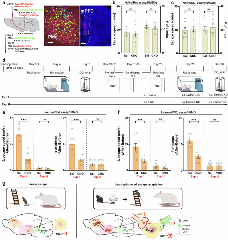
Suppressing the prefrontal input during threat experience prevents a clear subsequent escape enhancement; suppressing it during testing after experience also weakens the enhancement. It participates in both learning and subsequent expression of stronger action.
In mice without the relevant learning, however, enhancing prefrontal input alone does not increase escape speed or number. These findings in adult male mice show how experience can retune innate escape, with prefrontal input, local lipid feedback and inhibitory connections jointly shaping response intensity.
Source: Chai R et al. · Nature communications · CC BY-NC-ND
Original figure and English caption ↗