How does bile acid signaling influence mood-related circuits? A pathway from lateral hypothalamus to hippocampus
A study in male mice follows TGR5 bile acid receptor signaling in the lateral hypothalamus, linking potassium channel regulation and a hippocampal–septal circuit to depression-like behavior.
Published in Neuron on March 21, 2024, this study by Su-Su Tang and Hao Hong's group at China Pharmaceutical University and Hao Wang's group at the Affiliated Mental Health Center of Zhejiang University School of Medicine (with Xu-Yi Li and Shi-Ya Zhang as co-first authors) demonstrates that chronic stress downregulates Takeda G protein-coupled receptor 5 (TGR5) in lateral hypothalamic area (LHA) GABAergic neurons via glucocorticoid receptor (GR)-nGRE transcriptional repression and reduced bile acid levels. Using in vivo fiber photometry and optogenetics (with Inper 200 μm, 0.37 NA optical fibers, Inper Studio, and Inper Data Process software) alongside viral tracing and patch-clamp electrophysiology, the authors show that LHA GABAergic TGR5 suppresses ERK phosphorylation to restore Kv4.2-mediated A-type K+ currents, dampening LHA GABAergic hyperexcitability and disinhibiting the downstream dCA3CaMKIIα-to-DLSGABA circuit to alleviate depressive-like behaviors.
Chronic stress downregulates TGR5 in LHA GABAergic neurons via GR-mediated transcriptional repression and bile acid depletion, driving neuronal hyperexcitability through elevated p-ERK and reduced Kv4.2-mediated A-type K+ currents; activating LHA GABAergic TGR5 restores Kv4.2 function and relieves monosynaptic inhibition onto the dCA3CaMKIIα-to-DLSGABA pathway to reverse depressive-like behaviors.
Contents · 7 sections
Chronic Stress Downregulates TGR5 in LHA GABAergic Neurons via Bile Acid Depletion and GR-nGRE Repression
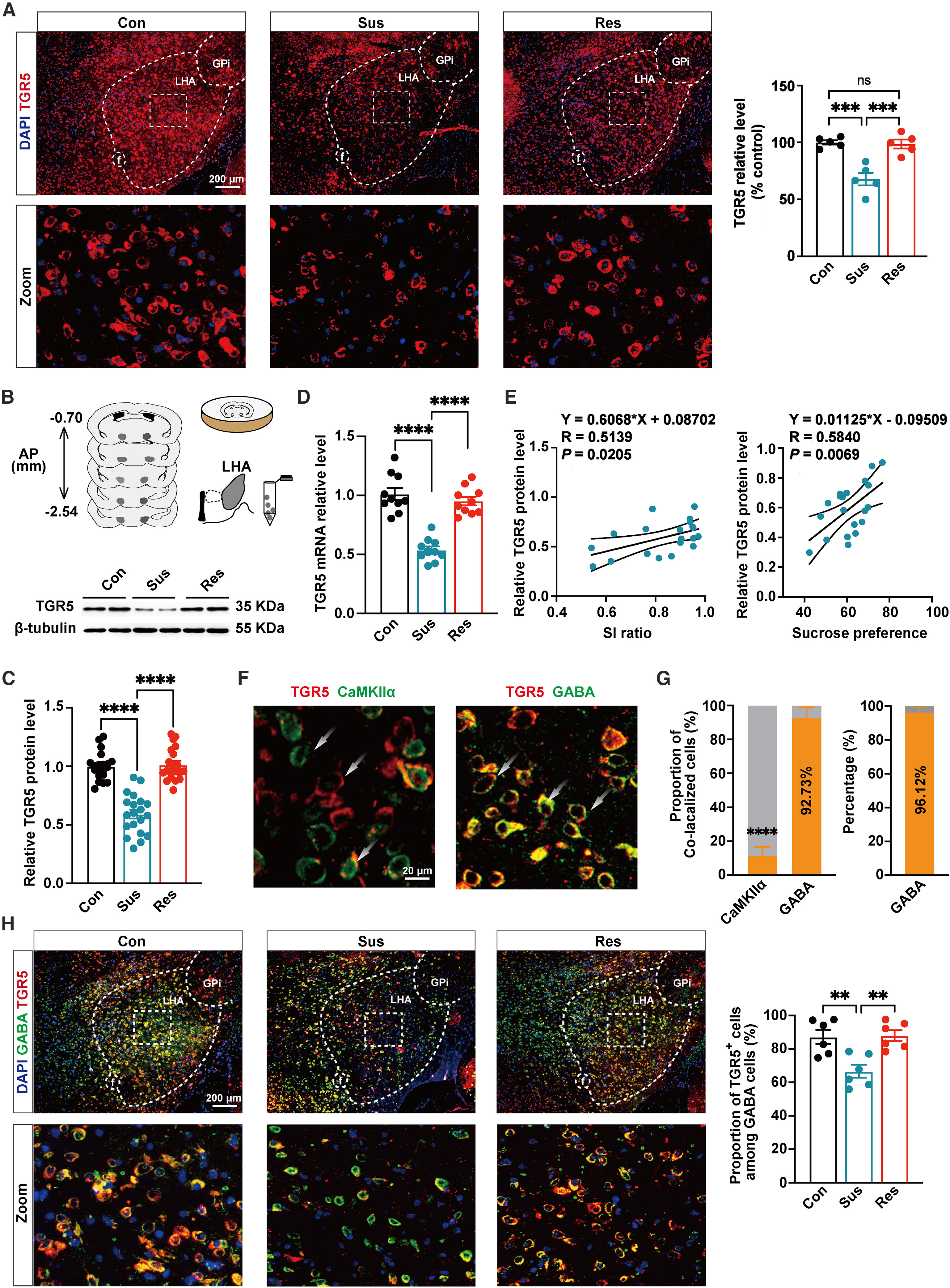
In 10-day chronic social defeat stress (CSDS) and 21-day chronic restraint stress (CRS) mouse models, LHA TGR5 protein and mRNA levels were significantly reduced in CSDS-susceptible and CRS mice and correlated positively with social interaction (SI) ratio and sucrose preference. Dual immunofluorescence revealed that 92.73% ± 6.30% of TGR5+ cells in the LHA were GABA+ whereas only 11.34% ± 5.31% were CaMKIIα+, with chronic stress selectively reducing TGR5 in LHA GABAergic neurons.
UPLC-MS/MS profiling showed decreased blood and hypothalamic bile acids (including CA and CDCA) in stressed mice, while exogenous CA or CDCA supplementation restored LHA TGR5 and alleviated depressive-like behaviors. Concurrently, stress-elevated corticosterone drove glucocorticoid receptor (GR) nuclear translocation in the LHA; ChIP assays confirmed direct GR binding to negative glucocorticoid response elements (nGRE1/nGRE2) on the TGR5 promoter to repress transcription, an effect reversed by the GR antagonist RU486.
Source: et al. · Neuron · Original figure reproduced for academic literature review under fair scholarly commentary; copyright remains with the authors/publisher.
Original figure and English caption ↗
LHA GABAergic Neurons Exhibit Stress-Induced Hyperactivity and Bidirectionally Regulate Depressive-Like Behaviors
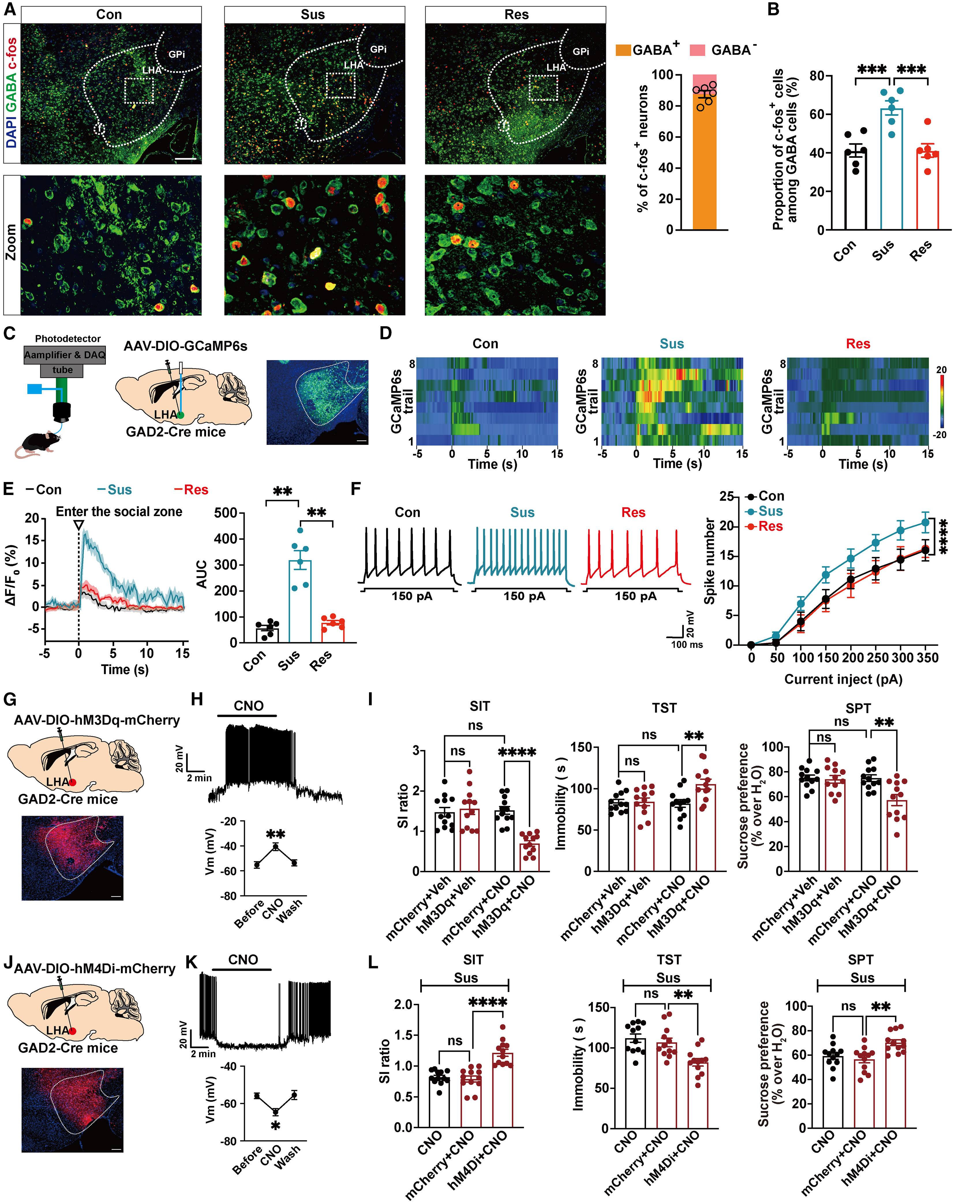
Brain-wide c-Fos mapping revealed elevated c-Fos specifically in LHA GABAergic neurons of CSDS-susceptible mice. In vivo fiber photometry in GAD2-Cre mice expressing AAV-DIO-GCaMP6s (recorded and analyzed using Inper Studio and Inper Data Process software) demonstrated robust Ca2+ transients (increased 0–15 s AUC) in LHA GABAergic neurons when susceptible mice entered the CD1 social interaction zone, and whole-cell current-clamp recordings confirmed higher evoked firing frequencies in both CSDS-susceptible and CRS mice.
Chemogenetic activation of LHA GABAergic neurons (AAV-DIO-hM3Dq-mCherry + CNO) in naive GAD2-Cre mice directly induced social avoidance, prolonged tail suspension test (TST) immobility, and reduced sucrose preference, whereas chemogenetic inhibition (AAV-DIO-hM4Di-mCherry + CNO) in CSDS-susceptible or CRS mice significantly reversed these depressive-like behaviors.
Source: et al. · Neuron · Original figure reproduced for academic literature review under fair scholarly commentary; copyright remains with the authors/publisher.
Original figure and English caption ↗
TGR5 in LHA GABAergic Neurons Exerts Antidepressant Effects by Suppressing Neuronal Hyperexcitability
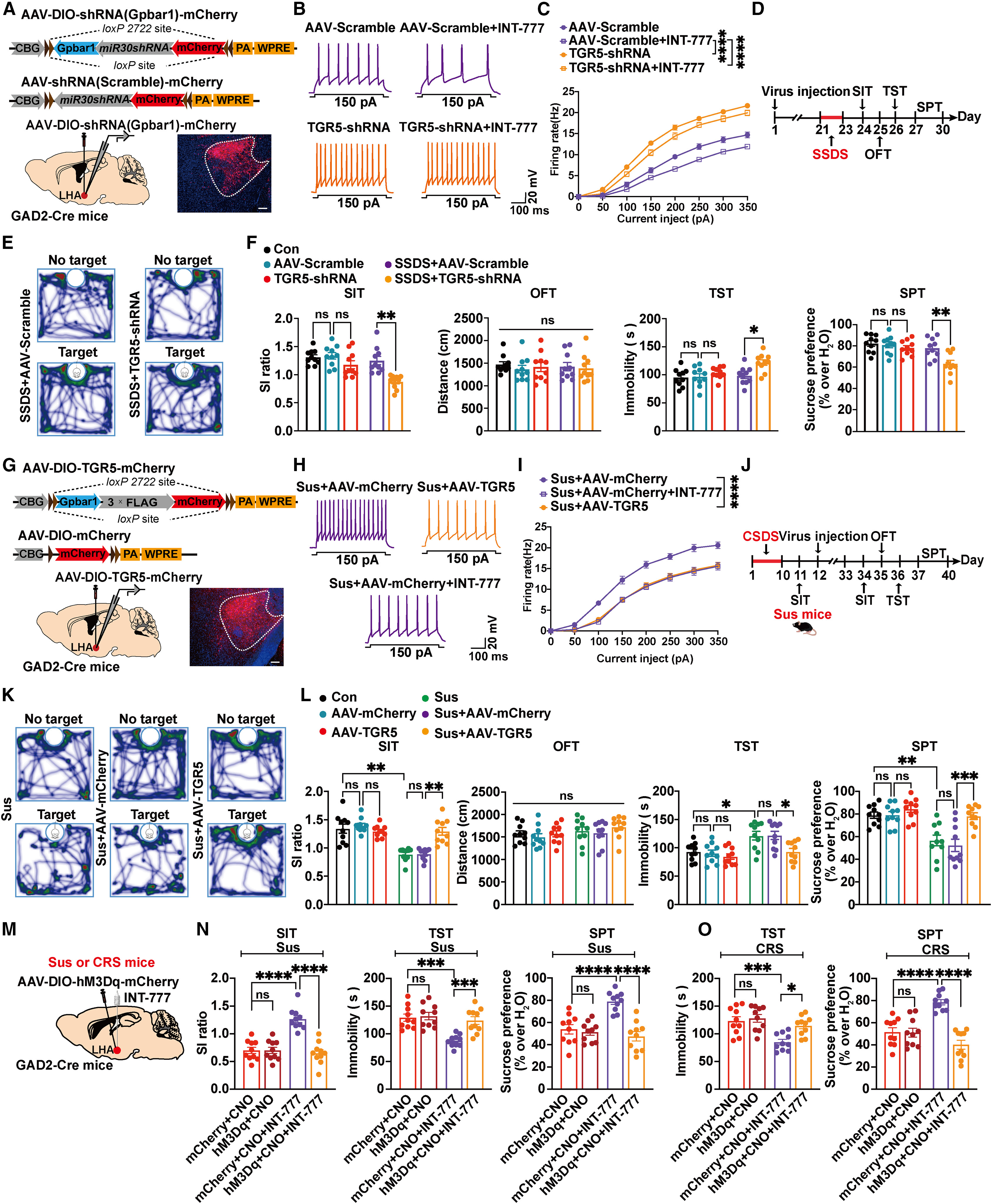
Knockdown of TGR5 in LHA GABAergic neurons (AAV2/9-DIO-shRNA(Gpbar1)-mCherry in GAD2-Cre mice) or application of the TGR5 antagonist SBI-115 increased action potential firing frequency and social-zone Ca2+ transients, blocked the inhibitory effect of the TGR5 agonist INT-777 (10 μM), and promoted depressive-like behaviors under subthreshold social defeat stress (SSDS) or subthreshold restraint stress (SRS).
Conversely, overexpressing TGR5 in LHA GABAergic neurons (AAV-DIO-TGR5-mCherry) or intra-LHA microinfusion of INT-777 (1.5 μg/side) suppressed LHA GABAergic hyperexcitability and reversed depressive-like behaviors in CSDS and CRS mice, whereas overexpressing TGR5 in Vglut2-Cre LHA glutamatergic neurons had no antidepressant effect, and chemogenetic activation of LHA GABAergic neurons abolished INT-777's behavioral rescue.
Source: et al. · Neuron · Original figure reproduced for academic literature review under fair scholarly commentary; copyright remains with the authors/publisher.
Original figure and English caption ↗
TGR5 Suppresses ERK Phosphorylation to Restore Kv4.2-Mediated Transient A-Type Potassium Currents
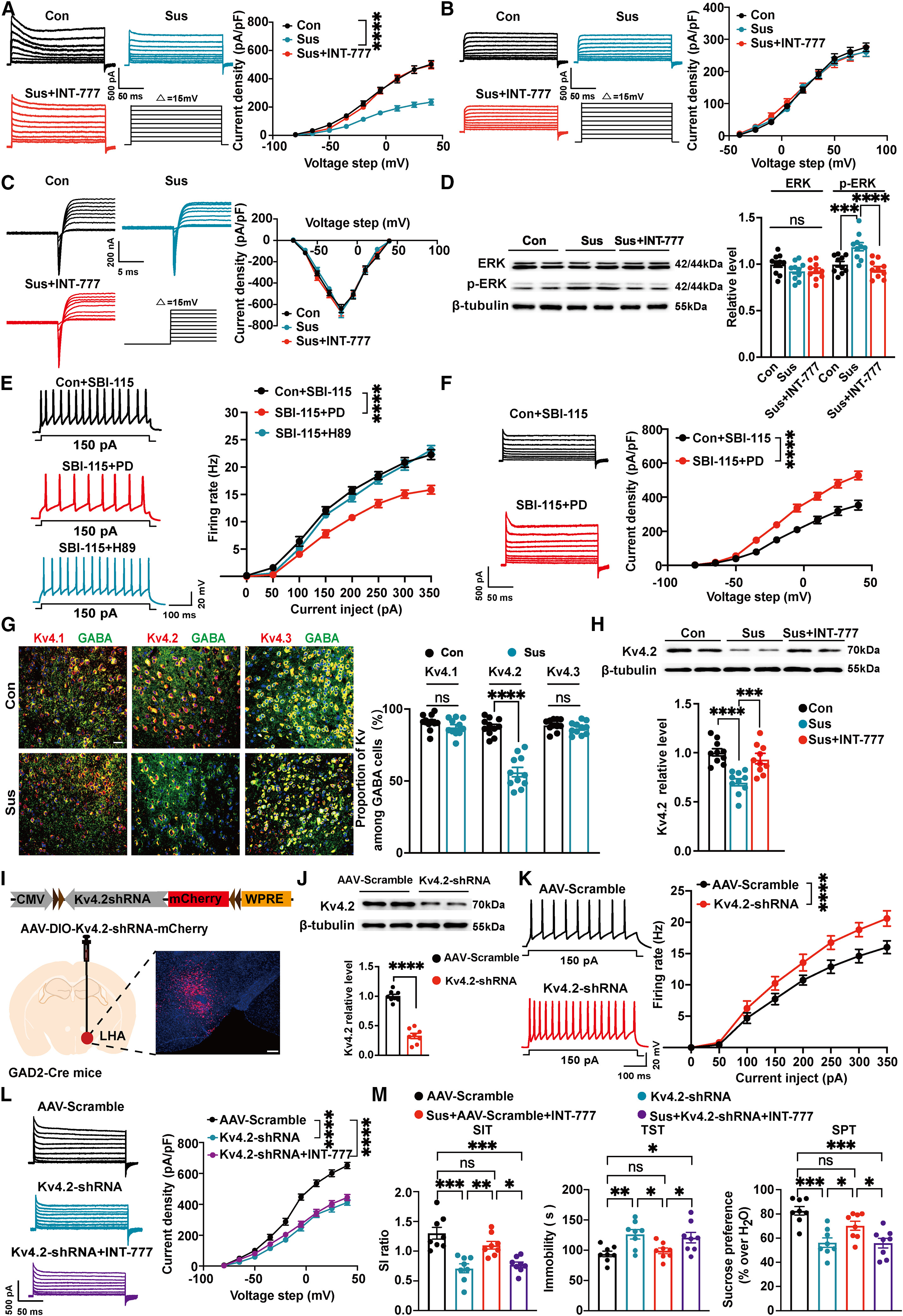
Voltage-clamp recordings revealed that INT-777 or TGR5 overexpression selectively increased transient outward A-type K+ current density in LHA GABAergic neurons without altering delayed rectifier K+ or voltage-gated Na+ currents, whereas TGR5 knockdown reduced A-type K+ currents. Chronic stress elevated phosphorylated ERK (p-ERK, but not p-PKA or p-AKT) in the LHA, which was reversed by TGR5 activation; the ERK inhibitor PD98059 (but not PKA inhibitor H89) blocked SBI-115-induced A-type K+ current suppression and hyperexcitability.
Among Kv4 channels (Kv4.1–Kv4.3), Kv4.2 was selectively downregulated in LHA GABAergic neurons of CSDS-susceptible mice and restored by INT-777. Specific knockdown of Kv4.2 (AAV-DIO-Kv4.2-shRNA-mCherry in GAD2-Cre mice) prevented INT-777 from restoring A-type K+ currents and completely abolished its antidepressant behavioral effects.
Source: et al. · Neuron · Original figure reproduced for academic literature review under fair scholarly commentary; copyright remains with the authors/publisher.
Original figure and English caption ↗
LHA GABAergic Neurons Monosynaptically Inhibit dCA3 CaMKIIα Neurons to Regulate Depressive-Like Behaviors
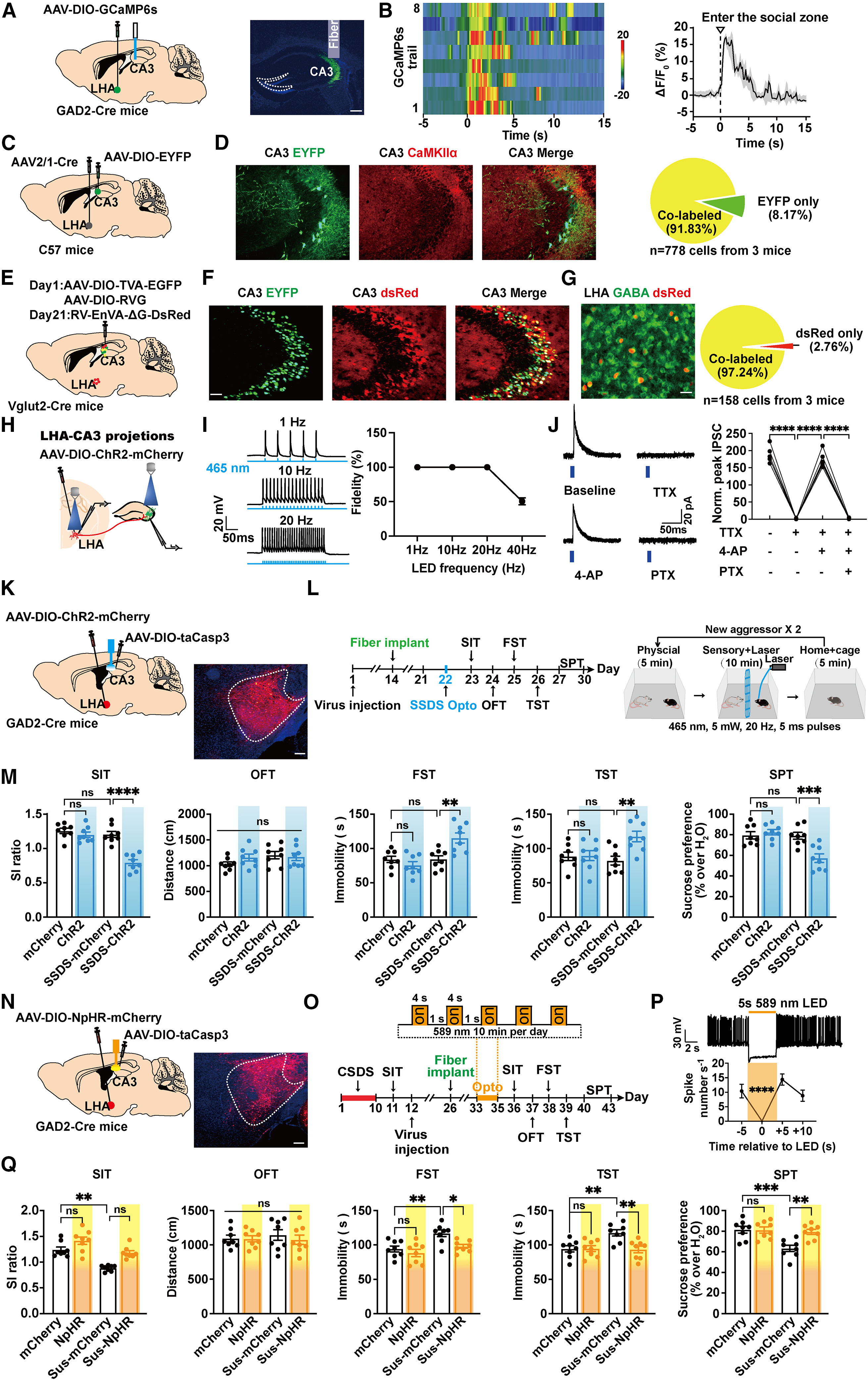
Axonal terminal fiber photometry across five LHA GABAergic projection targets (dCA3, CPU, Acb, LPO, PAG) revealed that only dCA3 terminals exhibited elevated Ca2+ signals when susceptible mice entered the social zone. Anterograde trans-synaptic AAV2/1-Cre tracing (91.83% of postsynaptic dCA3 cells were CaMKIIα+), monosynaptic rabies tracing in Vglut2-Cre mice (97.24% of upstream LHA cells were GABA+), and slice recordings (TTX + 4-AP-resistant, picrotoxin-sensitive light-evoked IPSCs) confirmed a direct monosynaptic LHAGABA→dCA3CaMKIIα inhibitory pathway.
After ablating local dCA3 GABAergic interneurons with AAV-DIO-taCaspase3, optogenetic stimulation (465 nm, 20 Hz via implanted Inper optical fibers) of LHAGABA→dCA3CaMKIIα terminals induced real-time place aversion and promoted depressive-like behaviors under SSDS, whereas 589 nm yellow-light NpHR inhibition rescued depressive-like behaviors for at least 2 weeks by reducing presynaptic release probability (increased PPR). By contrast, activating LHAGABA→dCA3GABA terminals did not induce depressive-like behavior.
Source: et al. · Neuron · Original figure reproduced for academic literature review under fair scholarly commentary; copyright remains with the authors/publisher.
Original figure and English caption ↗
LHA-Recipient dCA3 Glutamatergic Neurons Project to Dorsolateral Septum GABAergic Neurons to Mediate Antidepressant Effects
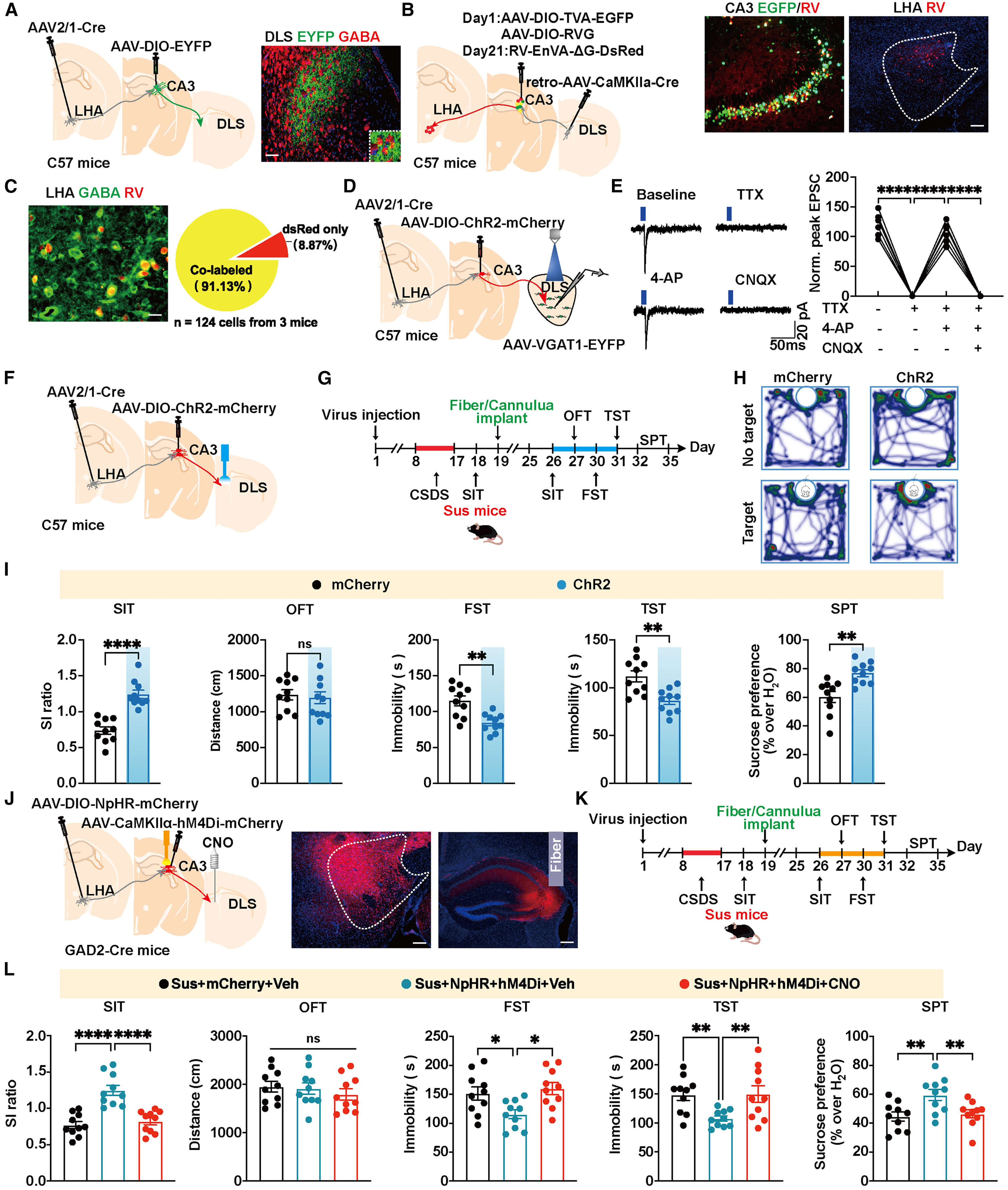
Injecting anterograde trans-synaptic AAV2/1-Cre into the LHA and DIO-EYFP into the dCA3 revealed dense axonal projections to the dorsolateral septum (DLS) surrounding DLS GABAergic neurons. Projection-specific rabies tracing (retro-CaMKIIα-Cre in DLS + DIO-TVA/RVG/RV in dCA3) and slice recordings (TTX + 4-AP-resistant, CNQX-sensitive monosynaptic eEPSCs in DLS GABAergic neurons) established the trisynaptic LHAGABA→dCA3CaMKIIα→DLSGABA architecture.
Optogenetic activation of LHA-recipient dCA3→DLS terminals significantly alleviated depressive-like behaviors in CSDS-susceptible mice, whereas chemogenetic inhibition (hM4Di + intra-DLS CNO) of DLS-projecting dCA3 CaMKIIα neurons completely abolished the antidepressant effects of optogenetically inhibiting LHAGABA→dCA3 terminals.
Source: et al. · Neuron · Original figure reproduced for academic literature review under fair scholarly commentary; copyright remains with the authors/publisher.
Original figure and English caption ↗
Intra-LHA TGR5 Activation Remodels Dendritic Spines and Exerts Antidepressant Effects via the LHA-dCA3-DLS Circuit
Finally, the authors validated the circuit mechanism of pharmacological TGR5 activation across the trisynaptic pathway. Three-day intra-LHA microinfusion of the TGR5 agonist INT-777 (1.5 μg, 200 nL/side) reversed the CSDS-induced increase in sIPSC frequency onto LHA-recipient dCA3 CaMKIIα+ neurons without altering sEPSCs. Projection-specific Supernova sparse reconstruction (retro-AAV-GAD1-Cre in dCA3 plus AAV-DIO-YPet-mGFP in LHA) further revealed that CSDS increased total and mature mushroom-shaped dendritic spine density on dCA3-projecting LHA GABAergic neurons, which was normalized by intra-LHA INT-777 infusion.
In behavioral occlusion experiments, either chemogenetically activating LHA->dCA3 GABAergic terminals (AAV-DIO-hM3Dq-mCherry in GAD2-Cre LHA with intra-dCA3 CNO infusion) or chemogenetically inhibiting downstream dCA3->DLS glutamatergic projections (AAV-DIO-hM4Di-mCherry in Vglut2-Cre dCA3 with intra-DLS CNO infusion) completely abolished the antidepressant-like behavioral effects of intra-LHA INT-777, establishing the LHA TGR5 -> dCA3 -> DLS trisynaptic circuit as an essential substrate for antidepressant regulation.