Exercise and oxytocin: a brain connection in stressed mice
Treadmill exercise during four weeks of stress preserves oxytocin-related responses linking hypothalamus and nucleus accumbens. Suppressing projection-associated cells weakens several behavioral benefits, identifying a circuit contribution to exercise-related adaptation.
How does the brain respond to a moving body? Exercise engages many signals, including oxytocin. A PNAS study follows behavior and brain responses in male mice receiving treadmill exercise during four weeks of chronic restraint stress. Stress weakens responses in paraventricular-nucleus, or PVN, oxytocin cells and NAc-projecting cells; exercise preserves them. Manipulating this projection-associated population changes several behavioral outcomes, connecting physical activity with specific brain cells.
Exercise during stress accompanies preserved PVN–NAc-associated oxytocin responses. Activating projecting cells improves stress-related behavior; suppressing them weakens several exercise benefits. Sucrose preference and immobility do not change together in every intervention.
Contents · 6 sections
Exercise during stress protects several behaviors
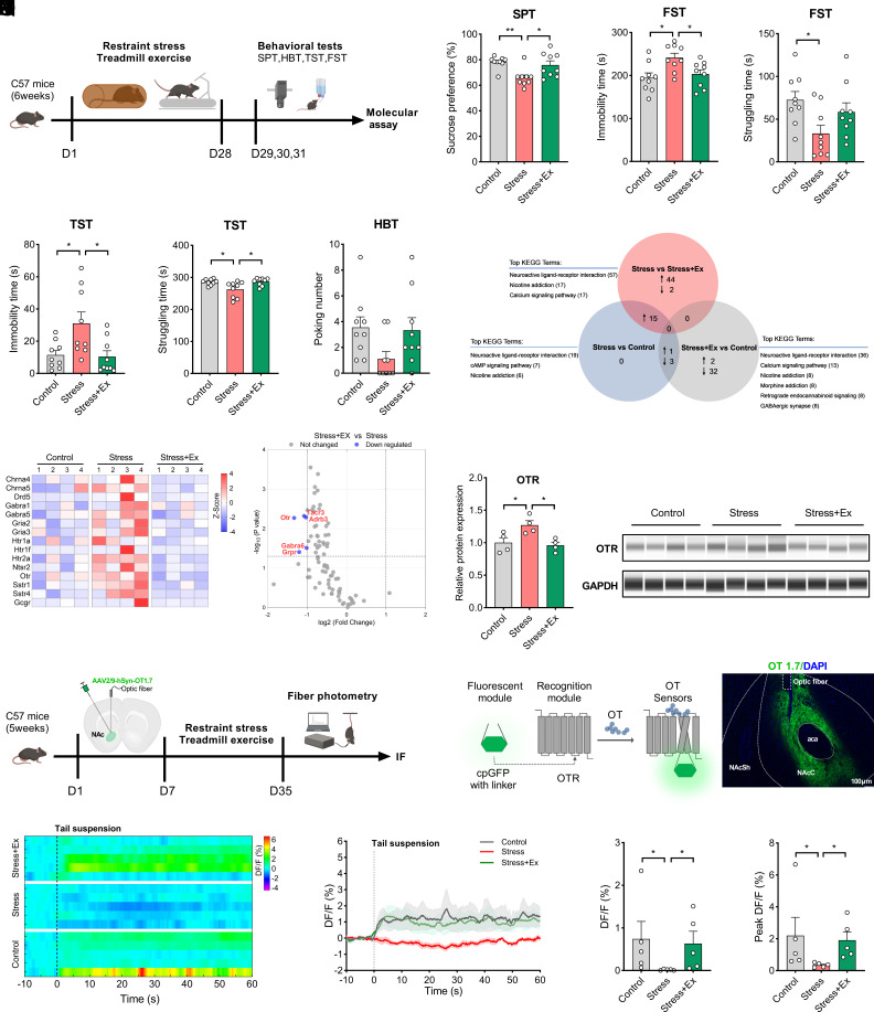
Stress lowers sucrose preference and raises tail-suspension and forced-swim immobility. Concurrent exercise raises preference, reduces both immobility measures and increases tail-suspension struggling. Hole-board poking does not significantly differ.
Nucleus accumbens contributes to reward and motivation. Its c-Fos activity marker decreases with stress and improves with exercise. Receptor-gene screening highlights oxytocin receptors, motivating examination of local peptide responses.
Source: Xia J et al. · Proceedings of the National Academy of Sciences of the United States of America · CC BY-NC-ND
Original figure and English caption ↗
More receptor expression accompanies weaker oxytocin responses
Stress increases accumbal oxytocin-receptor protein but weakens the local oxytocin-sensor response to tail suspension. Exercise raises the response and brings receptor expression nearer control levels. Receptor abundance and ligand-related responses move differently.
The authors propose compensatory receptor upregulation in response to insufficient oxytocin input. This interpretation connects static receptor expression with stimulus-responsive signaling, extending the exercise-related change beyond receptor abundance alone.
Hypothalamic oxytocin cells regain responsiveness
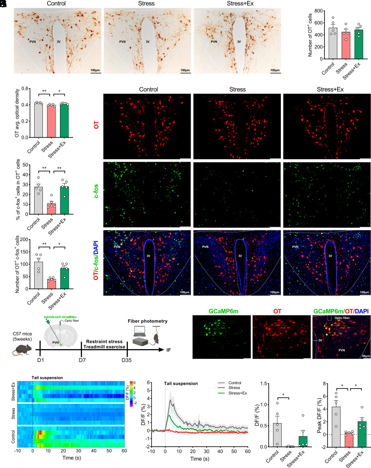
PVN is an important source of brain oxytocin projections. Stress reduces oxytocin staining intensity and activity labeling, both of which recover with exercise. Total oxytocin-positive cell numbers do not significantly change.
During tail suspension, population calcium responses are stronger in control and exercise groups and weaker with stress. They complement local accumbal transmitter responses, adding dynamics to a hypothalamic connection with a reward-related region.
Source: Xia J et al. · Proceedings of the National Academy of Sciences of the United States of America · CC BY-NC-ND
Original figure and English caption ↗
Following the projection identifies an exercise-responsive population
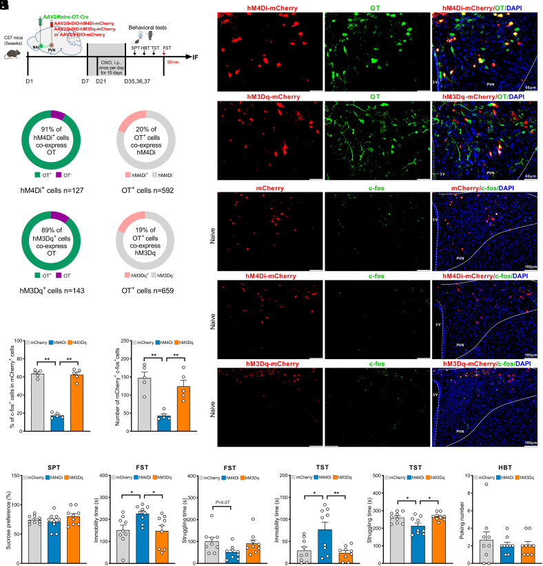
Selecting NAc-projecting PVN oxytocin-related cells identifies lower activity labeling and weaker tail-suspension calcium after stress, with stronger responses maintained by exercise. Projection identity defines a route for further functional tests.
Suppressing the population without chronic stress raises tail-suspension/forced-swim immobility and reduces tail-suspension struggling. Sucrose preference and poking are unchanged, revealing selective rather than uniform behavioral effects.
Source: Xia J et al. · Proceedings of the National Academy of Sciences of the United States of America · CC BY-NC-ND
Original figure and English caption ↗
Activation improves outcomes; suppression weakens exercise benefits
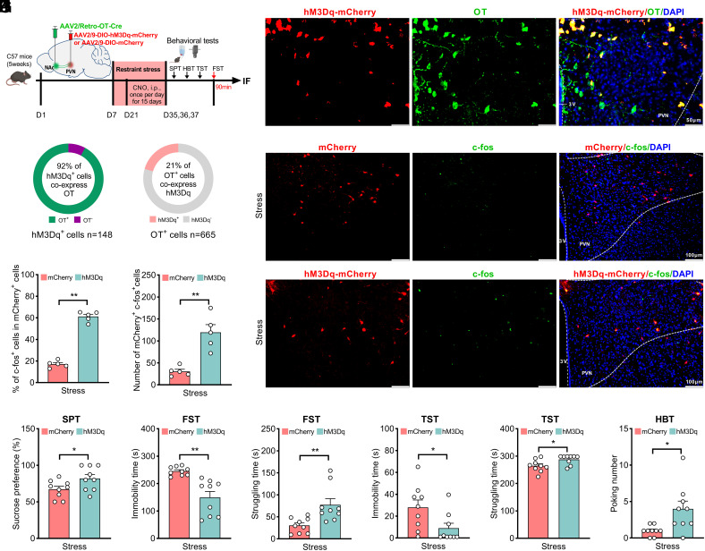
Activating these projecting cells during stress raises sucrose preference, reduces both immobility measures and increases struggling and poking. Manipulation reproduces several directions of exercise-related improvement, showing a functional contribution.
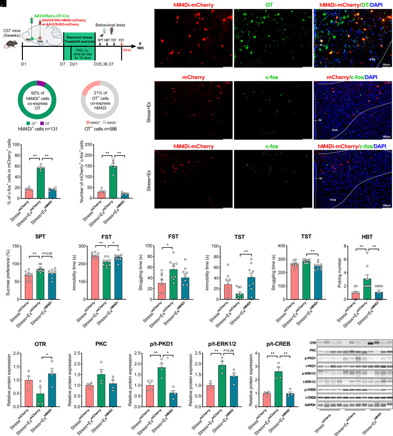
Suppressing them during stress plus exercise weakens several benefits: immobility increases while tail-suspension struggling and poking decline. Sucrose preference does not significantly fall with suppression, separating components of the exercise effect.
Combining exercise with cell activation does not clearly outperform exercise alone. Similar beneficial directions do not necessarily produce further gain when combined.
Source: Xia J et al. · Proceedings of the National Academy of Sciences of the United States of America · CC BY-NC-ND
Original figure and English caption ↗
Exercise-related brain adaptation involves cellular connections
Accumbal PKD1, ERK and CREB phosphorylation also changes. Suppressing projecting cells weakens exercise-associated PKD1 and CREB changes, linking molecular, cellular and behavioral observations and motivating further study of local adaptation.
In these male mice, physical activity meets a defined brain projection. Stress changes responsiveness, exercise preserves it, and cell manipulation changes selected behaviors. The findings connect exercise-related adaptation with particular cells and conditions.
Source: Xia J et al. · Proceedings of the National Academy of Sciences of the United States of America · CC BY-NC-ND
Original figure and English caption ↗