How does stress reach immunity? A red nucleus–lymph node connection
A blood CCL5 clue in stress-exposed people leads to rat experiments linking red nucleus activity, cervical lymph-node responses and anxiety-like behavior.
Stress changes how we feel and behave, while the immune system may quietly join the response. What signals connect the two? A study published in Nature Communications on October 30, 2023 starts with people exposed to stressful work, follows the chemokine CCL5 in blood, and takes the question into rats. The evidence converges on the midbrain red nucleus and cervical lymph nodes: reduced activity in an excitatory-related red nucleus population accompanies less exploration of exposed spaces and altered lymph-node and blood CCL5. A region familiar from motor control thus reveals another role in brain–immune communication.
In mechanistic experiments mainly in male rats, inhibiting red nucleus populations increases CCL5 and anxiety-like behavior, whereas activation attenuates selected stress-related behaviors and lowers CCL5. Cervical lymph nodes contribute, while the intervening neural route remains unresolved.
Contents · 6 sections
A blood signal after stressful work
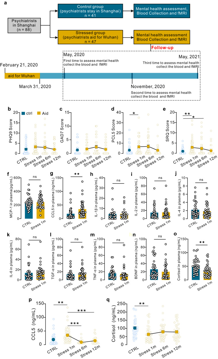
The study first examined psychiatrists who worked on the Wuhan front line in 2020. Questionnaires involved 47 stress-exposed participants and 41 controls. About one month after returning, the exposed group had higher post-traumatic stress-related and general psychological symptom scores, while depression and generalized-anxiety scales did not significantly differ between groups. The measures capture different aspects of the stressful experience.
Blood samples from 43 exposed participants and 38 controls showed higher CCL5 but lower cortisol in the exposed group; several other immune-related measures did not significantly differ. CCL5 fell during follow-up. This was a selective molecular clue rather than a uniform inflammatory rise. Because blood was first collected after the stressful work, individual pre-exposure blood baselines were unavailable.
Source: Shi DD et al. · Nature communications · CC BY
Original figure and English caption ↗
Why do cervical lymph nodes enter the story?
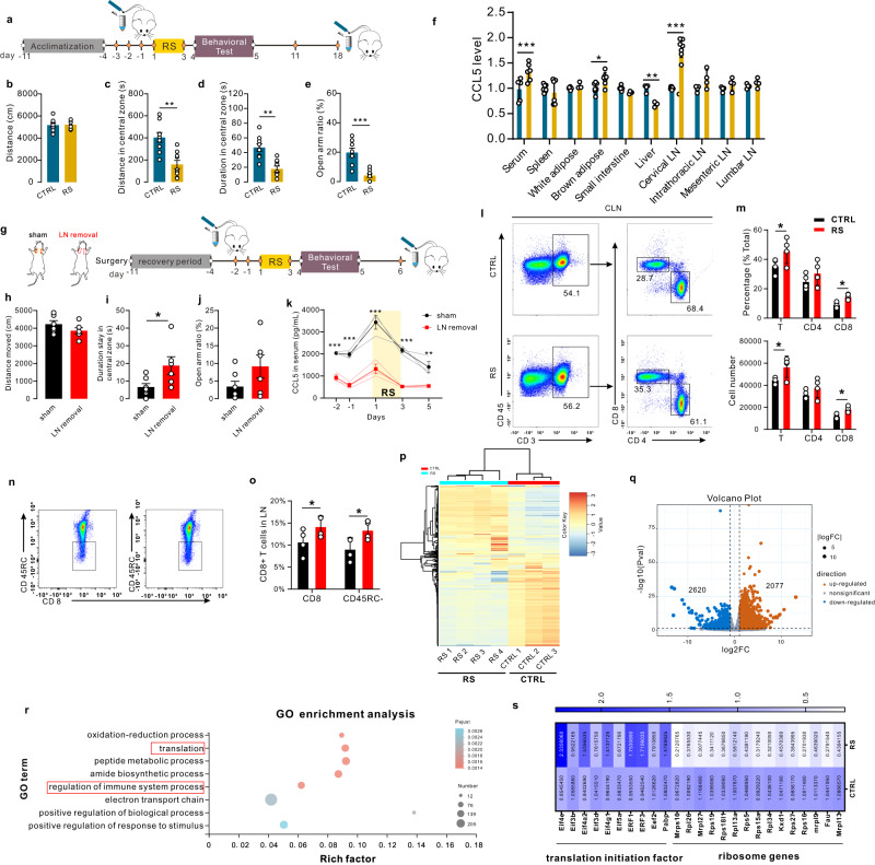
After brief restraint stress, male rats showed no significant change in overall distance traveled but explored the open-field center less and spent less time in exposed maze arms. Cervical lymph nodes showed particularly prominent CCL5 induction among the tissues tested, along with increased proportions and numbers of memory CD8 T cells, revealing tissue- and cell-associated immune changes.
Removing these nodes increased center time after stress and reduced the rise in blood CCL5. The open-arm trend was not significant, and a residual CCL5 rise remained. The nodes were therefore important contributors rather than the entire explanation. Female rats also showed increased CCL5 after stress without the same anxiety-like behavioral changes, so subsequent mechanistic work mainly used males.
Source: Shi DD et al. · Nature communications · CC BY
Original figure and English caption ↗
A familiar motor region reveals another role
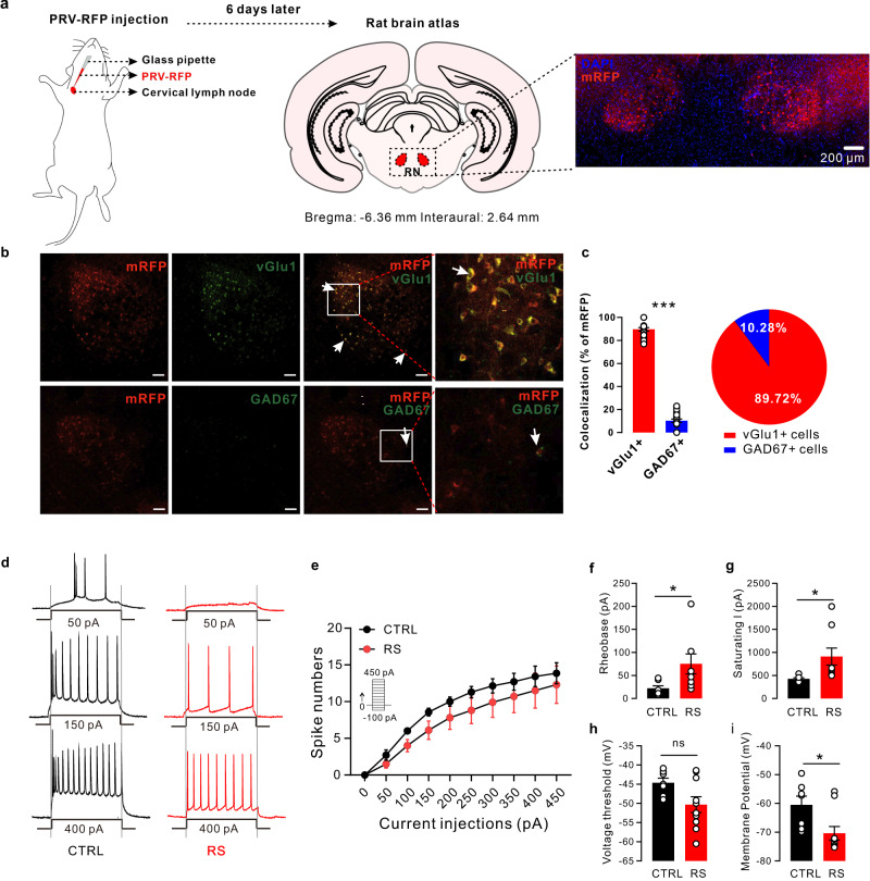
Tracing upstream networks associated with cervical lymph nodes identified the red nucleus, usually discussed in motor coordination. About nine in ten labeled cells there also carried a glutamatergic marker. The transsynaptic tracing established a network association and raised a new question: does stress alter how these cells work?
Recorded excitatory cells in stressed rats fired fewer spikes under current stimulation, required more current to begin firing, and had more negative resting membrane potentials. These findings point to reduced excitability. The node-to-brain labeling may involve multiple relays; it does not establish a single direct nerve connecting the red nucleus to lymph nodes.
Source: Shi DD et al. · Nature communications · CC BY
Original figure and English caption ↗
These cells respond during exploration
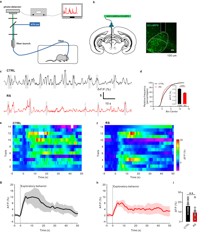
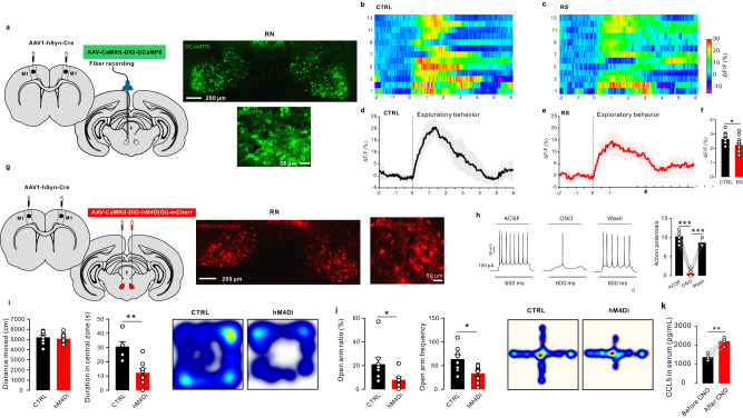
In freely moving rats, red nucleus population calcium signals changed with exploration. Control animals showed increased activity during exploration associated with the open-field center. After restraint stress, average fluorescence and exploration-related responses were lower, linking altered exploration to a weaker activity signal.
The recordings connect cellular state to particular behavioral moments. They show an association between population activity and exploration; whether changing these cells also alters behavior and peripheral immune signals is addressed by the interventions that follow.
Source: Shi DD et al. · Nature communications · CC BY
Original figure and English caption ↗
Changing activity shifts behavior and immune signals
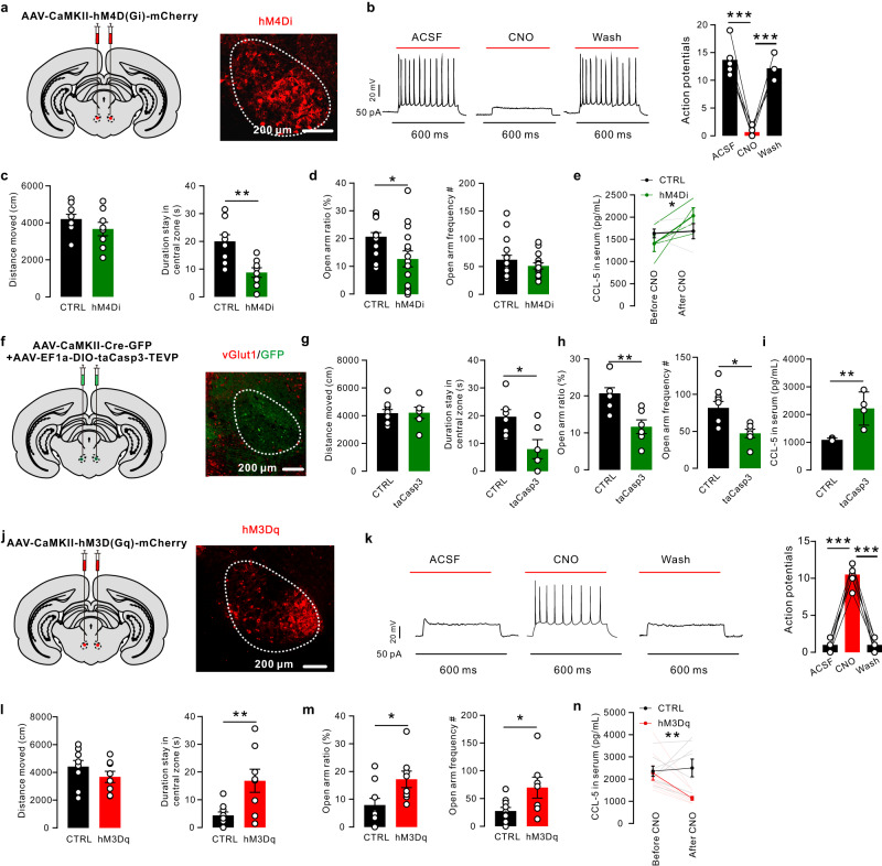
Inhibiting excitatory-related red nucleus cells reduced center time and open-arm ratio while increasing blood and lymph-node CCL5. Overall locomotion and open-arm entry frequency did not significantly change. A separate cell-ablation experiment also increased anxiety-like behavior and CCL5, strengthening the link between this population and peripheral responses.
Conversely, activating the red nucleus population in stressed animals increased center and open-arm exploration and lowered CCL5. Supplementary experiments found that systemic CCL5 neutralization attenuated selected behavioral changes induced by red nucleus inhibition. Neural activity and an immune signal thus enter the same causal investigation, without establishing cervical nodes as the only CCL5 source or CCL5 as the sole determinant of stress responses.
Source: Shi DD et al. · Nature communications · CC BY
Original figure and English caption ↗
What relays connect a motor network to immunity?
Primary motor cortex M1 also enters the network. Red nucleus cells labeled through M1 input relationships responded during exploration, with weaker responses after stress. Inhibiting these recipient cells reduced center and open-arm exploration and increased CCL5 without significantly changing overall locomotion, connecting a motor-associated network with exploratory and immune changes.
From a molecule in human blood to coordinated changes in rat brain and immune tissue, the study extends the story of stress across brain and body. The downstream relays from red nucleus to lymph nodes and the contributions of different immune cells remain to be resolved. Human observations provide the starting clue, while animal interventions outline a testable connection.
Source: Shi DD et al. · Nature communications · CC BY
Original figure and English caption ↗