How can gentle electroacupuncture calm post-infarction arrhythmias? A cortex-to-brainstem pathway
In a mouse model of post-myocardial infarction arrhythmia, low-intensity electroacupuncture modulates a motor cortex–zona incerta–medulla pathway to restrain sympathetic tone.
Myocardial infarction (MI) frequently triggers premature ventricular complexes (PVCs), which exacerbate ventricular remodeling and elevate the risk of sudden cardiac death. While sympathetic overactivation is a well-established driver of post-MI arrhythmias and clinical studies suggest that acupuncture at the Shenmen (HT7) acupoint can alleviate ischemic cardiac dysfunction, the central neural circuits linking somatic acupoint stimulation to cardiac sympathetic tone remain poorly understood. On December 18, 2025, Qing Yu, Rong-lin Cai, and colleagues at Anhui University of Chinese Medicine, together with Zhi Zhang's team at the University of Science and Technology of China, published a study titled "Electroacupuncture suppresses premature ventricular complexes occurring post-myocardial infarction through corticothalamic circuit" in Cell Reports (with Fan Zhang as first author). Combining trans-synaptic viral tracing, in vivo multichannel electrophysiology, fiber photometry, miniature microscope single-cell calcium imaging, and optogenetic/chemogenetic manipulations, the authors demonstrate that low-intensity (0.5 mA) electroacupuncture (EA) at HT7 suppresses hyperactive layer 5 glutamatergic neurons in the primary motor cortex (M1L5Glu), disinhibits ventral zona incerta GABAergic neurons (ZIvGABA) via an intrazonal microcircuit, and dampens rostral ventrolateral medulla (RVLM) sympathetic outflow to reduce post-MI PVCs.
Low-intensity (0.5 mA), but not high-intensity (3 mA), electroacupuncture at HT7 suppresses MI-induced hyperactivity of M1L5 glutamatergic neurons, acting through an M1L5Glu → dorsal zona incerta non-GABAergic (ZIdnon-GABA) → ventral zona incerta GABAergic (ZIvGABA) → RVLM pathway to lower cardiac sympathetic tone and norepinephrine levels, thereby suppressing PVCs and improving post-MI survival.
Contents · 5 sections
Low-Intensity (0.5 mA) Electroacupuncture at HT7 Reduces Sympathetic Tone and Suppresses Post-MI PVCs
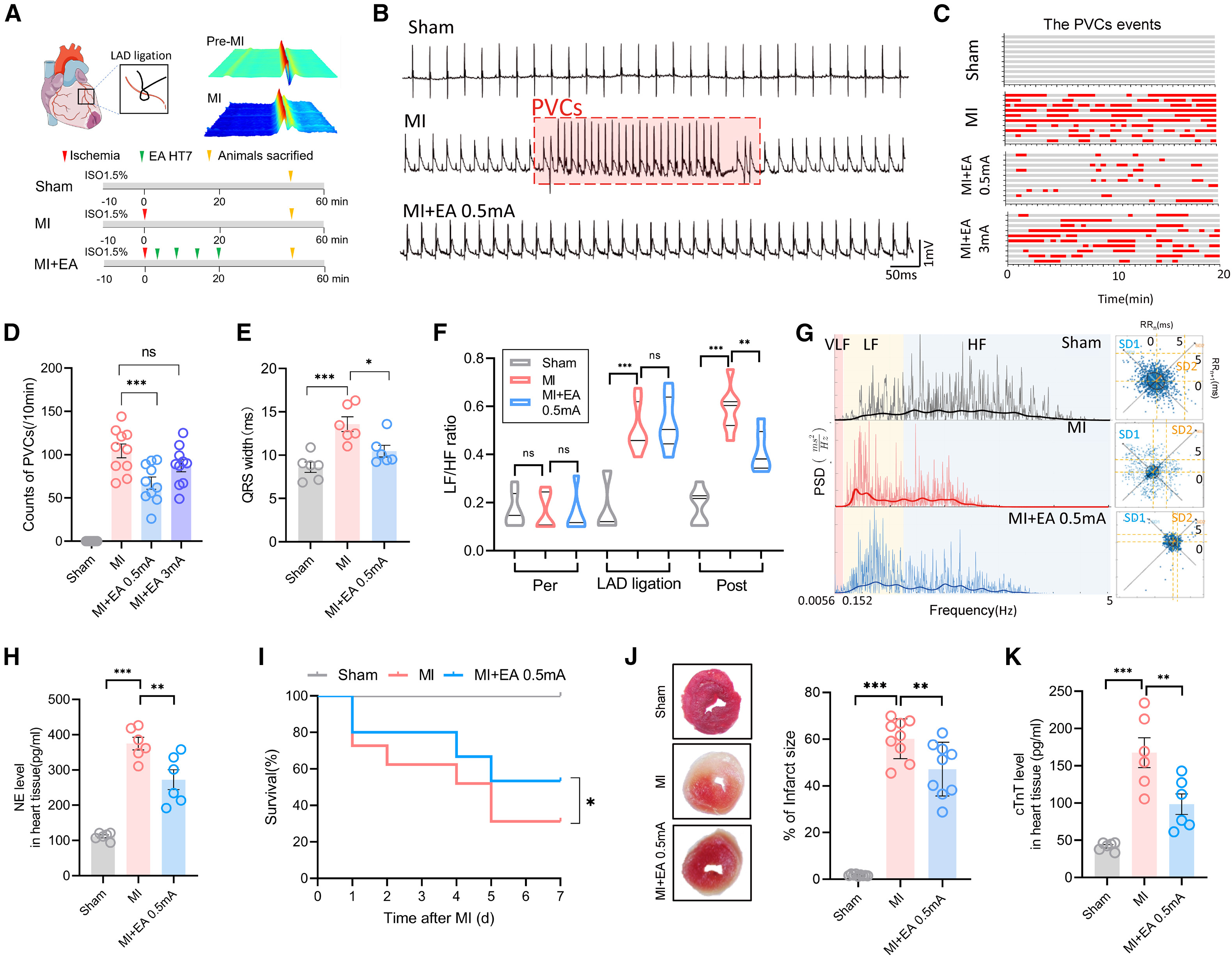
The authors first evaluated the anti-arrhythmic effects of 2 Hz electroacupuncture (EA) at the HT7 (Shenmen) acupoint—located on the ulnar side of the palmar wrist crease adjacent to the ulnar nerve—in male C57BL/6J mice subjected to left anterior descending (LAD) coronary artery ligation. In the acute paradigm (Figure 1A), mice received a single 20-minute EA session immediately following LAD ligation (0–20 min) and were sampled at 50 min post-ligation, while survival cohorts were monitored over 7 days. Electrocardiographic (ECG) recordings showed that LAD ligation induced frequent premature ventricular complexes (PVCs) and QRS widening. Low-intensity EA at 0.5 mA markedly reduced PVC incidence and counts per 10 min and normalized QRS duration, whereas sham EA (needle insertion without electrical current) or high-intensity 3 mA EA failed to effectively suppress PVCs.
Heart rate variability (HRV) power spectral density and Poincaré plot (SD1/SD2) analyses further revealed that the low-frequency to high-frequency power ratio (LF/HF)—an index of cardiac sympathovagal balance—and myocardial norepinephrine (NE) levels were markedly elevated in MI mice; both were significantly reduced by 0.5 mA EA at HT7 alongside improved systolic blood pressure (Figure S1F). Across the 7-day survival study, 0.5 mA EA maintained an 80.0% survival rate during days 1–3 and increased day-7 post-MI survival from ~31% in untreated MI mice to ~53.3%, while significantly reducing TTC-stained myocardial infarct size and cardiac troponin T (cTnT) levels.
Source: Fan Zhang et al. · Cell Reports · © Elsevier Inc.
Original figure and English caption ↗
Retrograde and Anterograde Trans-Synaptic Tracing Identifies Layer 5 Glutamatergic Neurons in M1
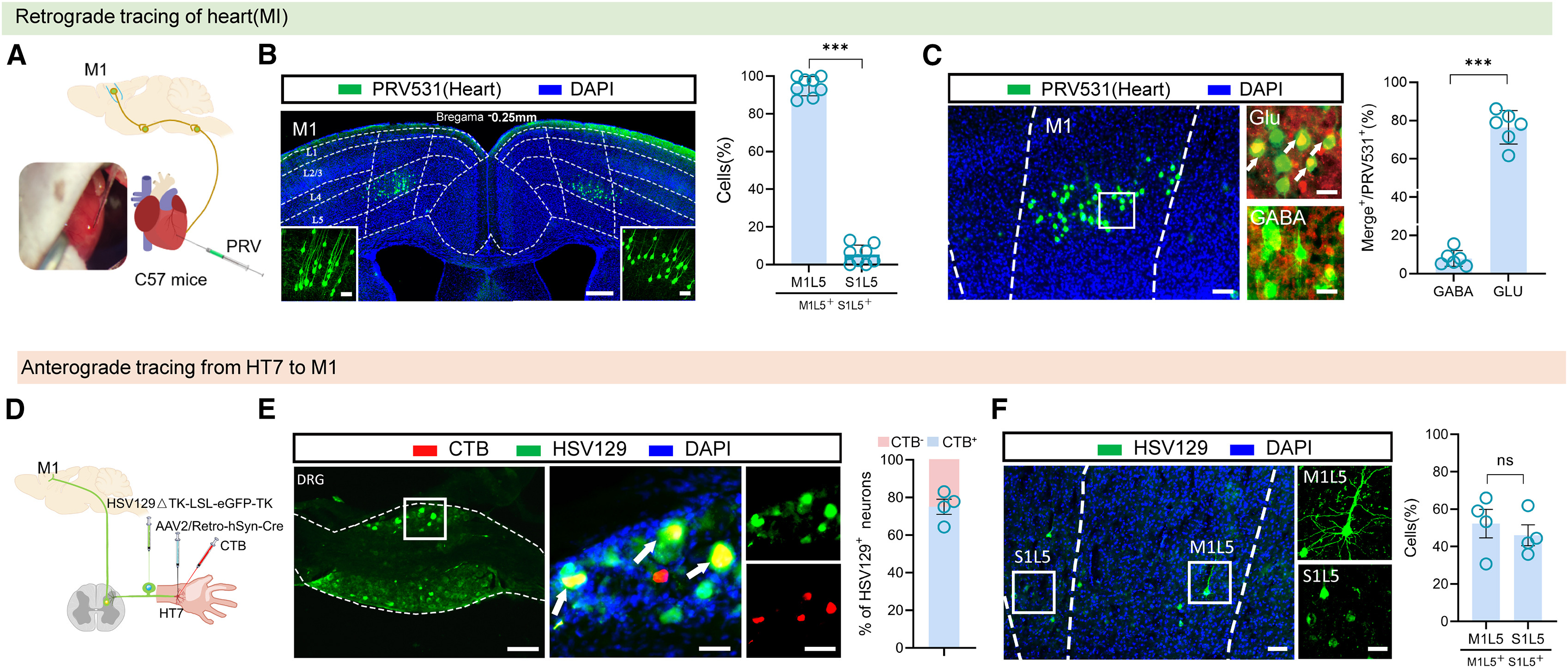
To identify cortical substrates linking cardiac autonomic control with HT7 stimulation, the researchers injected retrograde trans-multisynaptic pseudorabies virus (PRV531-EGFP) into the left ventricular wall. By day 6 post-injection, whole-brain mapping revealed dense EGFP-labeled neurons in the bilateral primary motor cortex (M1) in addition to canonical autonomic nuclei such as the RVLM, nucleus tractus solitarii (NTS), paraventricular nucleus (PVN), and periaqueductal gray (PAG). Cortical labeling was significantly higher in M1 than in the primary somatosensory cortex (S1), with approximately 95% of M1 EGFP+ neurons residing in layer 5 (M1L5). 3D Imaris reconstruction and immunofluorescence co-labeling confirmed that these heart-upstream M1L5 neurons exhibited pyramidal morphology and predominantly co-localized with glutamate (Glu) rather than GABA.
Conversely, to trace ascending pathways from the HT7 acupoint, cholera toxin subunit B (CTB) injection into HT7 predominantly labeled sensory neurons in the T3 dorsal root ganglion (DRG) and laminae I–IV of the T3 spinal cord. Injecting AAV2/retro-hSyn-Cre into HT7 followed by Cre-dependent anterograde trans-multisynaptic herpes simplex virus (HSV129ΔTK-LSL-EGFP-TK) into the T3 DRG labeled recipient pyramidal neurons in M1L5 and S1L5, establishing M1L5 glutamatergic neurons as an anatomical convergence hub for HT7 afferent input and cardiac descending control.
Source: Fan Zhang et al. · Cell Reports · © Elsevier Inc.
Original figure and English caption ↗
Electroacupuncture Suppresses PVCs by Inhibiting MI-Induced Hyperactivity of M1L5 Glutamatergic Neurons
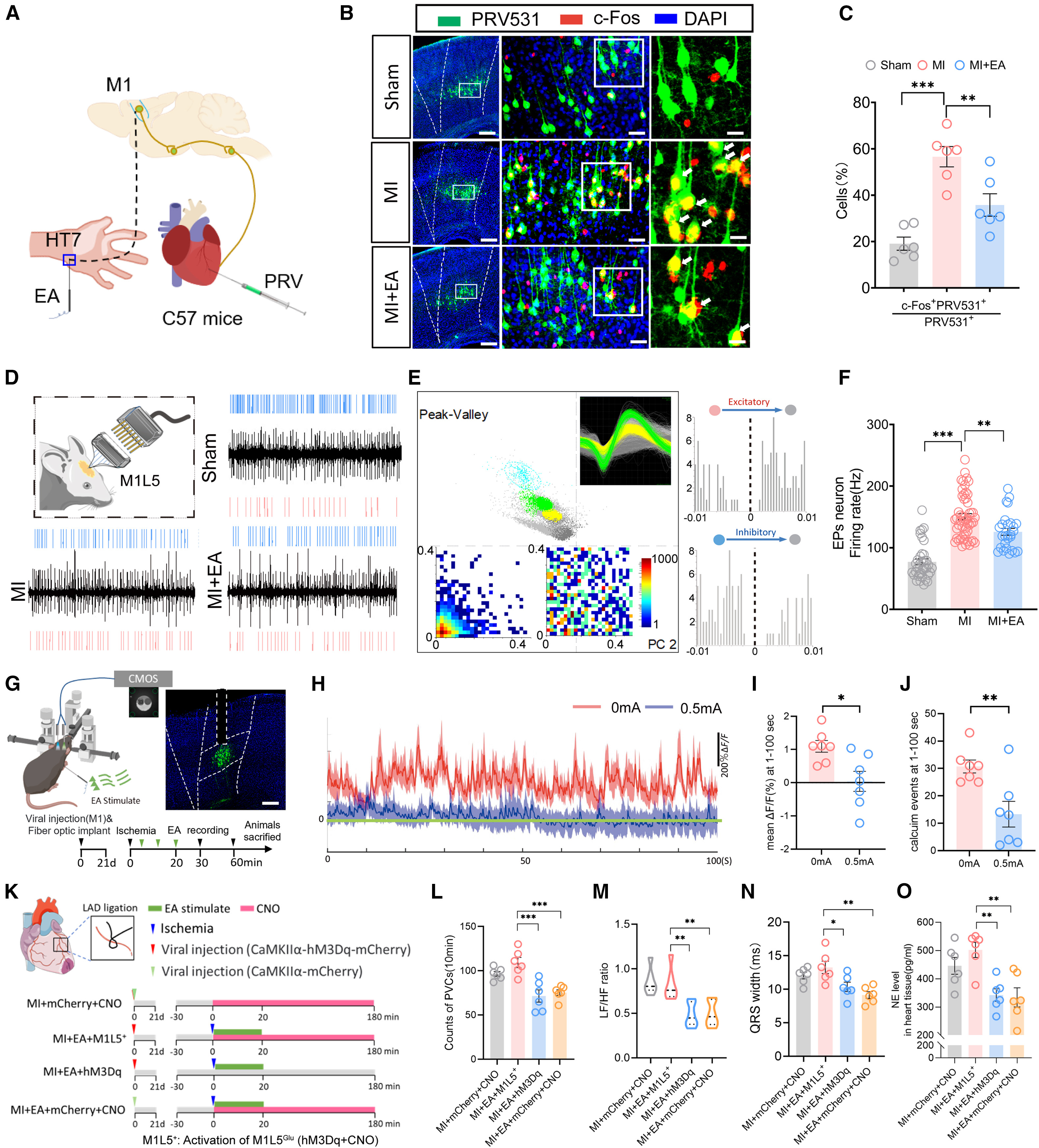
Further experiments showed that MI markedly increased c-Fos expression in tyrosine hydroxylase-positive (TH+) sympathetic premotor neurons in the RVLM, which was reversed by 0.5 mA EA at HT7; in naive mice, chemogenetic (hM3Dq) or optogenetic (ChR2, 20 Hz) activation of M1L5Glu neurons increased RVLM c-Fos expression, heart rate, LF/HF ratio, and cardiac NE levels (Original Figure 3). In MI mice, PRV531-labeled heart-upstream M1L5 neurons exhibited elevated c-Fos co-localization. Both in vivo multichannel electrophysiological recordings (154 putative excitatory pyramidal neurons, Eps) and AAV-CaMKIIα-GCaMP6m fiber photometry demonstrated that MI significantly increased M1L5Glu spike firing rates and calcium activity, which were suppressed by 0.5 mA—but not 3 mA—EA at HT7.
To test whether M1L5Glu suppression is required for the anti-arrhythmic effect of EA, the authors expressed AAV-CaMKIIα-hM3Dq-mCherry bilaterally in M1L5 and administered CNO prior to 0.5 mA EA. Chemogenetic activation of M1L5Glu neurons completely abolished EA-mediated reductions in PVC frequency, LF/HF ratio, QRS duration, and cardiac NE content in MI mice, demonstrating that suppression of M1L5Glu hyperactivity is necessary for the cardioprotective action of HT7 EA.
Source: Fan Zhang et al. · Cell Reports · © Elsevier Inc.
Original figure and English caption ↗
M1L5 Glutamatergic Projections Inhibit Ventral Zona Incerta GABAergic Neurons via Dorsal Non-GABAergic Interneurons
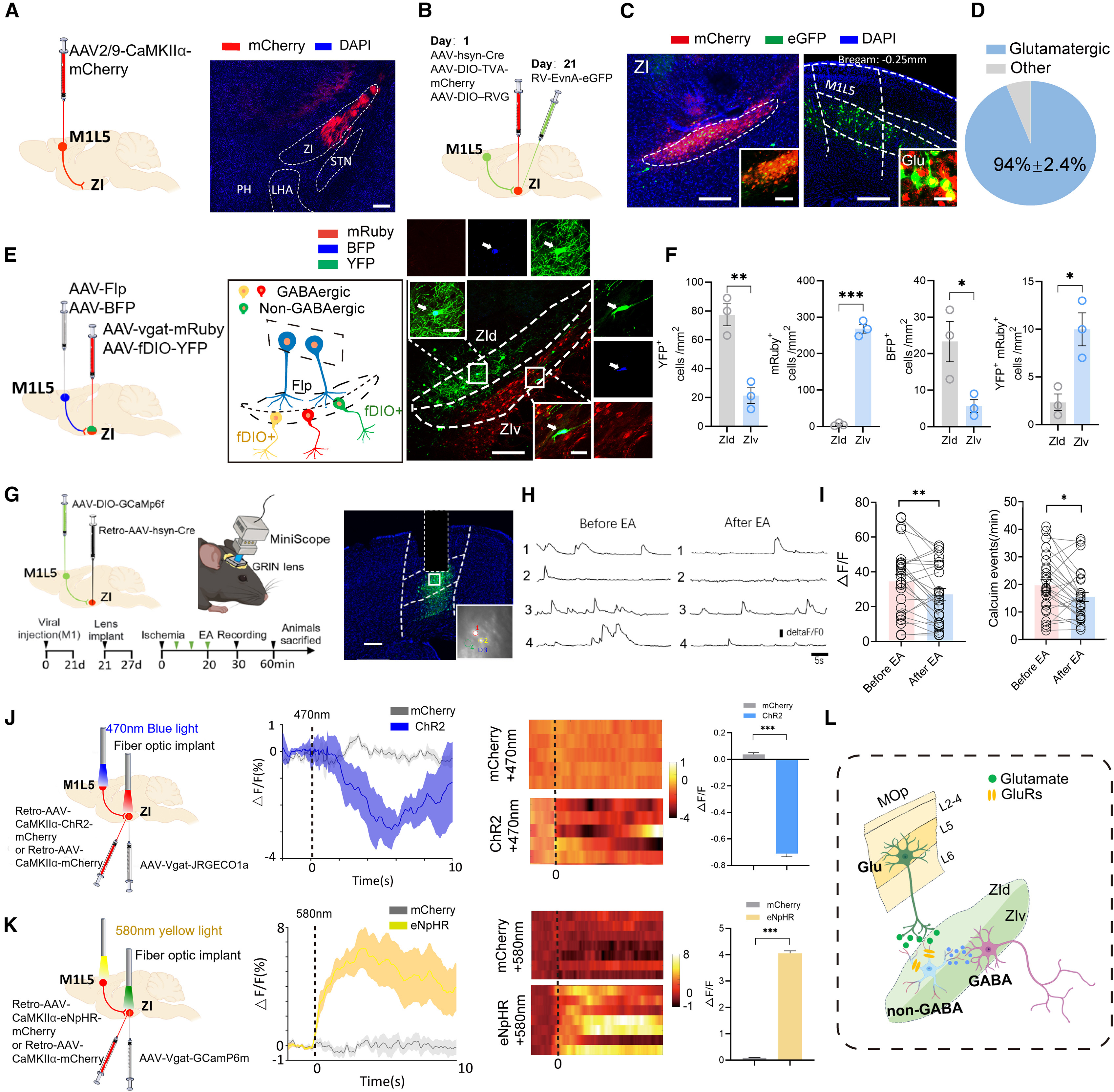
How do M1L5Glu neurons regulate the medullary RVLM? Anterograde axonal tracing (AAV-CaMKIIα-ChR2-mCherry) and monosynaptic rabies tracing (RV-EnvA-ΔG-EGFP) revealed that M1L5Glu neurons do not project directly to the RVLM, but instead send dense projections to the zona incerta (ZI) and ventrolateral periaqueductal gray (vlPAG). Optogenetic terminal stimulation showed that activating M1L5Glu→ZI projections—unlike M1L5Glu→vlPAG projections—markedly increased RVLM c-Fos expression, cardiac LF/HF ratio, and myocardial NE content (Figure 5A–C).
Anatomically, the ZI comprises a dorsal division enriched in non-GABAergic neurons (ZId) and a ventral division rich in GABAergic neurons (ZIv). Monosynaptic rabies tracing and anterograde trans-synaptic labeling (AAV1-hSyn-Flpo in M1L5 plus AAV-fDIO-EGFP and AAV-mDlx-mCherry in ZI) demonstrated that M1L5Glu axons preferentially innervate ZId non-GABAergic neurons (ZIdnon-GABA) that project locally to ZIvGABA neurons (Figure 5D–F). Single-cell calcium imaging using a head-mounted miniature microscope (tracking n = 26 ZI-projecting M1L5 neurons) confirmed that MI elevated calcium activity in ZI-projecting M1L5 neurons whereas 0.5 mA EA at HT7 suppressed their activity (Figure 5G–I). Moreover, fiber photometry showed that optogenetic activation (ChR2) of M1L5Glu→ZI terminals suppressed ZIvGABA JRGECO1a calcium signals, whereas optogenetic inhibition (eNpHR, 580 nm) of M1L5Glu→ZI terminals disinhibited and elevated ZIvGABA GCaMP6m calcium signals (Figure 5J–L).
Source: Fan Zhang et al. · Cell Reports · © Elsevier Inc.
Original figure and English caption ↗
ZI GABAergic Projections to the RVLM Mediate Sympathoinhibition and Rescue EA Efficacy
Anterograde monosynaptic HSV129-ΔTK tracing and retrograde monosynaptic rabies tracing from the RVLM demonstrated that ZIvGABA neurons project directly to the RVLM. Fiber photometry (AAV-Vgat-GCaMP6m) and activity-dependent Fos-TRAP labeling (in which 87.77% ± 2.97% of EA-trapped mCherry+ neurons co-localized with GABA) confirmed that 0.5 mA EA at HT7 robustly activated ZIvGABA neurons in MI mice; chemogenetic inhibition of ZIvGABA neurons abolished EA's anti-PVC effect, whereas activating the ZIvGABA→RVLM pathway mimicked cardioprotection (Original Figure 6).
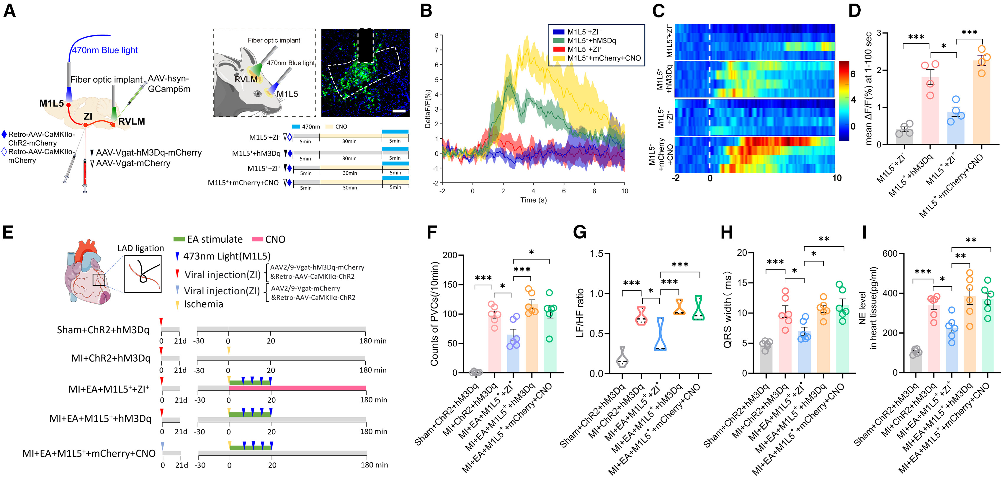
To establish causal necessity across the complete trisynaptic pathway, the authors combined optogenetic stimulation of M1L5Glu→ZI terminals with simultaneous chemogenetic activation of ZIGABA neurons (AAV-Vgat-hM3Dq-mCherry) and fiber photometry recording of RVLM GCaMP6m calcium signals in MI mice receiving 0.5 mA EA. Optogenetic activation of M1L5Glu→ZI elevated RVLM neuronal calcium activity and abrogated EA-induced reductions in PVCs, LF/HF ratio, QRS width, and cardiac NE; crucially, concurrent chemogenetic activation of ZIGABA neurons suppressed RVLM calcium signals and restored the anti-arrhythmic and sympathoinhibitory effects of EA, establishing the M1L5Glu → ZIdnon-GABA → ZIvGABA → RVLM circuit as a central pathway mediating electroacupuncture suppression of post-MI PVCs.
Source: Fan Zhang et al. · Cell Reports · © Elsevier Inc.
Original figure and English caption ↗