Pulmonary signals in pain modulation: a conversation between cells and vagal neurons
Mesenchymal stromal cells reduce nerve-injury-associated sensitivity in mice. Researchers trace their effects to pulmonary Npy2r sensory neurons and a midbrain network involved in pain modulation.
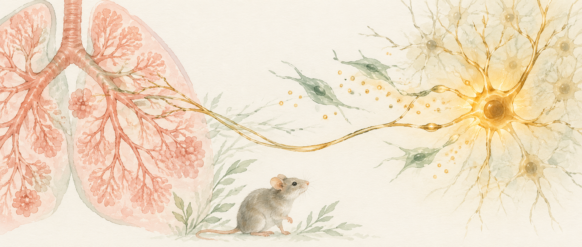
Changes in pain can begin somewhere other than the injured site. A study in Advanced Science finds that human-derived mesenchymal stromal cells delivered to mice interact with pulmonary sensory nerves, while a brain region involved in pain modulation responds. Alongside gas exchange, the lung can serve as an interface carrying cellular signals into body–brain sensory regulation.
Pulmonary Npy2r sensory neurons contribute to the pain-reducing effects of stromal cells. PANX1-associated ATP release and neuronal purinergic responses provide a molecular link for this lung–brain communication.
Contents · 6 sections
Responses to touch and heat change
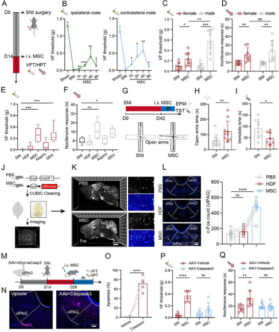
In the spared nerve injury model, mice have low mechanical withdrawal thresholds. These thresholds rise after stromal cells are delivered, and response latency to heat increases. A fibroblast control does not produce the same clear effect. The study examines responses over hours and follows their subsequent course.
Improvement is evident during the first few days but is no longer significant by day seven. Mechanical improvement is greater in males than females, whereas heat-response improvement is comparable. Motor coordination is not measurably impaired, and some anxiety-like and immobility readouts also improve. The effects have a time window, with sex differences in particular sensory readouts.
Supplementary observations also show improved sensory responses in chemotherapy-associated neuropathy and inflammatory pain models. Similar directions across models raise the possibility that the cells engage an existing pain-modulating system. The subsequent circuit analysis focuses on nerve injury to trace the source of this effect.
Source: Huang J et al. · Advanced science (Weinheim, Baden-Wurttemberg, Germany) · CC BY
Original figure and English caption ↗
A pain-modulating region becomes active
Responses occur in several brain regions, with the ventrolateral periaqueductal gray, or vlPAG, particularly prominent. This midbrain region contributes to descending pain modulation. Neuronal activity markers and population calcium signals increase after stromal cells. Removing its neurons eliminates the mechanical and thermal improvement, identifying an important node in the effect.
An explanation focused only on inflammation could miss this neural connection. At the later nerve-injury stage studied here, the measured serum inflammatory marker does not improve clearly. The team therefore asks how cells in the body can engage a distant midbrain response.
Vagal sensory nerves connect lung and brainstem
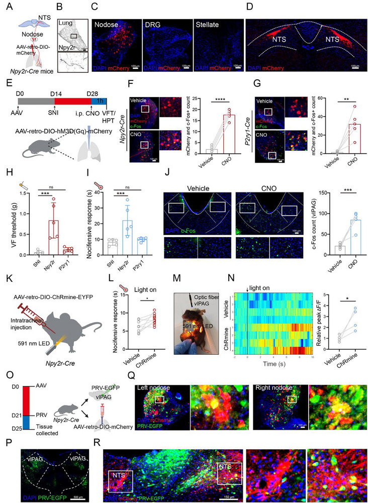
Tracking shows that delivered stromal cells mainly accumulate in the lungs, near sensory nerve fibers. Pulmonary signals can travel through vagal afferents to the nucleus tractus solitarius and connect with a network reaching the periaqueductal gray. Vagal sensory ganglia, the brainstem relay and the midbrain all show increased activity after stromal cells.
Pulmonary sensory nerves contain different populations. Increasing Npy2r neuron activity improves mechanical and heat responses and increases midbrain activity, whereas increasing P2ry1 neuron activity does not have the same effect. Which neurons carry bodily information matters for the resulting sensory change.
Source: Huang J et al. · Advanced science (Weinheim, Baden-Wurttemberg, Germany) · CC BY
Original figure and English caption ↗
Disrupting this link weakens the cellular effect
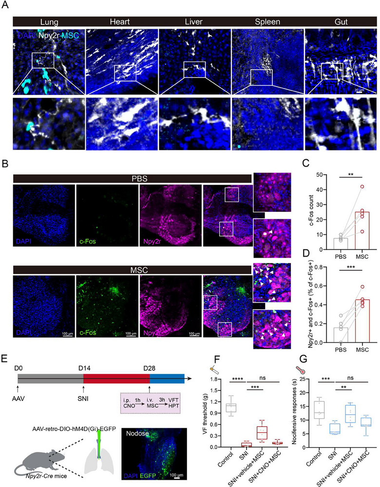
Stromal cells lie near pulmonary Npy2r fibers, while activity markers and calcium responses increase in the associated sensory neurons. Reducing the activity of these pulmonary neurons suppresses the mechanical and thermal improvement produced by stromal cells. Proximity, dynamic responses and behavioral changes connect the two.
When cells and neurons are observed together, neuronal calcium responses can persist for hours. The cells appear to signal into an existing network rather than replace injured neural tissue. The lung offers a meeting place where cellular signals can engage the nervous system.
Source: Huang J et al. · Advanced science (Weinheim, Baden-Wurttemberg, Germany) · CC BY
Original figure and English caption ↗
ATP links cellular signals to neuronal responses
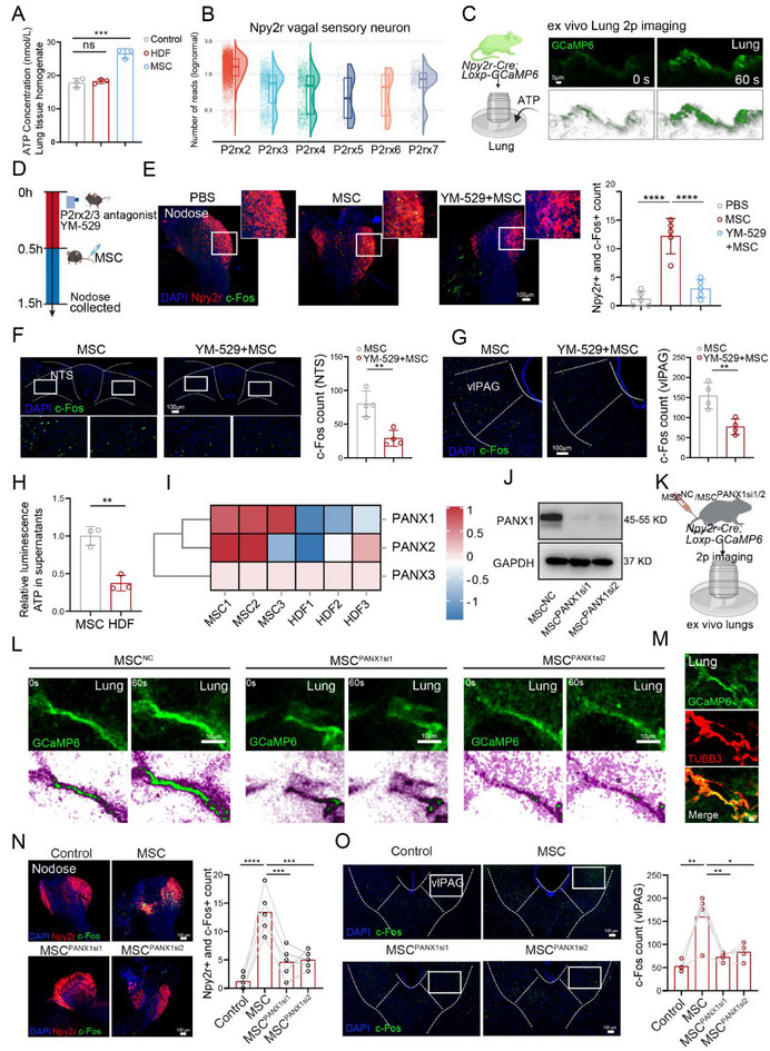
ATP is familiar as an energy carrier, but it also communicates outside cells. ATP levels in lung tissue increase after stromal cells, and ATP can increase calcium responses in Npy2r fibers. Reducing associated purinergic receptor signaling weakens activity increases in sensory ganglia, brainstem and midbrain.
The team then examines the PANX1 channel in stromal cells. Lowering its expression reduces ATP release, Npy2r responses and subsequent brain activity, and weakens the pain-reducing effects. Cellular release, neuronal reception and central responses form a converging set of findings.
Source: Huang J et al. · Advanced science (Weinheim, Baden-Wurttemberg, Germany) · CC BY
Original figure and English caption ↗
Pulmonary sensing can change distant sensory responses
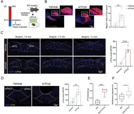
The study also examines the ATP analog ATPγS. In mice it increases activity in the associated sensory neurons and brain regions and improves mechanical and thermal responses after nerve injury. It also increases heat-response latency in uninjured mice. The finding moves the question from which cells produce an effect to which signals can engage the network.
The new insight is that cell–neuron communication in the lung can participate in sensory regulation: chemical signals at one bodily site alter stimulus responses elsewhere. The evidence comes from mice, mostly over short observation windows, and includes sex differences. Lung–brain communication emerges as a traceable mechanism in pain biology.
Source: Huang J et al. · Advanced science (Weinheim, Baden-Wurttemberg, Germany) · CC BY
Original figure and English caption ↗