From one day to four weeks of pain: why does the spleen change direction?
After nerve injury in male mice, splenic TH2-associated cell proportions rise early and fall later. The study links these stages to distinct inputs from somatosensory cortex and central amygdala through a brainstem–spleen connection.
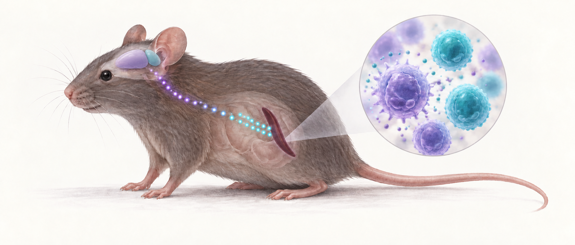
Pain may leave a story outside the nervous system. As a paw becomes more sensitive to light touch, immune-cell proportions in the distant spleen can change too. Intriguingly, some proportions move in opposite directions one day and four weeks after nerve injury. A study published in Nature Neuroscience on January 30, 2024 traces this timing difference into the brain: distinct inputs from somatosensory cortex and central amygdala converge on brainstem cells linked to the spleen, suggesting a changing brain–immune dialogue.
In this mouse model, early and later splenic TH2-associated responses are linked to enhanced cortical excitation and amygdalar inhibition, respectively. The inputs converge in the dorsal motor nucleus of the vagus and correspond to different stages of hypersensitivity.
Contents · 6 sections
Persistent sensitivity does not mean immune cells always move together
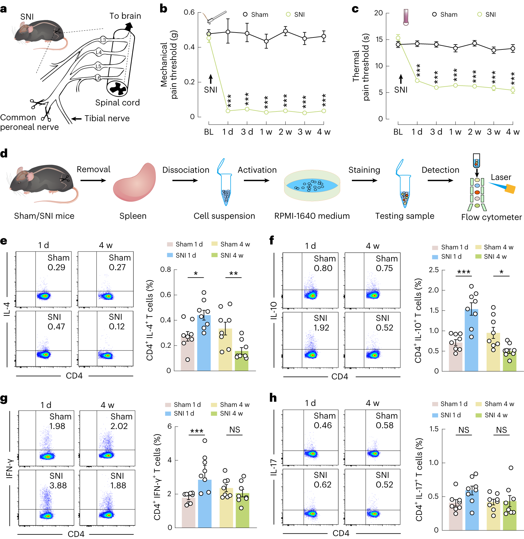
The team follows sensory responses and splenic cells in male mice after nerve injury. Mechanical withdrawal thresholds and withdrawal latencies from heat decrease early and remain reduced at four weeks. Lighter pressure and shorter heat exposure can therefore evoke responses throughout the period being compared.
Immune-cell proportions show a different pattern. CD4-positive cells with IL-4 or IL-10 markers, associated with the TH2 response examined here, increase at one day and decrease at four weeks. The early increase in IFN-γ-associated proportions is not significant in the four-week comparison, while IL-17-associated proportions show no significant change at either time. These are distinct cell-population responses, rather than uniform movement of the whole immune system.
Source: Xia Zhu et al. · Nature Neuroscience ·
Original figure and English caption ↗
Tracing back from the spleen reveals a brainstem relay
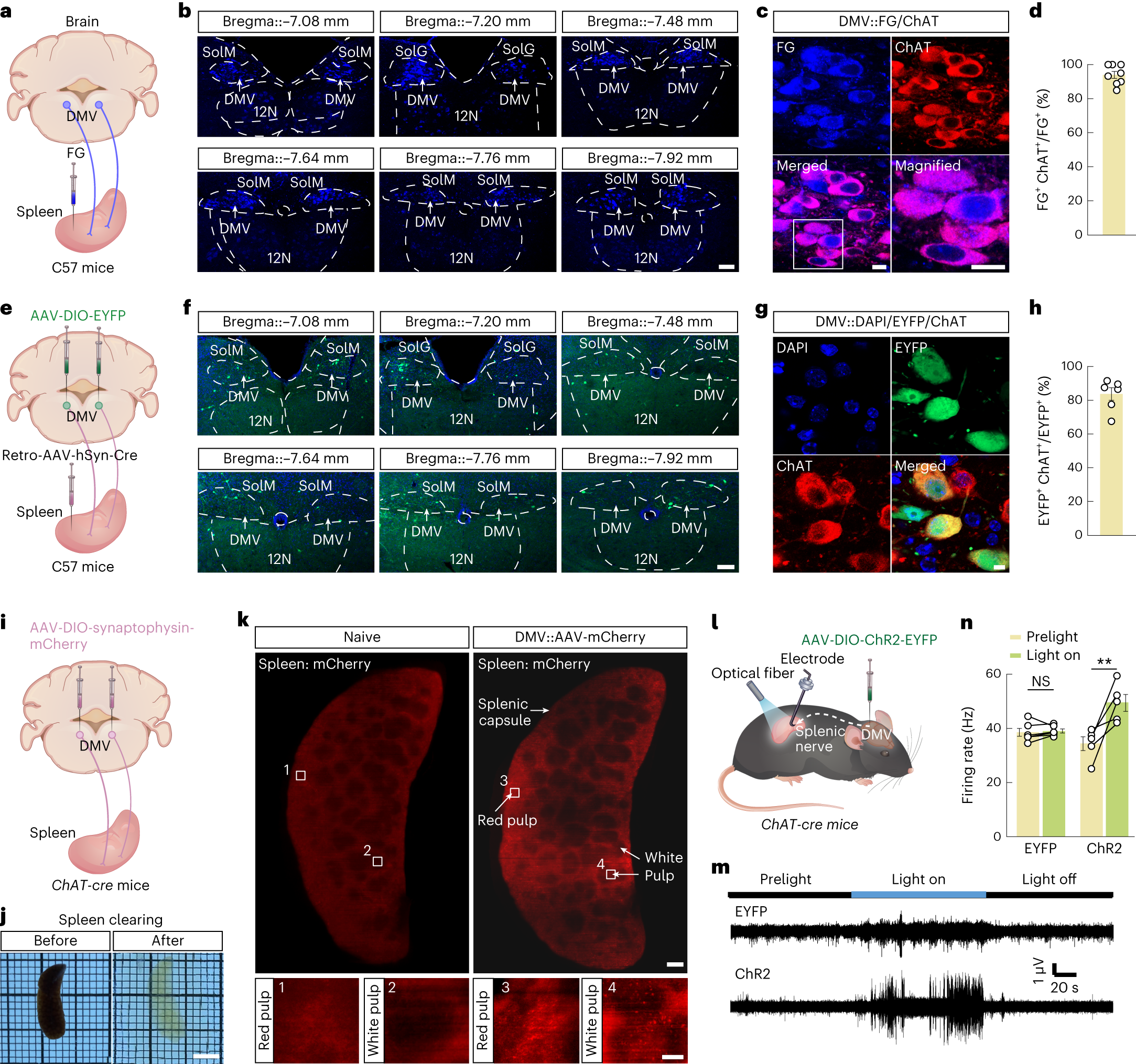
How might the brain influence the spleen? The study points to the dorsal motor nucleus of the vagus (DMV). Retrograde labeling from the spleen predominantly overlaps cholinergic markers in DMV, while labeling initiated in DMV appears in splenic tissue. Tracing in both directions makes the brainstem–organ connection more concrete.
The link also has a functional response: activating splenic terminals associated with DMV cholinergic cells increases recorded splenic-nerve activity, whereas the control group shows no corresponding significant change. The findings connect brainstem cells, peripheral neural activity and spleen, raising the question of how those cells behave at different pain stages.
Source: Xia Zhu et al. · Nature Neuroscience ·
Original figure and English caption ↗
At one day, the brainstem cells become more active
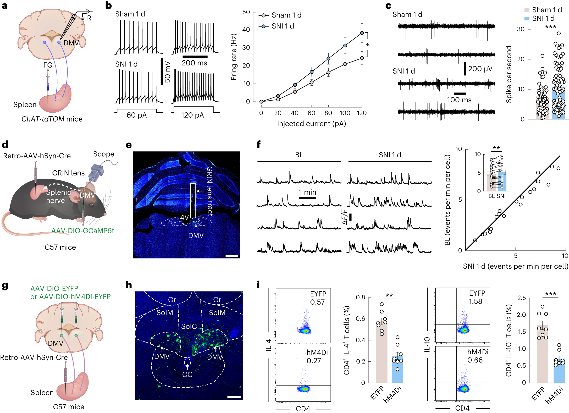
One day after injury, spleen-linked DMV cells fire more readily during current injection, and related cells recorded in freely moving mice also fire more. Calcium-event tracking of observable cells shows more frequent events. Different readouts thus converge on increased brainstem activity in the early stage.
Inhibiting spleen-related DMV projection cells lowers the splenic proportions of CD4/IL-4 and CD4/IL-10 double-labeled cells. Manipulation strengthens the link between the neural and immune changes. Changes in brainstem output can thus alter the cellular composition captured by these immune markers.
Source: Xia Zhu et al. · Nature Neuroscience ·
Original figure and English caption ↗
By four weeks, brainstem and splenic changes turn the other way
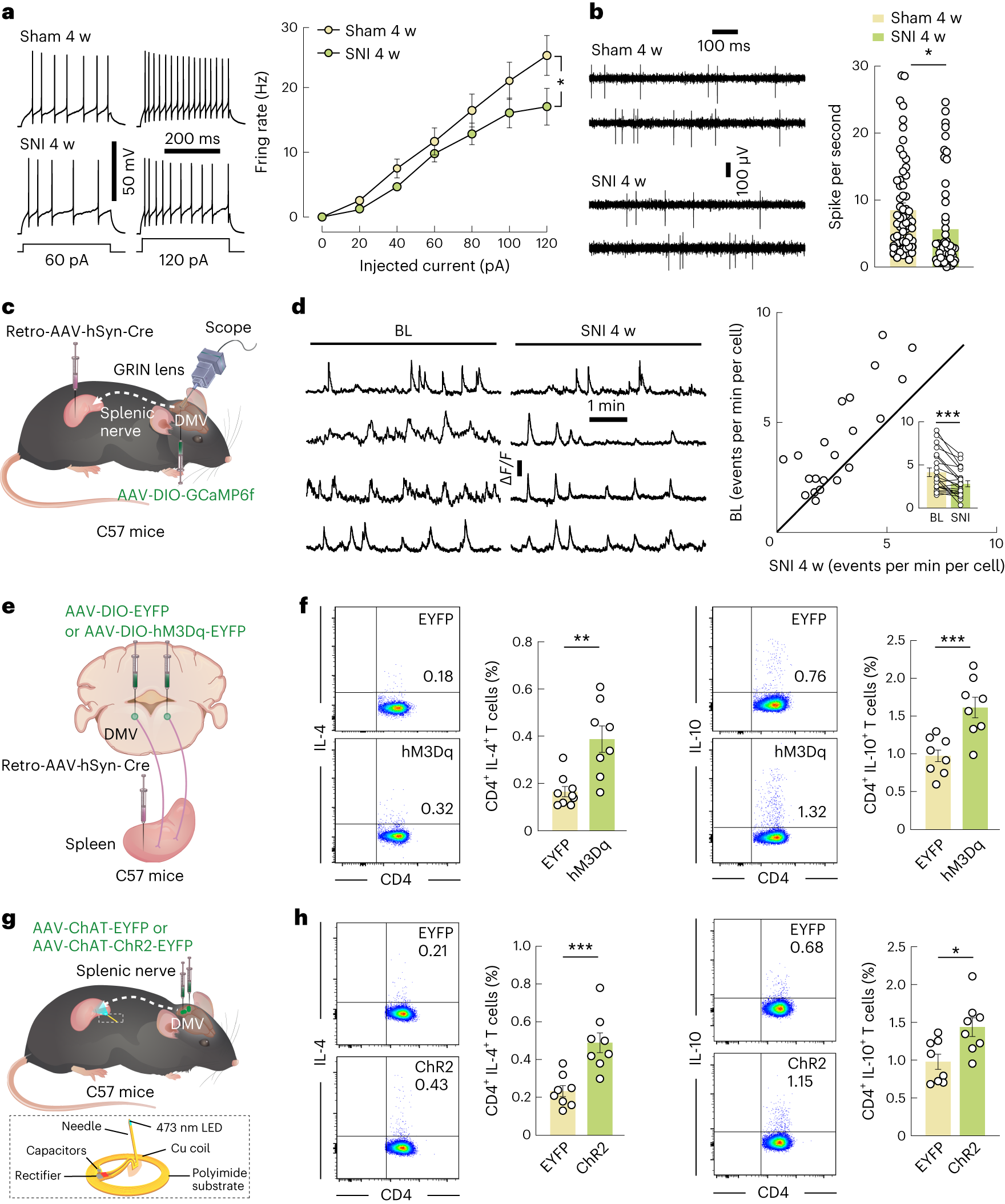
At four weeks, the same brainstem node moves in the opposite direction. Related DMV cells show less firing during current injection, lower in-vivo firing and fewer calcium events. This contrasts with the early active state and accompanies the change from increased to decreased TH2-associated splenic proportions.
Activating spleen-linked DMV projection cells now increases CD4/IL-4 and CD4/IL-10 double-labeled proportions. Local activation of associated cholinergic terminals in the spleen produces the same direction of change. Intervening at the cells and peripheral terminals supports adjustable output, even though hypersensitivity persists across the two stages.
Source: Xia Zhu et al. · Nature Neuroscience ·
Original figure and English caption ↗
Cortex supplies excitation and amygdala supplies inhibition
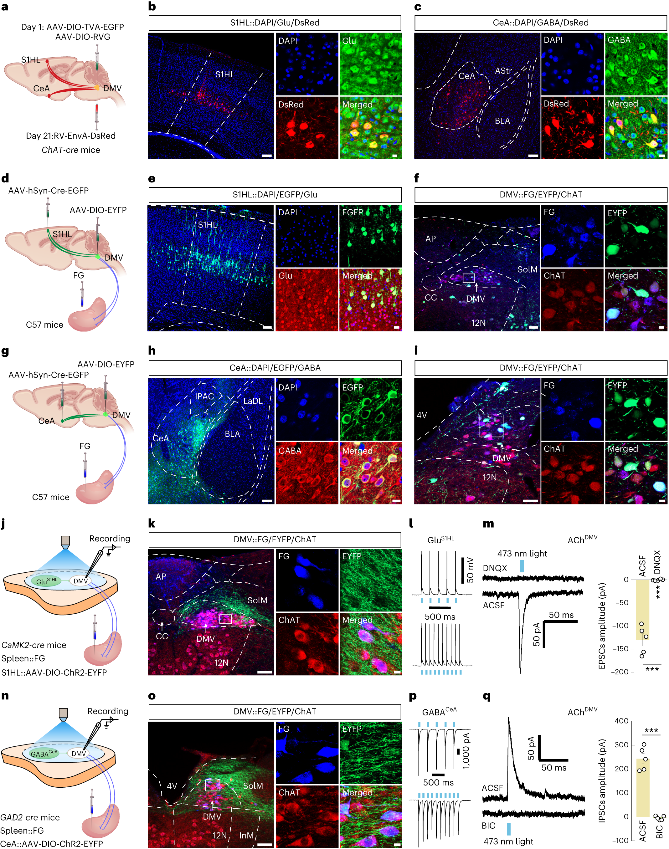
If DMV is the relay, which upstream signals change? The study identifies the hindlimb region of primary somatosensory cortex (S1HL) and central amygdala (CeA). Glutamate-associated S1HL cells supply excitation to DMV, while GABA-associated CeA cells supply inhibition. Labeling and local current responses support distinct influences on the same brainstem region.
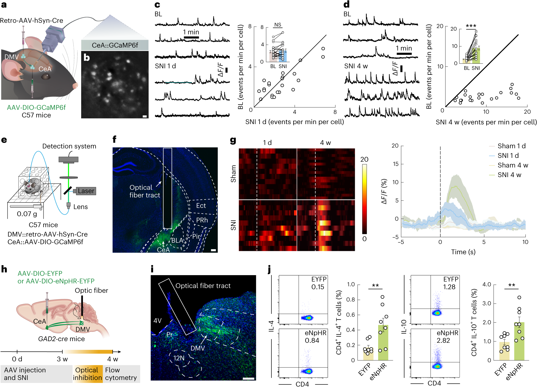
The pathways have different timing. Calcium signals in relevant S1HL projection cells increase at one day, while the corresponding four-week comparison is not significant. CeA projection-cell calcium-event comparisons are nonsignificant at one day and increase at four weeks. Timing becomes central to the circuit: more prominent early excitation and later inhibition can influence the shared output differently.
Source: Xia Zhu et al. · Nature Neuroscience ·
Original figure and English caption ↗
Releasing later inhibition changes splenic proportions
At four weeks, suppressing CeA terminals in DMV increases the two TH2-associated double-labeled proportions in the spleen. This agrees in direction with direct activation of DMV projection cells: reducing upstream inhibition or increasing relay output changes the measured splenic response. The pain-associated brain–organ connection is therefore adjustable.
The appealing insight is that the duration of pain matters to brain–immune mechanisms. The same nerve injury has different upstream inputs and peripheral cell proportions at different times, rather than a simple strengthening of every response. In male mice, the study links stages of hypersensitivity with upstream neural activity and selected immune-cell proportions.
Source: Xia Zhu et al. · Nature Neuroscience ·
Original figure and English caption ↗