How does prolonged sleep loss trigger systemic inflammation? Following brain-derived prostaglandin D2 to the periphery
A mouse study shows that prolonged sleep deprivation promotes efflux of brain-derived prostaglandin D2 across the blood-brain barrier via ABCC4, driving neutrophil accumulation and systemic inflammation.
Why is sleep indispensable for mammalian survival? Although experiments over a century ago showed that prolonged total sleep deprivation is lethal in animals, how sleep loss inside the brain crosses central boundaries to cause systemic failure has remained a central mystery. Conventional mouse sleep-deprivation setups are often blunted by strong homeostatic sleep rebound. In a study published online in Cell on November 27, 2023, Eric Erquan Zhang and colleagues at the National Institute of Biological Sciences (NIBS), together with Yulong Li and collaborators at Peking University, developed a curling prevention by water (CPW) paradigm for mice and uncovered a brain-to-periphery molecular pathway: prolonged sleep deprivation accelerates blood-brain barrier efflux of brain-derived prostaglandin D2 (PGD2) via the transporter ABCC4, activating DP1 signaling to trigger a neutrophil-driven cytokine-storm-like syndrome.
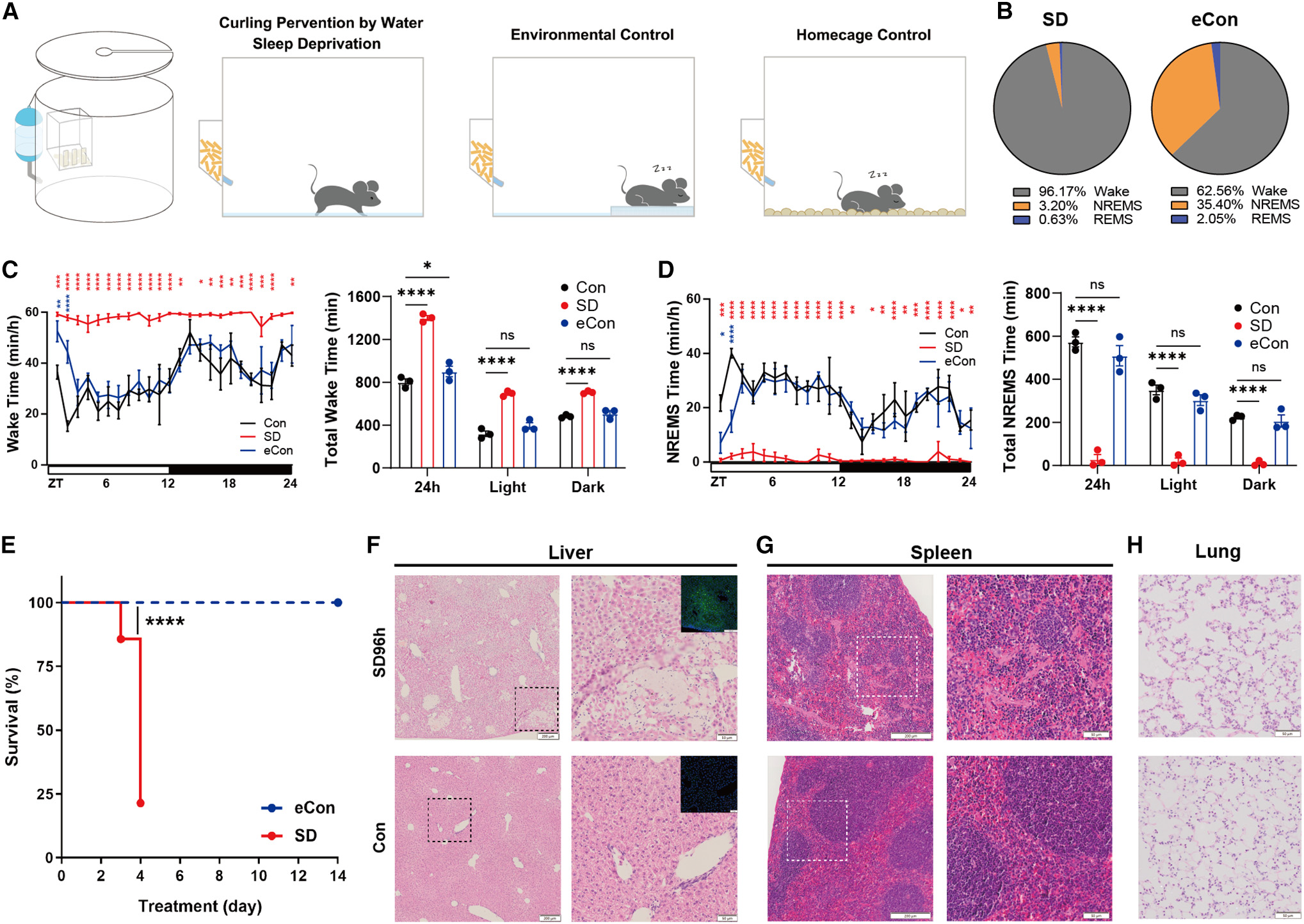
The curling prevention by water (CPW) paradigm maintains wakefulness for ~96.2% of the 24-hour cycle in mice; rather than acting through the sympathetic nervous system, prolonged sleep deprivation elevates brain PGD2 and enhances ABCC4-mediated blood-brain barrier efflux, activating peripheral DP1 receptors to drive circulating neutrophil accumulation and proinflammatory cytokine surges—effects markedly attenuated by Ptgds or Abcc4 deletion or DP1 blockade.
Contents · 5 sections
Maintaining 96% wakefulness: prolonged sleep deprivation triggers multi-organ neutrophil infiltration and cytokine storm
Mice typically adopt a curled, head-lowered posture when falling asleep. Exploiting this behavior, the authors designed the curling prevention by water (CPW) apparatus with an ~8-mm shallow layer of water that permits free locomotion and feeding while preventing curled sleep, alongside an environmental control (eCon) featuring a raised wet, gauze-covered platform and a homecage control (Con). Continuous EEG/EMG recordings confirmed that CPW mice remained awake for 96.17% of the 24-hour cycle (with only 3.20% NREMS and 0.63% REMS), whereas eCon mice experienced only a minimal daily sleep loss (99 min or 16%, restricted to ZT0-ZT1 at cage switching) with 24-hour NREMS and light/dark sleep architecture comparable to homecage controls.
When continuous sleep deprivation extended to days 3-4, mice suffered premature mortality accompanied by multiple organ dysfunction syndrome (MODS) and extensive neutrophil infiltration in the liver, spleen, and lungs, whereas all eCon mice survived. Serum profiling at 12-48 hours revealed surging proinflammatory cytokines and chemokines—including IL-6, IL-17A, CXCL1, and CCL20—and oral treatment with corticosterone or acetaminophen (APAP), or testing immunodeficient M-NSG mice lacking multiple cytokine pathways, significantly improved survival.
Source: Di Sang et al. · Cell · © 2023 Elsevier Inc.
Original figure and English caption ↗
Single-cell profiling pinpoints circulating neutrophil expansion as a key driver of lethality
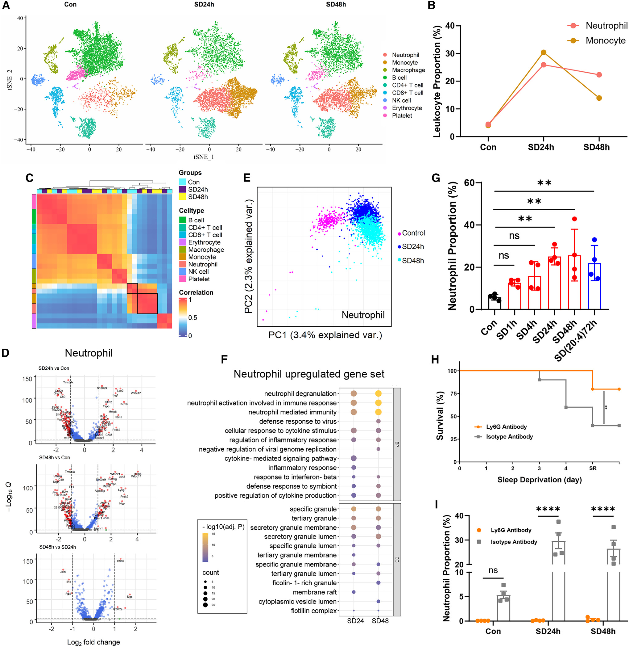
To identify which leukocyte populations drive this inflammatory cascade, the team performed single-cell RNA sequencing (scRNA-seq) on peripheral blood leukocytes after 24 and 48 hours of sleep deprivation. Unsupervised clustering and principal-component analysis revealed dramatic expansion and transcriptional remodeling of circulating neutrophils and monocytes: blood neutrophils rose from ~5% of CD45+ leukocytes in controls to ~25-30% during sleep deprivation, with upregulated genes enriched in neutrophil degranulation and cytokine production.
Testing causality directly, the authors depleted circulating neutrophils in vivo using an anti-Ly6G antibody. Anti-Ly6G treatment reduced blood neutrophil proportions to near zero at 24 and 48 hours of sleep deprivation and markedly improved mouse survival compared with isotype antibody controls, establishing circulating neutrophil accumulation as a critical contributor to sleep-deprivation lethality. As noted in the study limitations, because clodronate liposomes do not completely deplete the mononuclear phagocyte system, these experiments demonstrate a crucial pathogenic role for neutrophils while not fully excluding potential cooperative contributions from mononuclear phagocytes.
Source: Di Sang et al. · Cell · © 2023 Elsevier Inc.
Original figure and English caption ↗
Beyond sympathetic nerve signaling: enhanced blood-brain barrier efflux via ABCC transporters
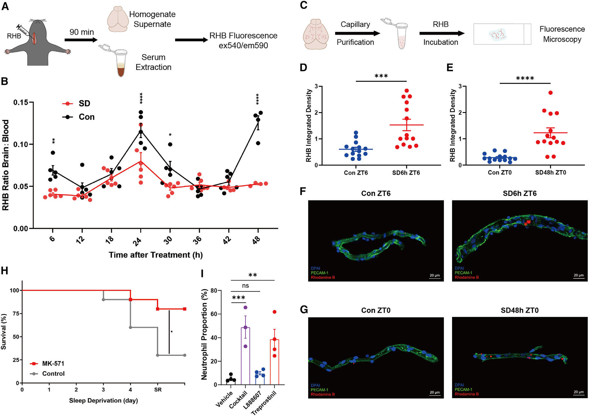
How does sleep loss in the brain ignite peripheral neutrophilia? The authors first performed chemical sympathectomy via intraperitoneal 6-hydroxydopamine (6-OHDA) administration and found that ablating sympathetic nerve signaling did not prevent sleep-deprivation-induced circulating neutrophil accumulation. This prompted them to examine direct humoral transport across the blood-brain barrier (BBB).
Using in vivo brain-to-blood rhodamine B ratios and ex vivo brain capillary transport assays, the team found that sleep deprivation significantly enhanced ABC-transporter-mediated efflux across the BBB. Oral administration of the broad-spectrum ABCC transporter inhibitor MK-571 markedly improved survival during sleep deprivation. Screening sleep-related brain small molecules further revealed that intravenous injection of the PGD2 receptor DP1 agonist treprostinil—but not a DP2 agonist—directly recapitulated peripheral blood neutrophil accumulation in non-sleep-deprived mice.
Source: Di Sang et al. · Cell · © 2023 Elsevier Inc.
Original figure and English caption ↗
Genetically encoded PGD2 sensors confirm rising brain PGD2 and peripheral metabolite accumulation
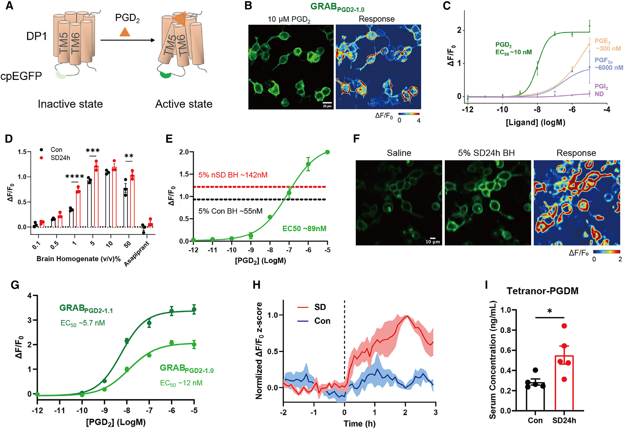
To directly monitor brain PGD2 levels during sleep deprivation, the authors engineered genetically encoded PGD2 fluorescent sensors—GRABPGD2-1.0 and the higher-sensitivity variant GRABPGD2-1.1—by coupling cpGFP to the DP1 receptor. In HEK293T cells, GRABPGD2-1.0 displayed ~10 nM affinity and high selectivity for PGD2. Applying brain homogenates to sensor-expressing cells revealed that 24 hours of sleep deprivation increased PGD2 concentration in 5% brain homogenate from ~55 nM in controls to ~142 nM, a response abolished by the DP1 antagonist asapiprant.
In vivo fiber photometry with GRABPGD2-1.1 in the preoptic area (POA) of freely moving mice further captured a rise in extracellular brain PGD2 fluorescence after sleep deprivation onset that peaked at ~2 hours and remained elevated above undisturbed control levels thereafter. Concurrently, serum levels of the stable PGD2 metabolite tetranor-PGDM rose significantly after 24 hours of sleep deprivation, corroborating brain-to-blood PGD2 spillover.
Source: Di Sang et al. · Cell · © 2023 Elsevier Inc.
Original figure and English caption ↗
Closing the PGD2-ABCC4-DP1 loop: blocking brain synthesis, efflux, or peripheral reception protects against lethality
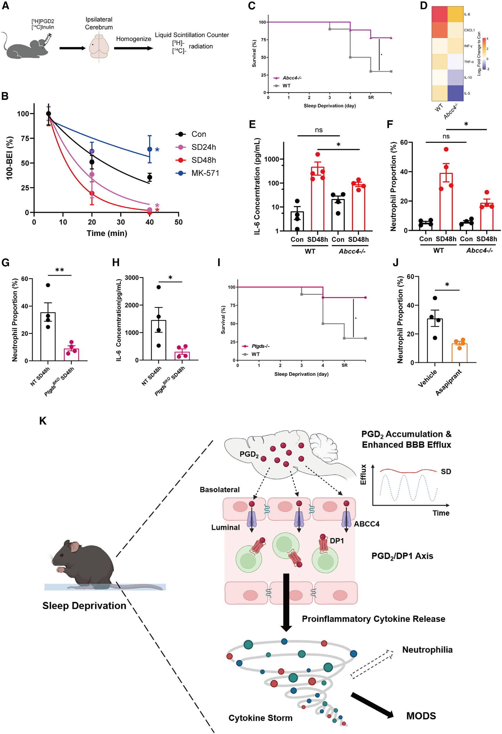
To close the causal loop, the authors measured the brain efflux index (BEI) of radiolabeled [3H]-PGD2, confirming accelerated clearance across the BBB after 24 and 48 hours of sleep deprivation that was slowed by MK-571. Testing mice lacking the prostaglandin efflux transporter ABCC4 (Abcc4-/-) showed markedly reduced serum IL-6, proinflammatory cytokines, circulating neutrophil proportions, and mortality after prolonged sleep deprivation compared with wild-type littermates.
Likewise, depleting prostaglandin D synthase (PTGDS) via germline knockout (Ptgds-/-) or adult brain-specific chimeric knockout (PtgdsBKO), or blocking the DP1 receptor pharmacologically with asapiprant, significantly blunted sleep-deprivation-induced neutrophil accumulation, IL-6 elevation, and/or mortality. While this establishes the brain PGD2-ABCC4-DP1 axis as a central bridge linking sleep loss to systemic inflammation, the authors note that the exact peripheral DP1-expressing immune or vascular cells that initiate the downstream cytokine cascade remain to be pinpointed.
Source: Di Sang et al. · Cell · © 2023 Elsevier Inc.
Original figure and English caption ↗