Cell signals in the lung engage the brain’s serotonin system
In male mice, peripheral mesenchymal stromal cells mainly lodge in the lung yet influence dorsal raphe activity through sensory-nerve-related connections and improve selected behaviors after chronic stress.

Why do brain neurons become more active when administered cells do not enter the brain? A study published in Nature Communications on November 16, 2023 follows a clue beginning in the lung. In male mice under chronic stress, peripheral mesenchymal stromal cells, MSCs, improve selected avoidance and immobility measures. The cells chiefly distribute to the lung, yet serotonergic neurons in the dorsal raphe nucleus, DRN, become engaged. The study follows pulmonary sensory nerves to examine how peripheral cells communicate with central networks.
The tested behavioral improvement associated with MSCs depends on DRN serotonin-cell activity. Pulmonary sensory nerves, the vagus and nucleus tractus solitarius contribute to the link, with MSC-derived BDNF and TrkB-related effects providing molecular evidence.
Contents · 7 sections
More exploration and less immobility
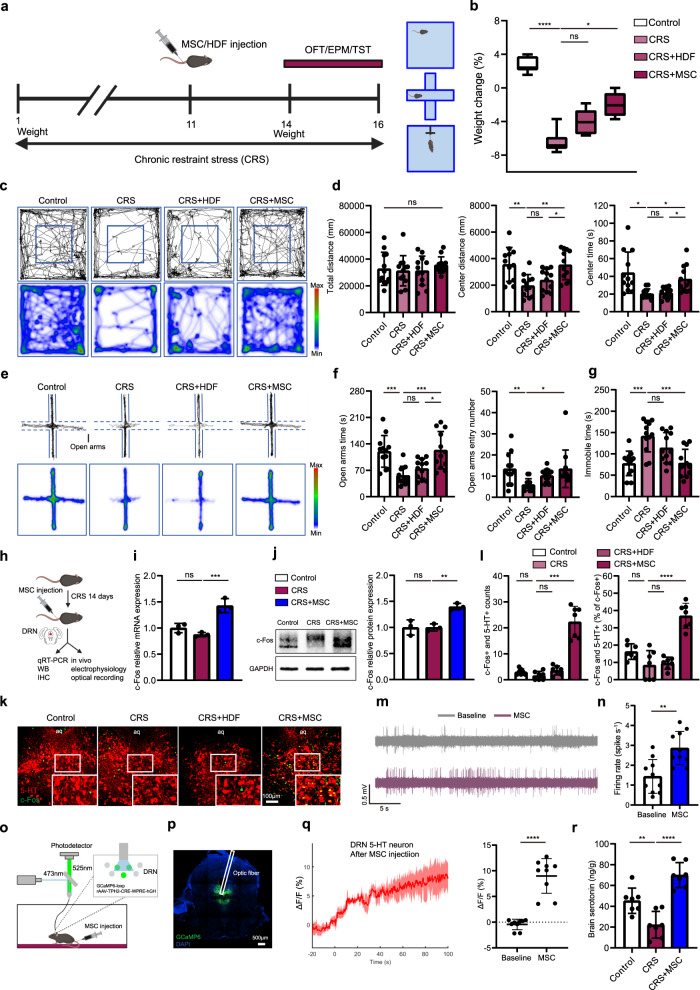
After chronic restraint, mice explore the open-field center and exposed maze arms less and spend more time immobile during tail suspension. MSCs increase central exploration and open-arm time and reduce immobility without a significant open-field total-distance change.
In a separate repeated-social-defeat model, MSCs also improve tested social avoidance and open-arm measures, unlike control dermal fibroblasts. Findings across stress contexts motivate a search for a shared link between the cells and behavior.
Source: Huang J et al. · Nature communications · CC BY
Original figure and English caption ↗
Chronic-stress improvement lacks the same inflammatory pattern
Because MSCs modulate immune responses, inflammation offers a plausible explanation. Brief stress raises IL-6, an effect attenuated by MSCs, while TNF-alpha lacks the same response to cell treatment. Stress duration changes the inflammatory setting.
At the tested chronic stage, IL-6, TNF-alpha and microglial activation do not show the same significant elevations, yet behavior improves. Dopamine-related measures and PVN CRH-related changes lack corresponding improvements, whereas DRN shows a clearer response.
Serotonin-cell activity contributes to behavioral improvement
DRN is a major source of brain serotonin, with outputs reaching cortical and limbic regions involved in different behavioral settings. After MSC administration, DRN serotonin cells show increased activity labeling and calcium responses, alongside higher recorded firing rates. Brain-homogenate serotonin also increases, linking peripheral administration to central cellular activity and tissue content.
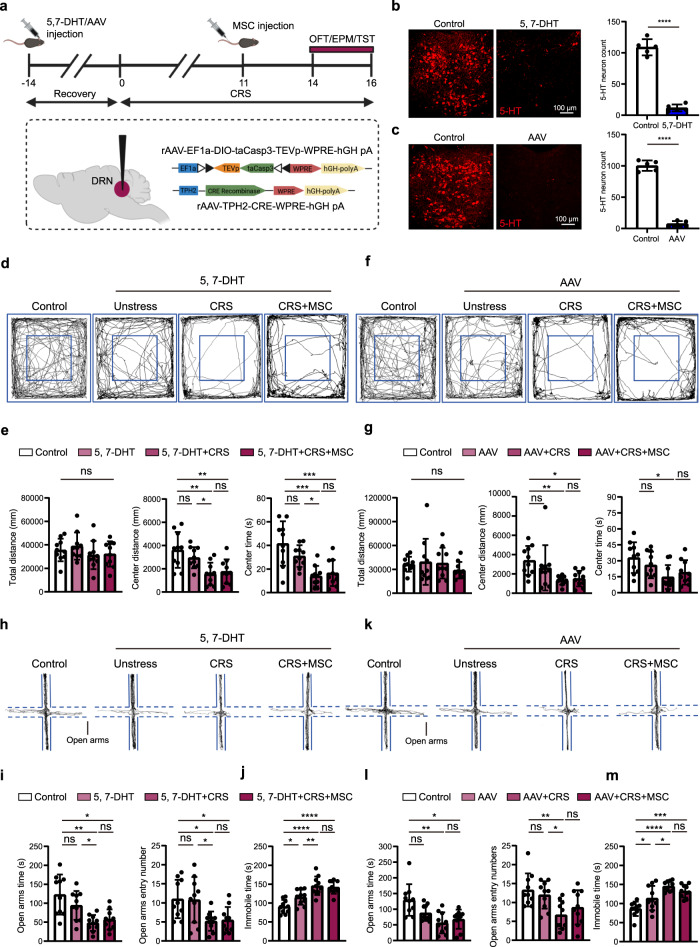
Removing these DRN cells prevents significant MSC-associated improvement in the tested center exploration, open-arm and tail-suspension measures. A separate silencing strategy also weakens improvement, moving from association toward dependence on serotonin-cell activity in this model.
Source: Huang J et al. · Nature communications · CC BY
Original figure and English caption ↗
Cells lodge mainly in the lung, where nearby nerves respond
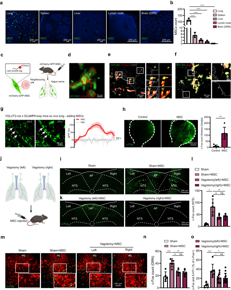
Tracking administered cells finds the most labeled MSCs in the lung and none detected in the brain. Cells and a secreted label lie near sensory fibers in lung tissue; related fibers in lung slices increase calcium in response to MSCs.
Activity labeling also increases in nodose ganglia, housing vagal sensory somata, and in the brainstem nucleus tractus solitarius, NTS. Unilateral vagotomy weakens ipsilateral NTS activation and reduces DRN activity labeling, connecting pulmonary events with an ascending neural link.
Source: Huang J et al. · Nature communications · CC BY
Original figure and English caption ↗
Tracing outlines links among lung, NTS and raphe
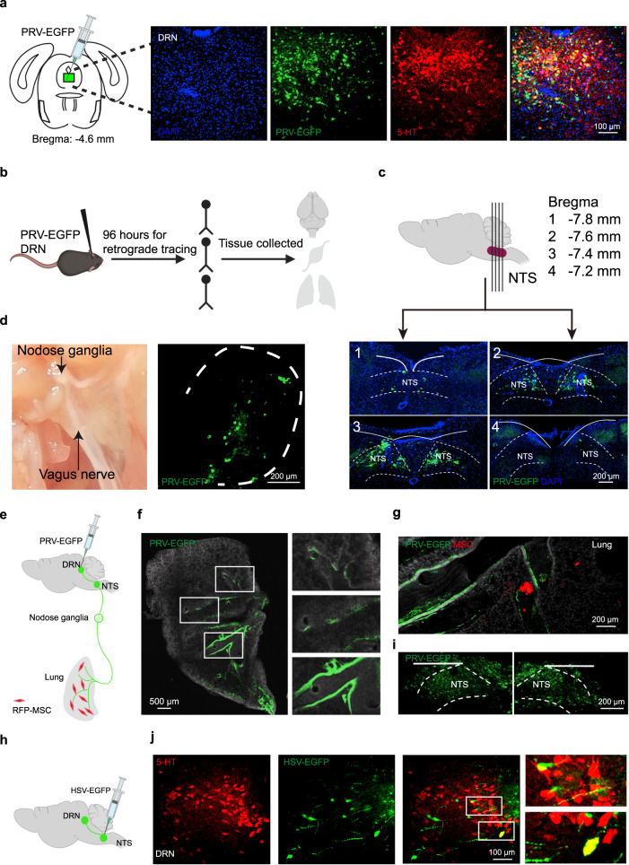
Multisynaptic retrograde tracing from DRN labels NTS, nodose ganglia and lung, with labeled pulmonary fibers near MSCs. Complementary multisynaptic anterograde tracing from NTS also reaches some serotonergic DRN cells.
The two tracing directions complement the vagotomy results and support a network from pulmonary afferents through brainstem circuitry to DRN. Multiple cells can intervene; the map does not resolve every step as a verified monosynaptic connection.
Source: Huang J et al. · Nature communications · CC BY
Original figure and English caption ↗
BDNF and TrkB connect cells with sensory nerves
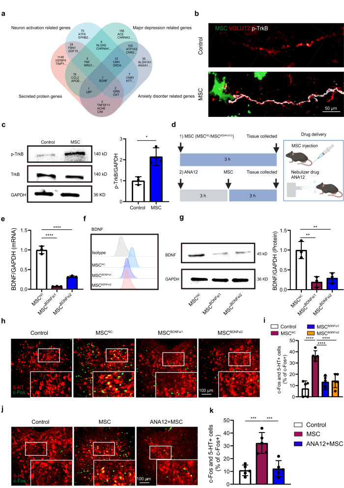
What makes nearby nerves respond to MSCs? Candidate analysis highlights brain-derived neurotrophic factor, BDNF. Lung-resident MSCs retain BDNF expression, and nearby sensory fibers show stronger phosphorylated TrkB labeling, supported by receptor-related tissue measurements.
Reducing MSC BDNF expression markedly weakens increased DRN serotonin-cell activity labeling and affects behavioral improvement. TrkB blockade likewise weakens DRN recruitment. Complementary interventions support BDNF–TrkB involvement in cell-to-nerve communication without requiring the administered cells to enter the brain.
Source: Huang J et al. · Nature communications · CC BY
Original figure and English caption ↗
Modulating the signal also changes tested behavior
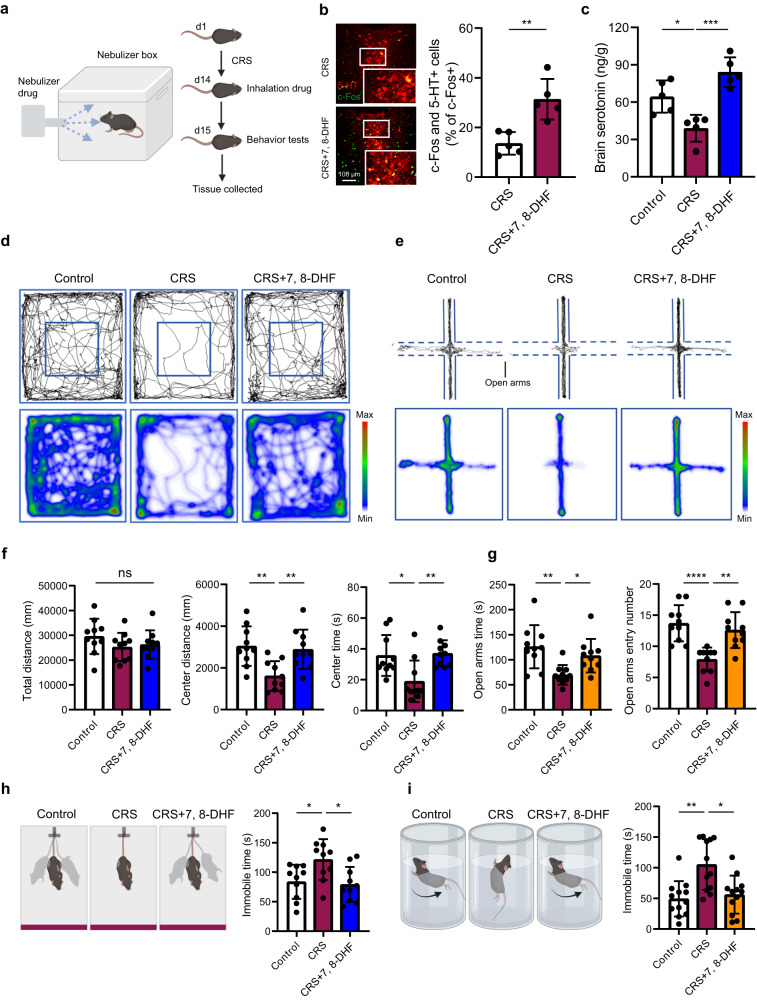
The team next examines TrkB-related effects of 7,8-DHF. Experiments involving pulmonary delivery increase DRN activity labeling and brain serotonin, improve center and open-arm exploration and reduce immobility in tail suspension and swimming, without significant total-distance changes.
Vagotomy weakens related improvements, while direct DRN administration lacks comparable significant effects. These male-mouse observations recast a cell-intervention question as one of body–brain communication: local cell signals can engage distant networks through sensory nerves. Precise relays and long-term effects remain unresolved, and human effects are not tested here.
Source: Huang J et al. · Nature communications · CC BY
Original figure and English caption ↗