Before food consumption: brain-wide links of tuberal SST neurons
Brain-wide mapping of mouse tuberal SST cells reveals broad inputs and diverse outputs; VTA-associated input and PAG-projecting cells regulate movement and food approach without significantly increasing intake.
Feeding includes finding, approaching and consuming food. Could feeding-promoting cells participate differently in these components? A Nature Communications study maps inputs and axonal outputs of tuberal-nucleus, or TN, somatostatin/SST neurons. Their brain-wide connections extend beyond a simple hunger switch, while functional experiments link one output-associated population with movement and food approach.
TN SST neurons have broad extra-hypothalamic inputs and diverse projection patterns. PAG-output or VTA-derived TN-terminal activation increases movement/approach; concurrent PAG-population inhibition removes these increases, without a significant intake rise.
Contents · 6 sections
Deep hypothalamic cells receive diverse brain inputs
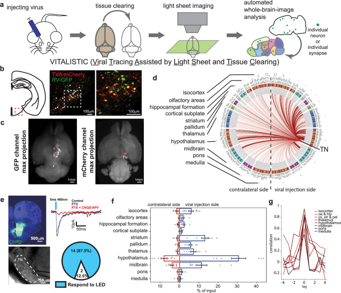
Tracing, clearing and whole-brain imaging quantify TN SST inputs. Hypothalamus is the largest source, alongside striatal, midbrain, olfactory and cortical inputs. Bilateral connections reveal broad convergence.
Prefrontal, motor and somatosensory areas contribute. Anterior-olfactory input evokes synaptic responses in TN SST cells, establishing functional connectivity for this long-range route and a potential entry for food-related environmental information.
Counting labeled inputs and comparing their distribution preserves hypothalamic prominence while bringing dispersed extra-hypothalamic connections into one map.
About sixty percent of recorded TN SST cells receive lateral-hypothalamic input, with glutamate-related, mixed and uncharacterized response types. One upstream region can supply varied signals within a marker-defined population.
Source: Senol E et al. · Nature communications · CC BY-NC-ND
Original figure and English caption ↗
Two feeding-promoting populations receive different input proportions
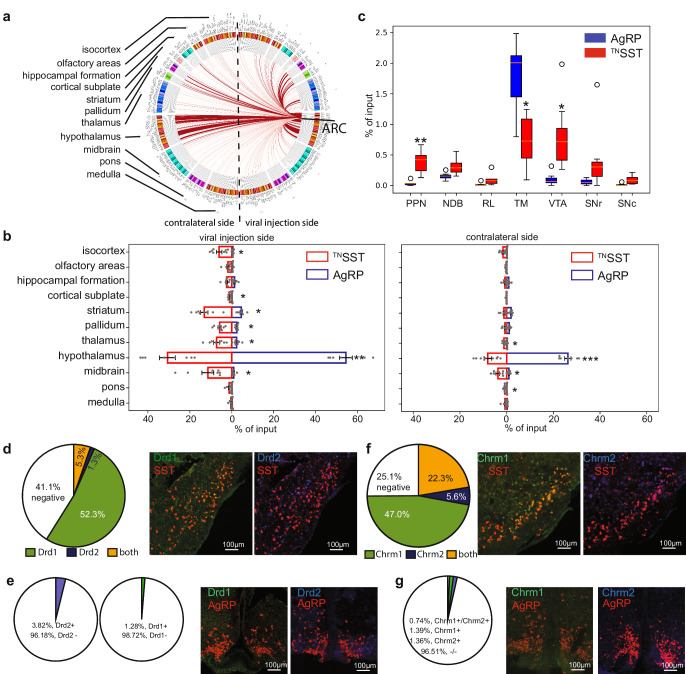
AgRP inputs are more concentrated within hypothalamus, whereas TN SST cells receive more extra-hypothalamic input. Lateral-hypothalamic responses occur in fewer recorded AgRP cells and have smaller amplitudes.
VTA and some other modulatory inputs are more prominent for TN SST, alongside receptor differences. More than half express Drd1, and Chrm1 is common, suggesting distinct access to state-related signals.
Similar feeding-promoting functions need not imply identical information sources. Nutrition-associated and environmental/state-processing regions converge in different proportions on the two populations.
Source: Senol E et al. · Nature communications · CC BY-NC-ND
Original figure and English caption ↗
Cells within one region have different axonal destinations
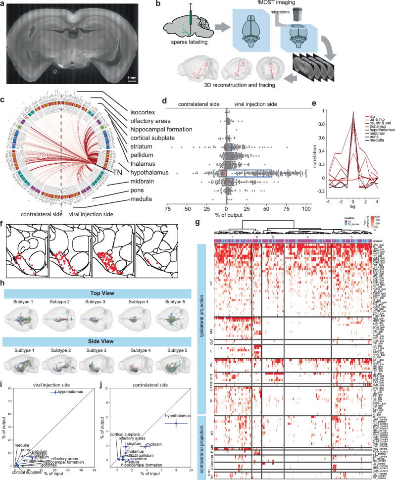
Reconstructions of 186 TN SST cells show mainly ipsilateral axons extending to multiple extra-hypothalamic regions. Five projection clusters differ in destinations including PAG, BNST, lateral septum and thalamus.
BNST is a common extra-hypothalamic target. Some BNST-projecting cells have little PAG projection, while others branch more widely. Axonal lengths and routes reveal heterogeneous outputs within TN SST.
Input/output comparisons reveal reciprocal links with some regions and chiefly afferent links with others. Lateral septum is more reciprocal, whereas accumbens and caudoputamen mainly provide input, adding directional organization to the network.
Source: Senol E et al. · Nature communications · CC BY-NC-ND
Original figure and English caption ↗
Output-associated populations show finer input differences
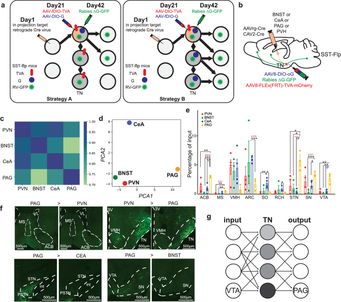
Input maps for TN SST cells projecting to PVN, BNST, CeA or PAG are broadly similar, especially PVN/BNST-associated populations, while PAG-associated inputs show some distinctive features.
The maps move from a regional average to output-associated populations. Broad inputs reach TN with different combinations across outputs, motivating functional tests of the VTA-associated input and PAG-associated population.
Source: Senol E et al. · Nature communications · CC BY-NC-ND
Original figure and English caption ↗
Output activation increases movement without a clear speed change
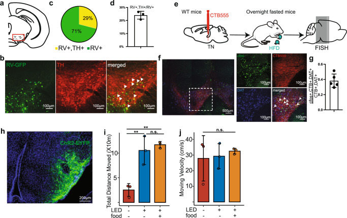
About thirty percent of VTA-associated inputs to PAG-projecting populations carry dopamine-associated TH markers. After high-fat food consumption, about forty percent of TN-projecting dopamine-associated cells carry c-Fos, linking input activity with a food context.
Activating TN SST terminals in PAG increases distance without significantly changing measured movement velocity, including without food. BNST-terminal stimulation does not significantly increase movement, distinguishing output-associated motor effects.
Source: Senol E et al. · Nature communications · CC BY-NC-ND
Original figure and English caption ↗
Recruiting PAG-projecting cells affects food approach
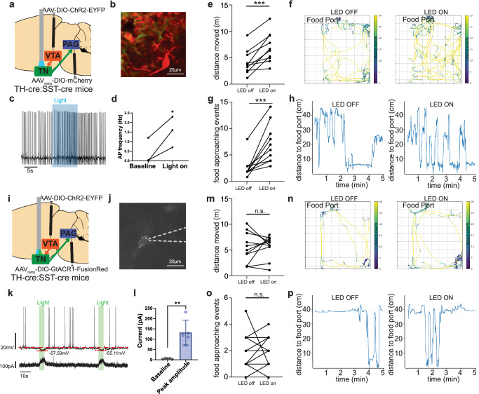
VTA dopamine-associated terminal activation increases firing in PAG-projecting TN SST cells. In vivo activation increases movement and food-port approaches without significantly increasing intake, separating approach from consumption outcomes.
Concurrent inhibition of PAG-projecting TN SST populations removes the movement and approach increases. This study including adult mice of both sexes turns a brain-wide map into a specific behavioral contribution within the components of feeding.
Source: Senol E et al. · Nature communications · CC BY-NC-ND
Original figure and English caption ↗