After sleep loss, oxytocin cells connect with cardiovascular responses
In male mice, chronic sleep deprivation lowers heart rate and raises blood pressure. Modulating hypothalamic paraventricular oxytocin-associated cells changes pressure and rate at different times, alongside altered blood-borne signals.
How does sleep loss affect the heart and blood vessels? The answer may involve both brain cells and signals in blood. A study in Current Issues in Molecular Biology follows adult male mice after a week of chronic sleep deprivation and focuses on oxytocin-associated cells in the hypothalamic paraventricular nucleus. Different ways of modulating this population produce different cardiovascular responses, and pressure and heart rate do not change together.
In sleep-deprived mice, acute cellular inhibition lowers elevated blood pressure without immediately changing heart rate. After a longer-lasting modulation, heart rate improves the next day, accompanied by changes in serum oxytocin and mPST.
Contents · 7 sections
Heart rate slows while pressure rises
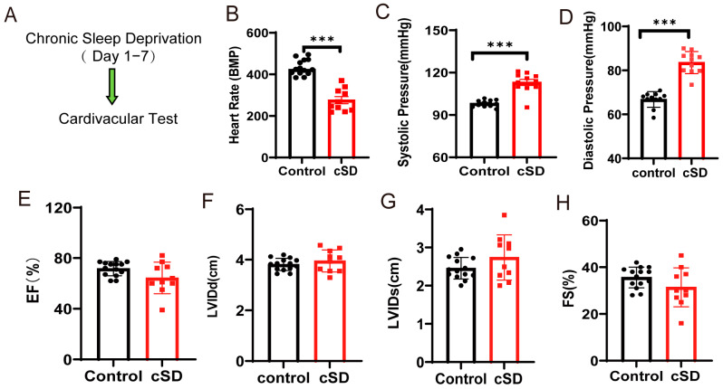
After a week of chronic sleep deprivation, heart rate decreases while systolic and diastolic pressures rise. Ejection fraction, fractional shortening, and ventricular dimensions do not differ significantly. These are changes in specific cardiovascular measures, without an overall decline in measured pumping performance at this stage. The echocardiographic assessments are made under anesthesia, so these group comparisons also have a specific measurement context.
Cardiac tissue shows changes in cellular arrangement and morphology, together with altered autophagy- and apoptosis-associated protein measures. Physiology and tissue-level responses describe different aspects of the post-deprivation state: tissue changes are apparent even when measured pumping function shows no significant difference.
Source: Zhang Y et al. · Current issues in molecular biology · CC BY
Original figure and English caption ↗
The response depends on the starting state
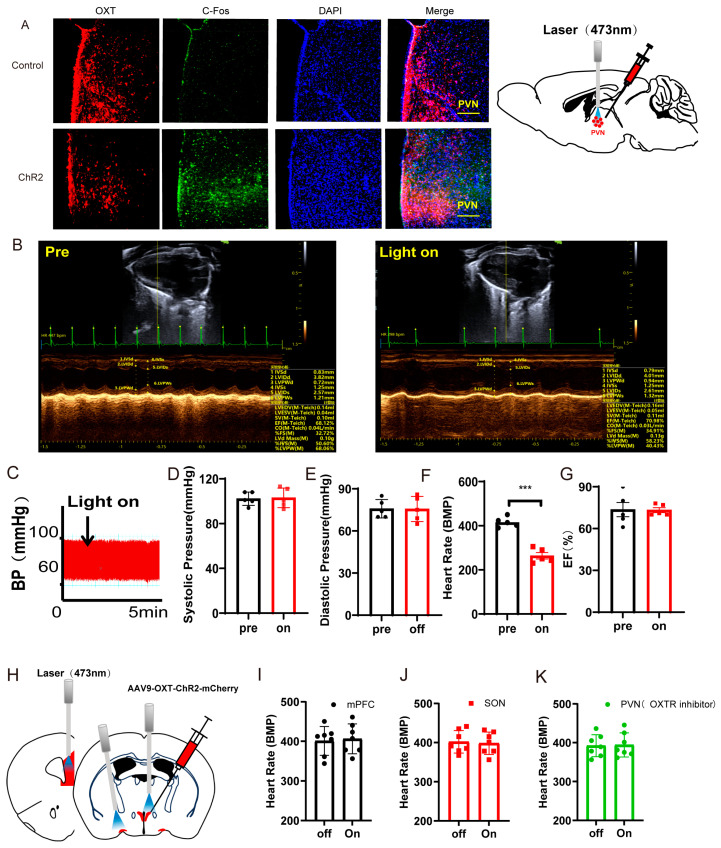
Paraventricular oxytocin cells have neural and endocrine functions and can influence the body through several routes. Activating oxytocin-associated cells lowers heart rate in normal mice without significantly changing pressure or ejection fraction. Activating projections to medial prefrontal cortex or supraoptic nucleus does not change heart rate.
In sleep-deprived mice, heart rate is already lower and decreases further during cellular activation; some animals exhibit intermittent cardiac arrest. Peripheral oxytocin-receptor blockade abolishes the heart-rate decrease in normal mice, implicating both the starting physiological state and peripheral receptor involvement.
Source: Zhang Y et al. · Current issues in molecular biology · CC BY
Original figure and English caption ↗
Pressure changes first; heart rate does not immediately follow
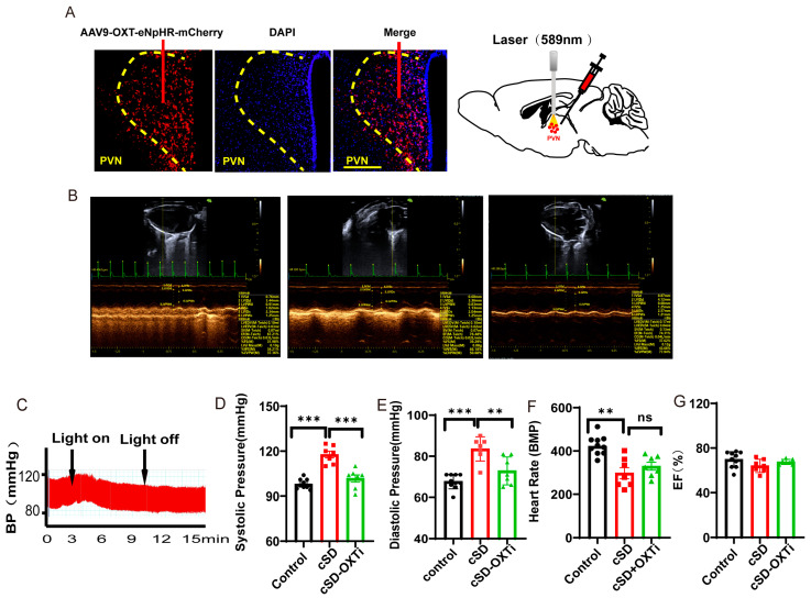
Reducing activity also has state-dependent effects. Heart rate, pressure, and ejection fraction do not change significantly in normal mice. Elevated systolic and diastolic pressures decrease in sleep-deprived mice, while heart rate remains low.
This separates the two physiological measures. Both change after sleep deprivation, but their regulation need not unfold at the same speed. Pressure improves over a short interval, whereas heart-rate recovery must be followed over a longer interval.
Source: Zhang Y et al. · Current issues in molecular biology · CC BY
Original figure and English caption ↗
Heart-rate improvement appears the next day
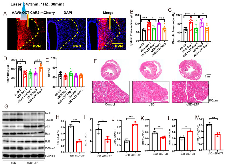
The team next uses a different, longer-lasting modulation associated with a lower cellular-activity marker. Pressure falls that day in sleep-deprived mice. Heart rate does not recover significantly that day but is higher the next day than in sleep-deprived mice without this modulation, whose low rate has not simultaneously returned to normal.
Tissue changes accompany this response: morphological abnormalities lessen and several autophagy- and apoptosis-associated protein measures move toward control values, while ejection fraction still shows no significant group difference. The changes span several levels, each retaining its own interpretation.
Source: Zhang Y et al. · Current issues in molecular biology · CC BY
Original figure and English caption ↗
From cellular modulation to circulating signals
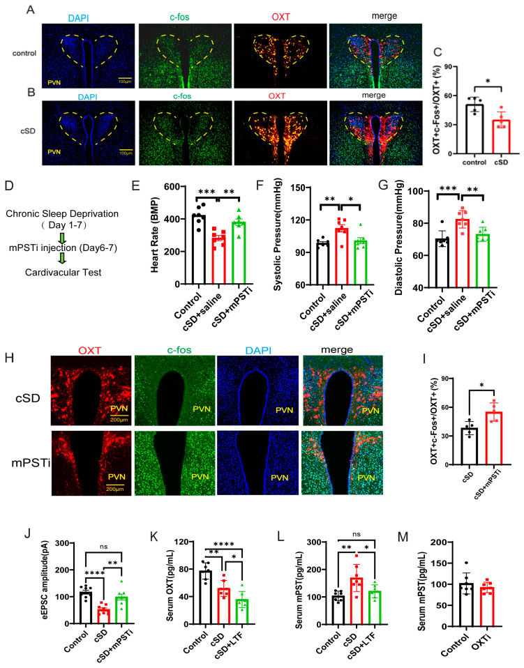
The study also examines whole-blood RNA expression. Sleep deprivation changes expression of genes associated with metabolism and cellular stress, including mPST, an enzyme contributing to hydrogen-sulfide production. Serum measurements likewise show higher mPST and lower oxytocin.
Whole-blood expression largely reflects blood-cell activity and is a different observation from serum protein measurement. Following both lets the team examine changes in circulation alongside central cellular responses.
After the longer-lasting cellular modulation, serum oxytocin decreases further while the mPST elevation is attenuated, and whole-blood expression changes as well. This extends the study from brain cells to circulation and suggests an association between oxytocin-related modulation and mPST changes.
Several measures change after mPST inhibition
When mPST is inhibited during the final stage before sleep deprivation ends, subsequent measurements show higher heart rate and lower pressure. Among paraventricular oxytocin-labeled cells, the c-Fos-positive fraction and evoked excitatory-current amplitude increase, linking peripheral molecular changes with central responses.
The results combine interventions and accompanying changes under different conditions, supporting a model of interactions between brain and circulating signals. The precise source of mPST, how it influences central cells, and the specificity of cellular identification remain unresolved parts of that model.
Source: Zhang Y et al. · Current issues in molecular biology · CC BY
Original figure and English caption ↗
The effect of a signal depends on state and timing
A striking finding is that the effects of oxytocin-associated cells do not follow one fixed direction. Normal and sleep-deprived states differ, acute and longer-lasting modulation differ, and pressure and heart rate respond at different times. Together, these distinctions reveal a fuller brain–body picture.
These changes in adult male mice add a neuroendocrine perspective to the sleep–cardiovascular relationship. Brain activity, circulating molecules, and organ state are intertwined, so the effect of a neural population is best understood alongside the body’s existing state.