Signals during running follow a brain route to prefrontal cortex
In male chronic-stress mice, running accompanies activity in a gracile–raphe–prefrontal route. Interventions alter the measured behavioral benefits, while sustained exercise accompanies synaptic changes.
Running changes more than limb position: sensory information from the body continually enters the nervous system. Which signals intersect with emotion-related behavior? A 2024 Advanced Science study examines running in two male mouse models of chronic stress and follows connections among the gracile nucleus, GR, dorsal raphe nucleus, DRN, and medial prefrontal cortex, mPFC. The study links bodily input, serotonin signaling and prefrontal activity, then tests how the populations contribute to behavioral improvement during exercise.
Running during stress improves measured sucrose preference, immobility and exposed-space exploration. Activating GR-associated DRN or DRN-associated mPFC populations produces similar directions, while their suppression weakens exercise benefits. DRN and mPFC also show synaptic labeling and current changes.
Contents · 7 sections
Two models show similar behavioral directions
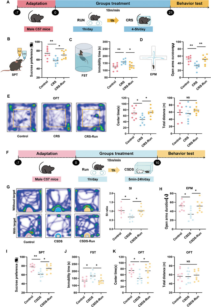
The team uses chronic restraint and chronic social defeat stress. With running during stress, mice show increased sucrose preference, reduced swim immobility, and more time in the open-field center and elevated-maze open arms. The social-defeat model also shows improved social interaction.
Total open-field distance does not change significantly in either model. The results therefore describe specific measured behaviors rather than simply greater activity after running. Exercise occurs during the stress period, and the study next identifies neural nodes recruited along the way.
Source: Lan T et al. · Advanced science (Weinheim, Baden-Wurttemberg, Germany) · CC BY
Original figure and English caption ↗
Connections link gracile nucleus with raphe
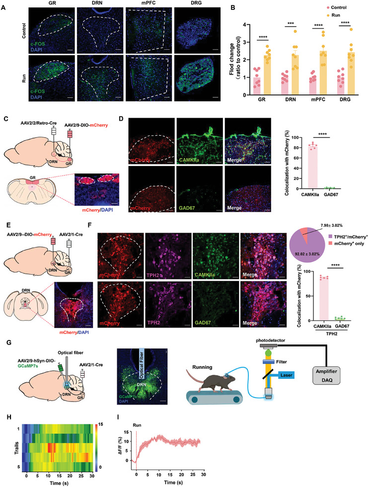
GR receives sensory information from the lower body and relays ascending input. Such input can convey bodily position and movement state. After exercise, dorsal root ganglia, GR, DRN and prefrontal cortex show increased c-Fos labeling. Tracing also identifies dorsal-root-ganglion input to GR and GR connections to DRN, providing an anatomical context linking bodily signals with brain activity.
Most DRN-projecting GR cells carry an excitatory CaMKII marker, and most GR-input-labeled DRN cells express the serotonin-synthesis marker TPH2. Calcium signals in this DRN population rise rapidly after running begins and maintain an elevated plateau.
Source: Lan T et al. · Advanced science (Weinheim, Baden-Wurttemberg, Germany) · CC BY
Original figure and English caption ↗
Serotonin and prefrontal cells respond during running
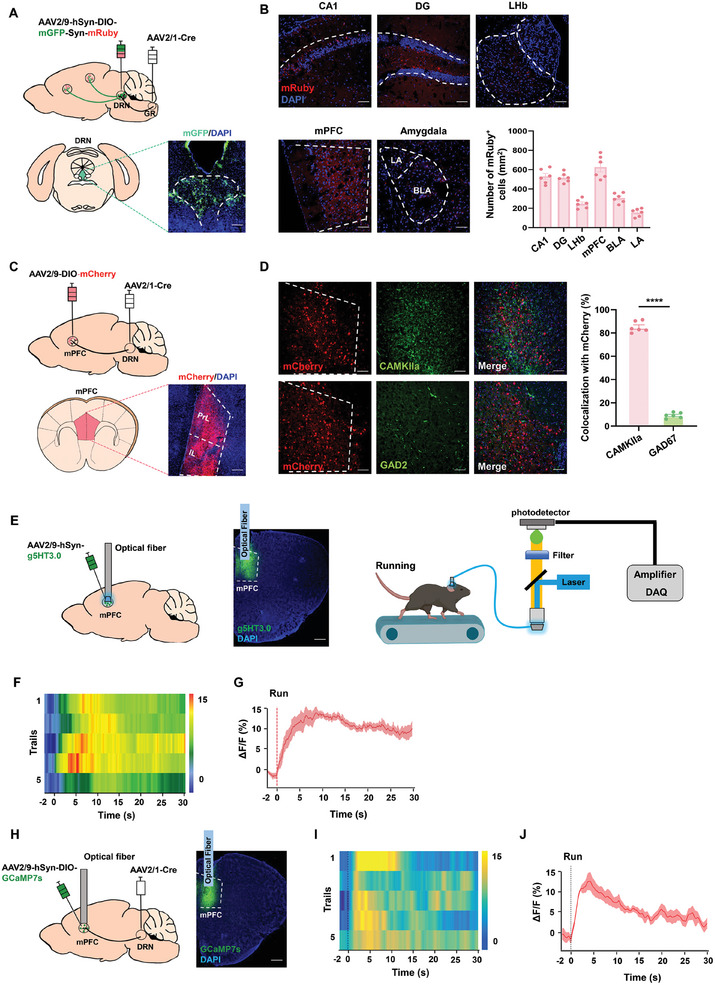
Outputs of GR-input-associated DRN cells reach multiple regions, including hippocampus, amygdala and lateral habenula. Labeling is prominent in mPFC. Further tracing of the DRN–mPFC relationship identifies predominantly excitatory-marked recipient prefrontal cells.
At running onset, mPFC serotonin-sensor signals rise, as do calcium signals in the DRN-input-associated mPFC population. One readout describes local transmitter changes and the other cellular responses, extending the investigation to prefrontal cortex beyond the sensory relay.
Source: Lan T et al. · Advanced science (Weinheim, Baden-Wurttemberg, Germany) · CC BY
Original figure and English caption ↗
Activating populations also alters measured behavior
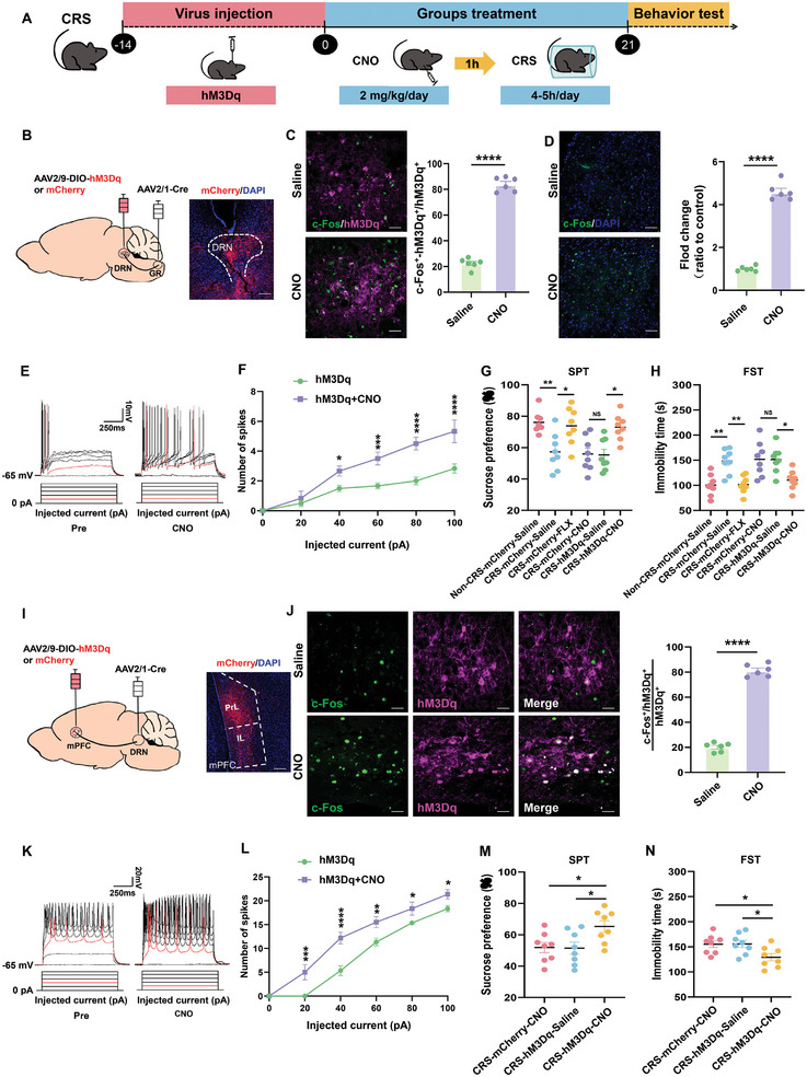
Increasing activity in GR-input-associated DRN cells during stress raises sucrose preference, reduces swim immobility and improves exposed-space exploration in supplementary experiments. Increasing activity in DRN-input-associated mPFC cells produces similar directions, with related benefits also observed in the social-defeat model.
These interventions directly change population activity and are separate from exercise itself. They demonstrate an ability to modulate the behaviors without isolating every individual output. Labeled cells can still project elsewhere, and mPFC is not DRN’s only recipient.
Source: Lan T et al. · Advanced science (Weinheim, Baden-Wurttemberg, Germany) · CC BY
Original figure and English caption ↗
Exercise continues while its benefits weaken
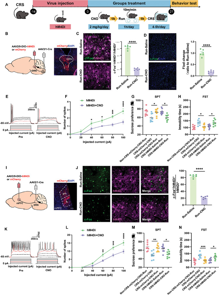
In the opposite experiment, suppressing GR-input-associated DRN cells during exercise and stress lowers the running group’s sucrose-preference benefit and increases immobility. Suppression of DRN-input-associated mPFC cells also weakens these benefits, with similar supplementary exploration findings.
Suppressing DRN-projecting GR cells also affects exercise-related improvement. Interventions at several nodes support involvement of this ascending connection in the measured benefits. They do not assign every bodily, rewarding or metabolic consequence of exercise to a single route.
Source: Lan T et al. · Advanced science (Weinheim, Baden-Wurttemberg, Germany) · CC BY
Original figure and English caption ↗
Sustained exercise accompanies input labeling and current changes
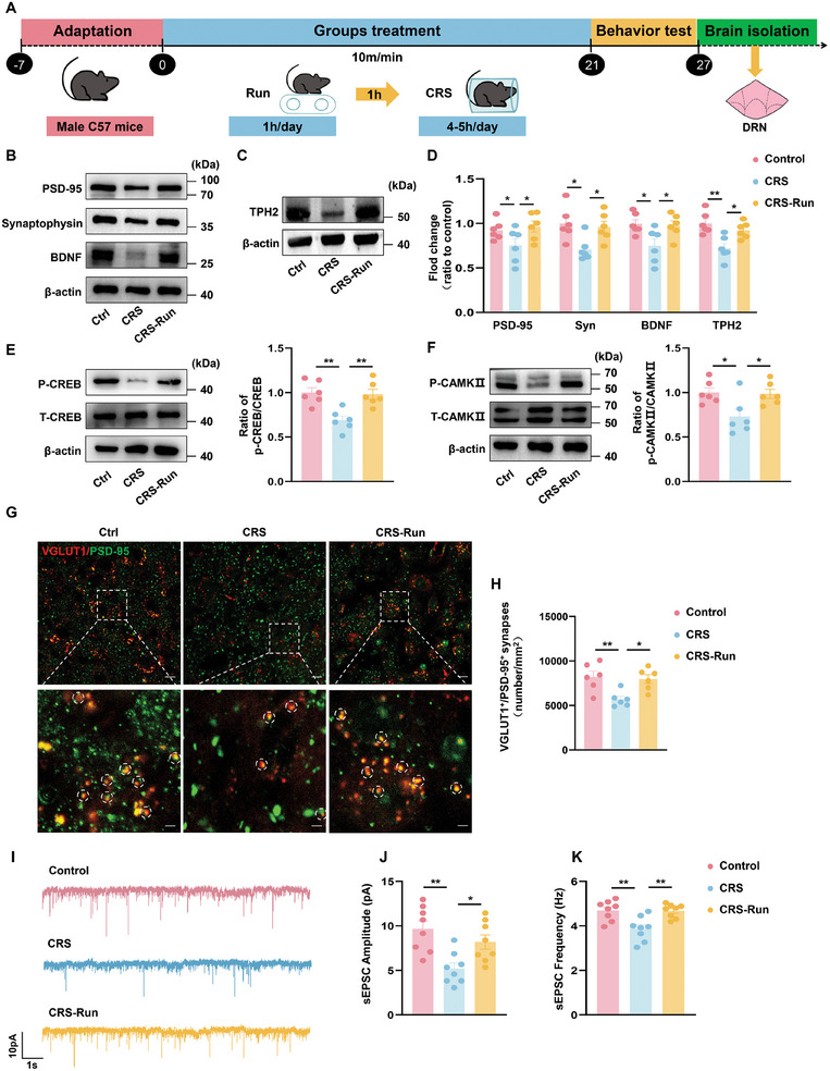
After sustained running, DRN BDNF, PSD95 and synaptophysin increase, CREB and CaMKII phosphorylation ratios rise, and TPH2 expression increases. More puncta jointly labeled for VGLUT1 and PSD95 suggest changes in excitatory-input-related structure.
Spontaneous excitatory-current amplitude and frequency also rise in slices, complementing the structural findings. These local currents do not specifically isolate GR input, and individual protein changes are not blocked to test their behavioral contribution. The findings describe synaptic changes following exercise.
Source: Lan T et al. · Advanced science (Weinheim, Baden-Wurttemberg, Germany) · CC BY
Original figure and English caption ↗
Bodily input connects with immediate and later changes
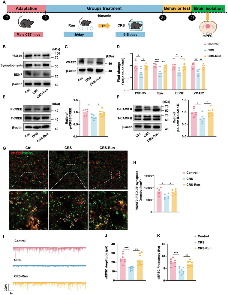
mPFC also shows changes in synaptic proteins, CREB and CaMKII phosphorylation, and spontaneous excitatory currents. Increased VMAT2 expression and VMAT2/PSD95 colabeling provide tissue clues concerning monoamine-associated synapses; the markers do not uniquely identify serotonergic input.
This study in young male mice connects immediate responses associated with bodily input during running with synaptic changes after sustained exercise. Several ascending nodes contribute to the measured behavioral benefits. Connections and cellular states together describe this interaction between movement and prefrontal recruitment.
Source: Lan T et al. · Advanced science (Weinheim, Baden-Wurttemberg, Germany) · CC BY
Original figure and English caption ↗