Why REM timing changes while its total amount stays similar
A mouse study links SLD-projecting orexin cells with subsequent REM intervals. Removing the population increases REM transitions after brief NREM and fragments sleep when REM need rises.

Orexin is familiar for promoting wakefulness, yet some of its cells also activate during rapid-eye-movement, or REM, sleep. What do they contribute? A study published in Research on April 30, 2024 follows orexin cells projecting to pontine sublaterodorsal tegmental nucleus, SLD. Their activity is associated with subsequent REM intervals. Removing them leaves total REM amount similar while altering its timing, revealing contributions of episode number, spacing and duration to sleep organization.
SLD-projecting orexin cells gradually activate during REM, with steady activity positively associated with subsequent REM intervals. Ablation increases sleep-onset REM-like transitions and produces more, shorter REM episodes when REM need is elevated.
Contents · 7 sections
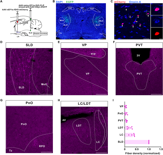
Dispersed cells preferentially reach one destination
Orexin cells lie in hypothalamus but send axons to many regions. Labeling by destination identifies a group targeting SLD: their somata are dispersed in lateral hypothalamus while fibers converge preferentially on SLD. Output destination reveals a distinction beyond soma location.
The cells also have collateral branches, with lower fiber density in locus coeruleus, laterodorsal tegmentum and other sampled regions. Their SLD preference motivates examining a particular contribution to REM regulation.
Source: Feng H et al. · Research (Washington, D.C.) · CC BY
Original figure and English caption ↗
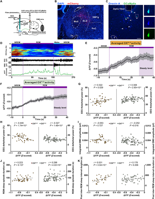
Activity in one episode relates to the following interval
After REM begins, calcium signals gradually rise and reach a relatively steady level until the episode ends. Higher steady activity is associated with greater relative theta power, without significant correlations with the current episode’s duration or integrated muscle activity.
More intriguingly, activity is positively associated with the subsequent REM-interval measure. PnO-projecting orexin cells have similar activity dynamics but correlate with current REM duration rather than the following interval. Different output-defined groups can relate to distinct timescales while both activate in REM.
Source: Feng H et al. · Research (Washington, D.C.) · CC BY
Original figure and English caption ↗
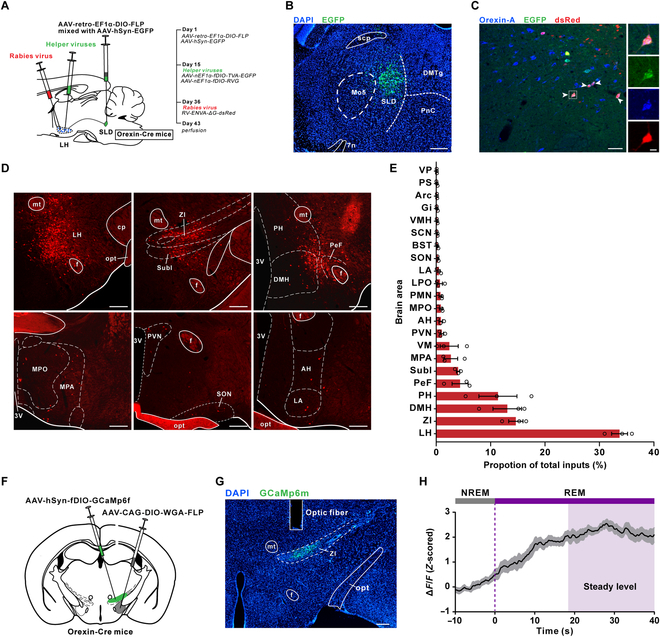
A focused output receives widespread input
The population has diverse upstream sources. Tracing reveals direct input from hypothalamic regions, zona incerta (ZI) and other areas. Orexin-projecting ZI cells also gradually activate during REM, without significant correlations with REM duration or subsequent interval.
Similar activity curves can accompany different functional associations. Multiple inputs may shape orexin-cell responses, with a particular output carrying them toward SLD. Tracing and activity recordings provide clues to this organization.
Source: Feng H et al. · Research (Washington, D.C.) · CC BY
Original figure and English caption ↗
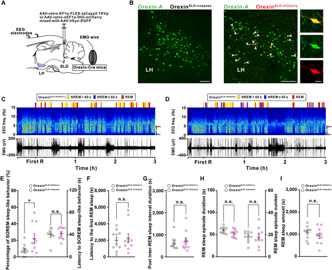
REM amount stays similar while transitions change
Ablating SLD-projecting orexin cells leaves many other orexin cells present. Under ordinary recording conditions, REM amount, episode number, mean duration, first-REM latency and average subsequent interval do not significantly change. Wake and non-REM amounts also do not significantly change in the same recording period. A particular transition becomes more frequent: REM following only a brief NREM period.
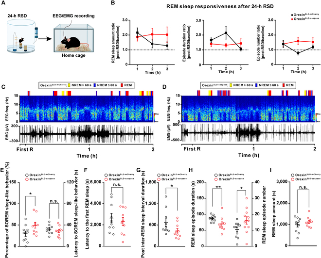
The fraction of these sleep-onset REM-like transitions increases from about 8% to 22%, without a significant change in their mean latency. The team links the pattern with insufficient relief of REM need, showing that similar sleep amounts can coexist with altered timing.
Source: Feng H et al. · Research (Washington, D.C.) · CC BY
Original figure and English caption ↗
Higher REM need reveals greater fragmentation
During recovery with elevated REM need, ablated mice show more sleep-onset REM-like transitions, shorter subsequent intervals, more episodes and shorter mean episode duration. Total REM still does not significantly differ between groups; its organization becomes more fragmented. First-REM latency remains similar, concentrating the changes on subsequent repetition and fragmentation.
Controls recover mainly through longer episodes, while ablated mice rely more on episode number and show delayed rebound. A REM episode can relate both to when the next one arrives and to how sleep need is met in an organized fashion.
Source: Feng H et al. · Research (Washington, D.C.) · CC BY
Original figure and English caption ↗
Patient records add a correlational observation
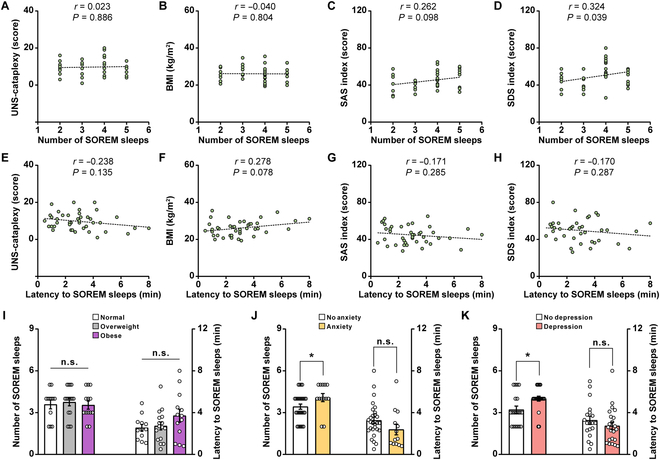
The team also reviews records from 41 patients with type-I narcolepsy. More sleep-onset REM episodes during daytime sleep testing correlate with a higher self-rated depression index, without significant linear correlations with cataplexy score, body-mass index or self-rated anxiety index.
Groups with elevated depression or anxiety scale indices have more sleep-onset REM episodes, while latency remains similar across groups. The records complement the animal timing findings through associations in existing patients, without direct measurements of their SLD-projecting orexin cells.
Source: Feng H et al. · Research (Washington, D.C.) · CC BY
Original figure and English caption ↗
Sleep organization also depends on timing
The study examines REM duration, spacing and stability separately. SLD-projecting orexin cells remain active during REM and relate to its subsequent timing. Losing the population can leave the total amount similar while changing transitions and recovery.
Activity–interval correlations and ablation effects support a division of labor within the orexin system in regulating relief of REM need. Patient records suggest a link between sleep timing and emotional symptoms, directing further questions toward specific populations, sleep organization and clinical states.