Active during wakefulness, yet acting as an arousal brake
A mouse study finds wake- and stress-active GAD2 cells in dorsal raphe that reduce wakefulness and increase NREM when activated. A thalamic projection contributes to constraining stress responses.
Stress-related sleeplessness often brings heightened arousal systems to mind. Yet a brake may also operate while the brain is awake. A mouse study published in Nature Communications on March 28, 2024 identifies GAD2 cells in dorsal raphe whose activity is higher during wakefulness than sleep, but whose activation shifts sleep toward non-rapid-eye-movement, or NREM, sleep. Following this apparent paradox, the team examines stress responses and thalamic connections to identify a pathway constraining arousal.
Dorsal raphe GAD2 cells activate during wakefulness and stress. Increasing their activity reduces wakefulness and increases NREM, while removing them prolongs wakefulness. PVT-projecting cells and their local terminals contribute to post-stress sleep and pupil-response regulation in separate experiments.
Contents · 7 sections
Sleep-promoting cells within an arousal-related region
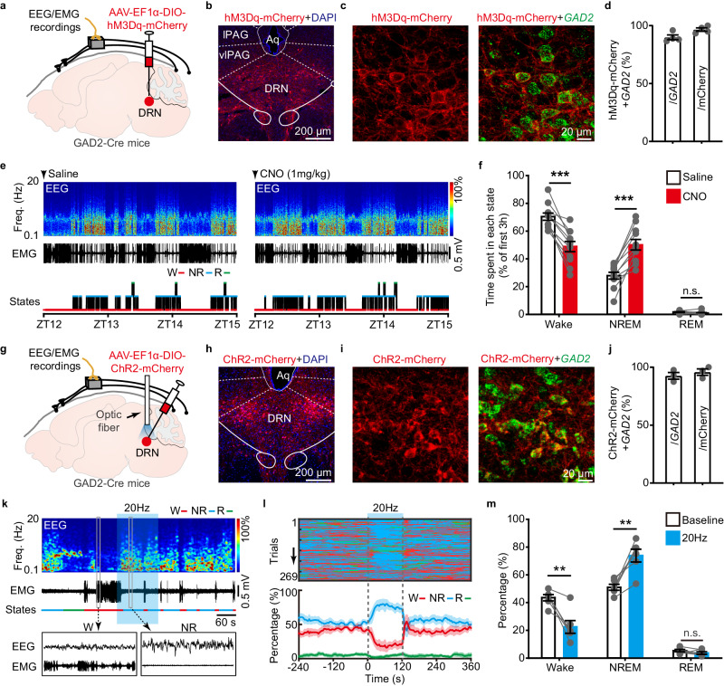
Dorsal raphe is familiar for serotonin neurons but contains several cell populations. The team focuses on GABAergic cells expressing GAD2. Increasing their activity reduces wakefulness and increases NREM during the first hours of the dark phase, while REM amount remains similar. The same direction appears during the light phase and with another activation approach.
Cells with overlapping markers within one nucleus can have different functions. GAD2 populations substantially overlap with those expressing another common GABAergic marker, Vgat. Wake-promoting effects in previous work and NREM promotion here point to the importance of output destinations and cellular composition. EEG and muscle recordings distinguish the sleep states rather than relying on immobility alone. Activation also reduces distance traveled in a novel environment, connecting this population with both sleep state and behavioral activity.
Source: Ren S et al. · Nature communications · CC BY
Original figure and English caption ↗
Removing the brake lengthens wake bouts
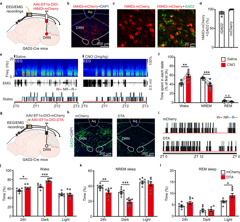
If the cells ordinarily restrain arousal, weakening them should shift behavior in the opposite direction. Inhibition during the light phase increases wakefulness and reduces NREM, with REM largely unchanged. Weeks after cell ablation, daily wakefulness also increases and NREM decreases, mainly during the normally active dark phase.
The change extends beyond totals: longer wake bouts become more common and the mice move more. Light-phase wakefulness and NREM amounts remain similar, while REM rises slightly. Sleep is reorganized differently across the daily cycle.
Source: Ren S et al. · Nature communications · CC BY
Original figure and English caption ↗
Most active when a brake may be needed
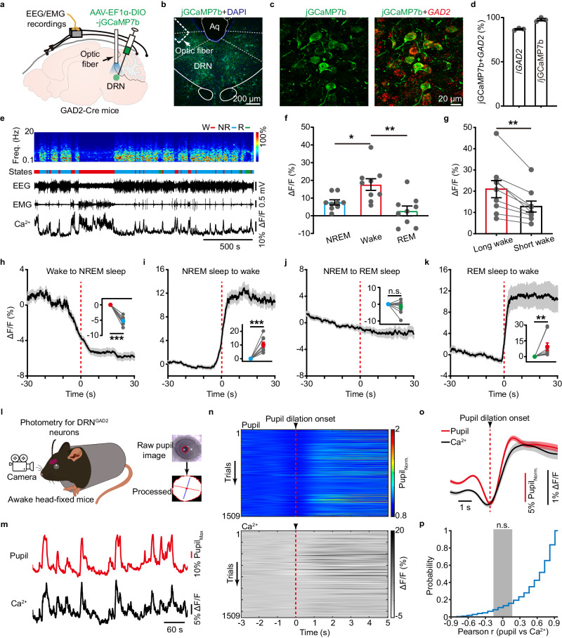
Natural recordings reveal a counterintuitive pattern: calcium signals are higher during wakefulness and higher in long than short wake bouts. Signals fall on entering NREM and rise on waking. Average signals do not change significantly at NREM-to-REM transitions.
These cells do not resemble a switch engaged only at sleep onset. Calcium signals also rise with pupil dilation. Together with activation and ablation results, this pattern motivates a negative-feedback model in which the population helps constrain rising arousal.
Source: Ren S et al. · Nature communications · CC BY
Original figure and English caption ↗
Returning to sleep after stress
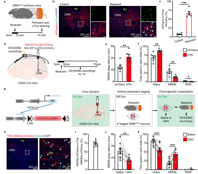
Restraint stress recruits these cells. After ablation, stressed mice take longer to enter their first NREM episode, spend more time awake and less time in NREM and REM. Restraining arousal becomes particularly relevant when stress occurs.
The team then labels GAD2 cells previously activated by stress and enhances that population. Following stress, mice enter NREM sooner, spend less time awake and more time in NREM. This links recruitment during stress with promoting sleep afterward in the same defined population.
Source: Ren S et al. · Nature communications · CC BY
Original figure and English caption ↗
The brake also constrains pupil dilation
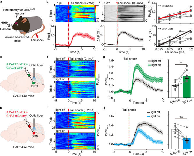
Brief tail stimulation evokes pupil dilation and a concurrent rise in GAD2-cell calcium signals; both responses grow with stimulus intensity. Inhibiting the cells strengthens pupil dilation for the tested stimulus, whereas activation weakens the pupil response in another tested stimulus condition.
Pupil measurements reveal changes over seconds, complementing sleep observations over minutes and hours. Constraining a rapid response and facilitating subsequent NREM provide evidence on different timescales for a role in stress-related arousal regulation.
Source: Ren S et al. · Nature communications · CC BY
Original figure and English caption ↗
Following inhibitory connections into thalamus
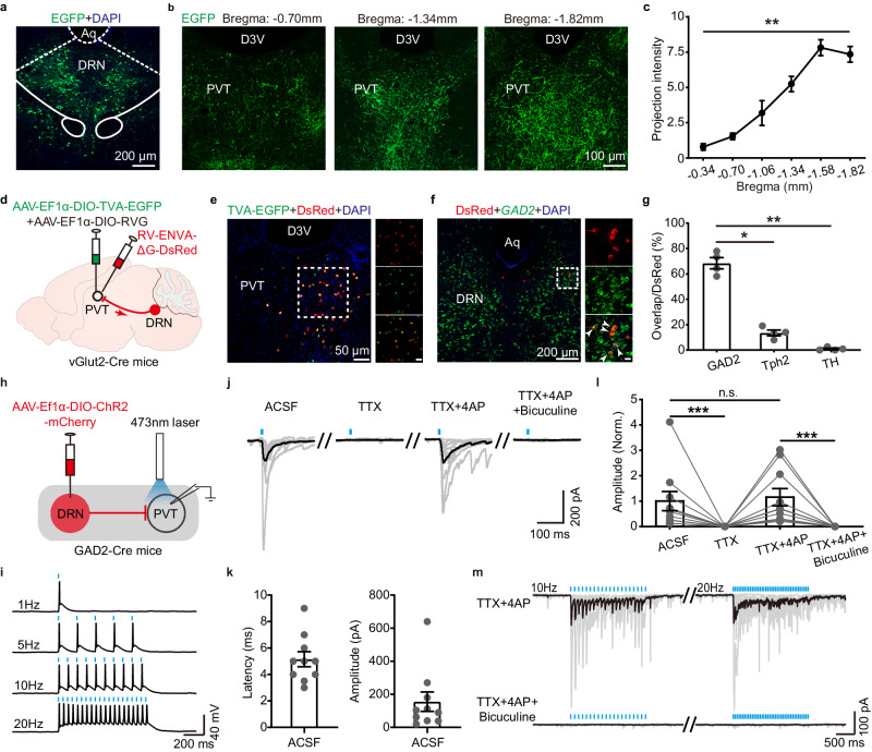
The cells send axons to several regions, including the paraventricular thalamic nucleus, PVT, with denser fibers posteriorly. Tracing backward from PVT glutamatergic cells labels dorsal raphe inputs predominantly bearing GAD2, with lower proportions of serotonin- and dopamine-related markers.
Slice recordings detect postsynaptic currents evoked by this input, which disappear with GABA-receptor blockade. Anatomy and electrophysiology identify an inhibitory pathway through which dorsal raphe cells can constrain PVT neuronal activity.
Source: Ren S et al. · Nature communications · CC BY
Original figure and English caption ↗
One branch links sleep with rapid stress responses
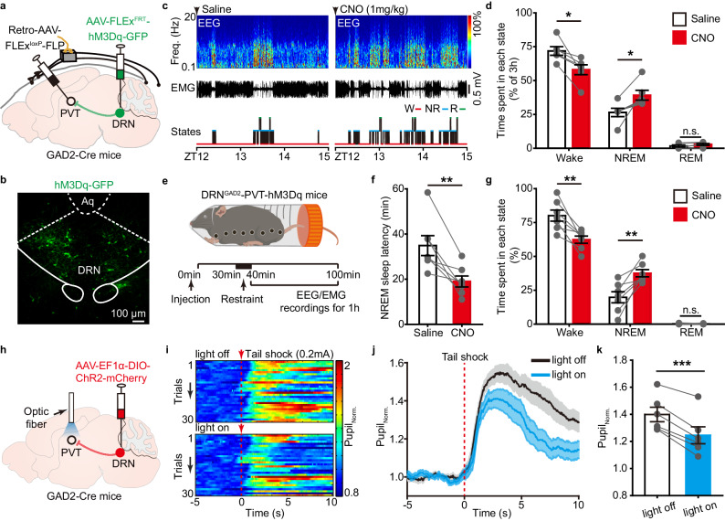
Activating the PVT-projecting GAD2 population reduces wakefulness and increases NREM at baseline, and brings earlier NREM onset after stress. This experiment manipulates projection-defined cell bodies in dorsal raphe, whose axons may also have collateral branches.
A separate experiment stimulates local terminals in PVT and reduces tail-stimulation-induced pupil dilation. These experiments connect the output with stress-related arousal at different levels. Conducted in adult mice under acute stress, the study proposes an arousal-brake model: cells active during wakefulness may also keep that state from being driven too far.
Source: Ren S et al. · Nature communications · CC BY
Original figure and English caption ↗