Why nap during an active night? A timed easing of arousal in mouse prefrontal cortex
NPAS2 acts through POU2F2 to regulate TH and local dopamine signals, lowering wake-promoting activity at a particular time and creating a window for a brief mouse nap.
For nocturnal mice, night is a busy time. Yet many pause late in the active phase for a nap lasting several tens of minutes. Why then? A Nature Communications study links the behavior to NPAS2, a circadian regulator in medial prefrontal cortex. Through a molecular relay, it creates a timed trough in local wake-promoting activity. Like briefly easing the accelerator, reduced arousal drive provides a window in which sleep can occur.
A local NPAS2–POU2F2–TH regulatory chain links molecular expression, neural activity and nap timing; reducing NPAS2 shrinks or removes the usual nap window.
Contents · 6 sections
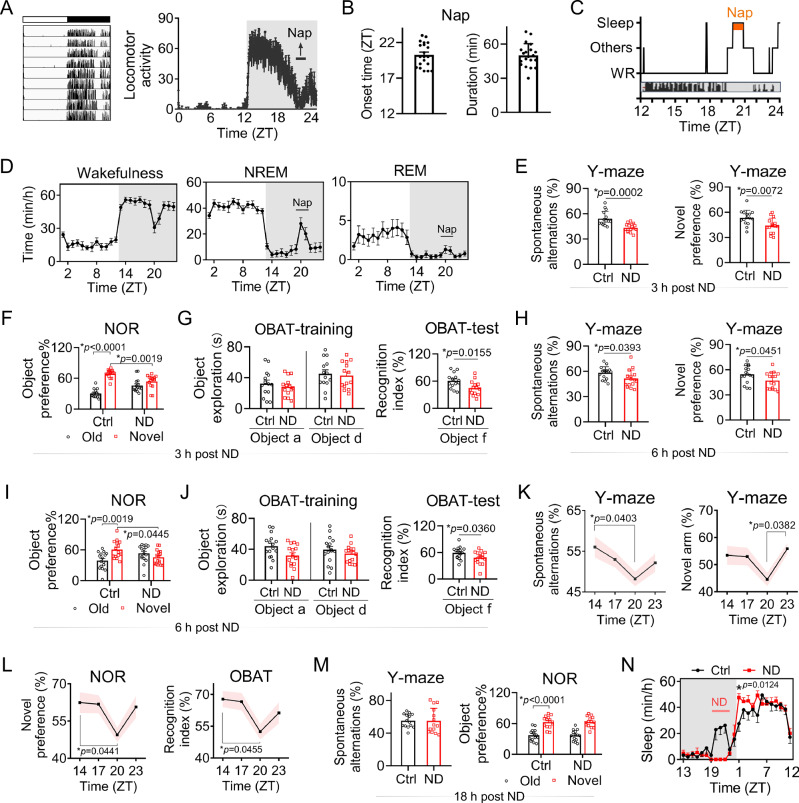
A pause in activity contains real sleep
The pause is more than sitting still. EEG and muscle recordings identify predominantly non-REM sleep with little REM. Naps persist in constant darkness without clear changes in duration or slow-wave intensity, and brief food deprivation does not abolish them. Immediate light and feeding cues do not fully explain the timing.
Preventing the nap reduces memory and object-attention performance over the following hours. By the next active phase, the measured changes are no longer significant, while compensatory rest-phase sleep increases. The brief nap relates to subsequent performance, and recovery timing matters: sleep lost in one window is followed by more sleep elsewhere.
Source: Guo L et al. · Nature communications · CC BY-NC-ND
Original figure and English caption ↗
Finding a clock factor for this nap
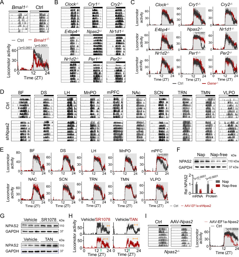
The study compares core clock genes. Bmal1 loss disrupts overall rhythmicity, whereas Npas2 loss removes this specific nap without equivalent sleep changes at other times. Several other single-gene deletions retain the nap. NPAS2 is therefore linked more specifically to one daily sleep window than to overall sleep quantity.
Regional comparisons point to medial prefrontal cortex (mPFC). Reducing local Npas2 diminishes naps, while restoring expression there in knockout mice restores them. Other tested regions do not show the same outcome. Nap-free wild-type individuals also have lower local NPAS2. Molecular differences, regional changes and behavior converge on one location.
Source: Guo L et al. · Nature communications · CC BY-NC-ND
Original figure and English caption ↗
Which cells need to slow down around nap time?
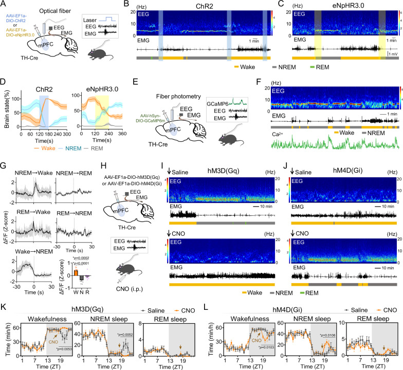
The study examines prefrontal TH-positive cells. TH is an enzyme involved in catecholamine synthesis, and these cells show stronger calcium activity during wakefulness. Increasing their activity raises wakefulness and reduces sleep; reducing it shifts outcomes oppositely and changes naps. Tested interventions in local glutamatergic populations leave sleep states largely unchanged, focusing the functional clue.
The population is heterogeneous. About four in ten TH-positive cells also express Ddc, associated with dopamine synthesis; increasing population activity raises local extracellular dopamine signals. The observations support a dopamine-associated subset. Around nap hours, changes chiefly involve wakefulness and NREM, with REM not significantly altered in every comparison.
Source: Guo L et al. · Nature communications · CC BY-NC-ND
Original figure and English caption ↗
Increasing one factor lowers another signal
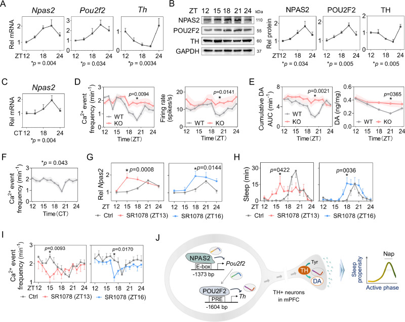
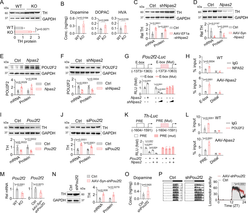
How does NPAS2 slow wake-promoting cells? POU2F2 is the intermediary: NPAS2 increases its expression, and POU2F2 represses Th transcription. Expression, promoter activity and binding support the chain. Reducing prefrontal Pou2f2 raises TH and dopamine and abolishes naps, linking opposite molecular changes with the behavioral window.
Without Npas2, local calcium activity, firing and dopamine are higher during nap hours. Reducing TH-positive activity restores nap propensity. Other tested dopamine-related genes do not change uniformly, and altering NPAS2 in ventral tegmental area does not reproduce the nap phenotype. The focus is local prefrontal regulation and its timing.
Source: Guo L et al. · Nature communications · CC BY-NC-ND
Original figure and English caption ↗
Scheduling a brief retreat of arousal drive
Late in the active phase, NPAS2 and POU2F2 rise while TH, firing and dopamine reach troughs around habitual nap hours. Some rhythms persist in constant darkness. Npas2 loss removes the trough; related pharmacological observations show shifted expression peaks alongside shifted nap and low-activity windows. Timing aligns across several levels.
The nap can be understood as an opportunity arising when wake-maintaining drive temporarily weakens. The work links molecular cycles to a cellular trough and then to sleep timing, as illustrated in the authors’ model. Specific downstream recipients remain unresolved, but the timing of reduced local arousal drive becomes a traceable mechanistic question.
Source: Guo L et al. · Nature communications · CC BY-NC-ND
Original figure and English caption ↗
A nap concerns when as well as how long
Nap prevalence does not differ significantly between male and female mice, while subsequent mechanistic work mainly uses males. The striking insight from these nocturnal animals is that sleep organization has a local timetable: a prefrontal molecular relay lowers wake-promoting signals at a particular time. A nap lasting several tens of minutes becomes linked with gene expression, cellular activity and its place in the day.