Restoring daily rhythm in the hypothalamus: how does the PVN influence aging in male mice?
A study in male mice follows circadian rhythm amplitude in the hypothalamic paraventricular nucleus, testing how rhythm restoration alters aging biomarkers and survival.
Aging is accompanied by progressive dampening and fragmentation of circadian rhythms alongside metabolic decline, chronic inflammation, and neuroendocrine dysregulation. While circadian aging has traditionally been attributed to decay of the suprachiasmatic nucleus (SCN) master pacemaker or peripheral clock desynchrony, whether pharmacologically or circuit-specifically rejuvenating a dedicated hypothalamic neuroendocrine hub in mid-to-late life can reverse systemic aging biomarkers and extend mammalian lifespan has remained unknown. On March 4, 2026, Eric Erquan Zhang, Dapeng Ju, Fengchao Wang, Haohong Li, and colleagues published a study titled "Restoring circadian rhythms in the hypothalamic paraventricular nucleus reverses aging biomarkers and extends lifespan in male mice" in Cell. The authors show that circadian-timed administration of cordycepin (3'-deoxyadenosine, 3dA), a small molecule targeting the clock component RUVBL2, restores behavioral and metabolic rhythms, lowers DNA methylation epigenetic age, and extends median lifespan by 11.84% in aged male mice, pinpointing the hypothalamic paraventricular nucleus (PVN) as the essential central hub mediating systemic rejuvenation.
Circadian-timed administration of the RUVBL2-targeting compound 3dA or timed chemogenetic activation of hypothalamic paraventricular nucleus (PVN) neurons at the light-to-dark transition rejuvenates central and endocrine circadian rhythms, reverses metabolic and epigenetic aging biomarkers, and extends lifespan in aged male mice.
Contents · 5 sections
Circadian-Timed 3dA Administration Restores Metabolic Rhythms and Extends Lifespan
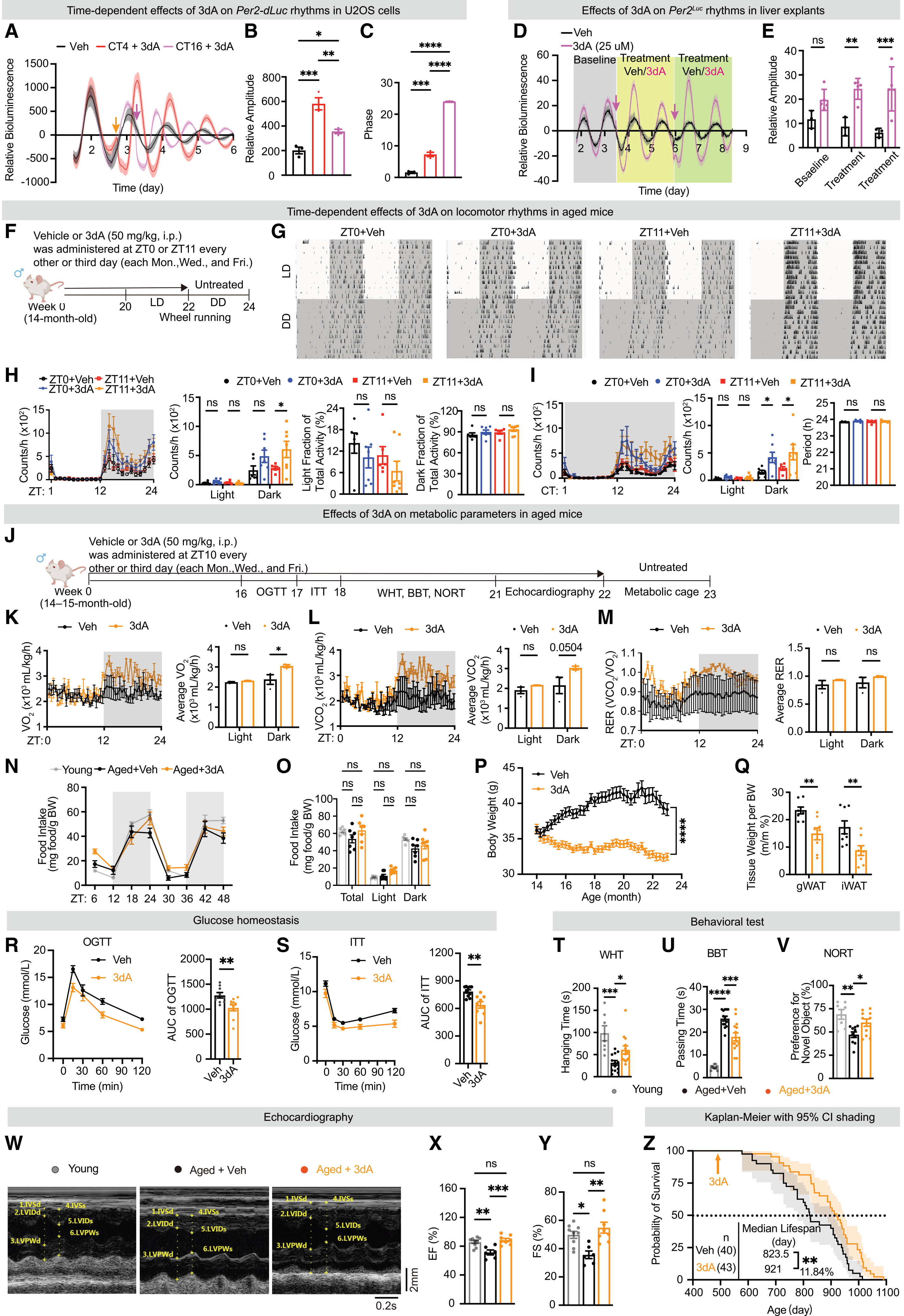
The authors first demonstrated that cordycepin (3dA) robustly amplifies clock gene oscillations in U2OS Per2-dLuc reporter cells and ex vivo liver explants from 18-month-old Per2Luc mice. Comparing dosing schedules (50 mg/kg i.p. every Monday, Wednesday, and Friday) in 14- to 15-month-old male mice revealed that administration just before dark onset (ZT10–ZT11)—unlike dosing at light onset (ZT0)—markedly restored dark-phase wheel-running activity and the diurnal amplitude of oxygen consumption (VO2) and carbon dioxide production (VCO2).
Chronic 3dA administration at ZT10–ZT11 restrained age-associated body weight and white adipose tissue gain, improved oral glucose and insulin tolerance (OGTT and ITT), and enhanced wire-hanging grip strength, balance-beam coordination, novel object recognition, and echocardiographic ejection fraction (EF%) and fractional shortening (FS%). Crucially, initiating 3dA treatment at 15 months of age extended median lifespan in male mice by 11.84%, from 823.5 days in vehicle controls to 921 days (p < 0.01).
Source: Haijiao Zhao et al. · Cell · © Elsevier Inc.
Original figure and English caption ↗
3dA Reverses Systemic Inflammation, Epigenetic Age, and Endocrine Rhythm Decay
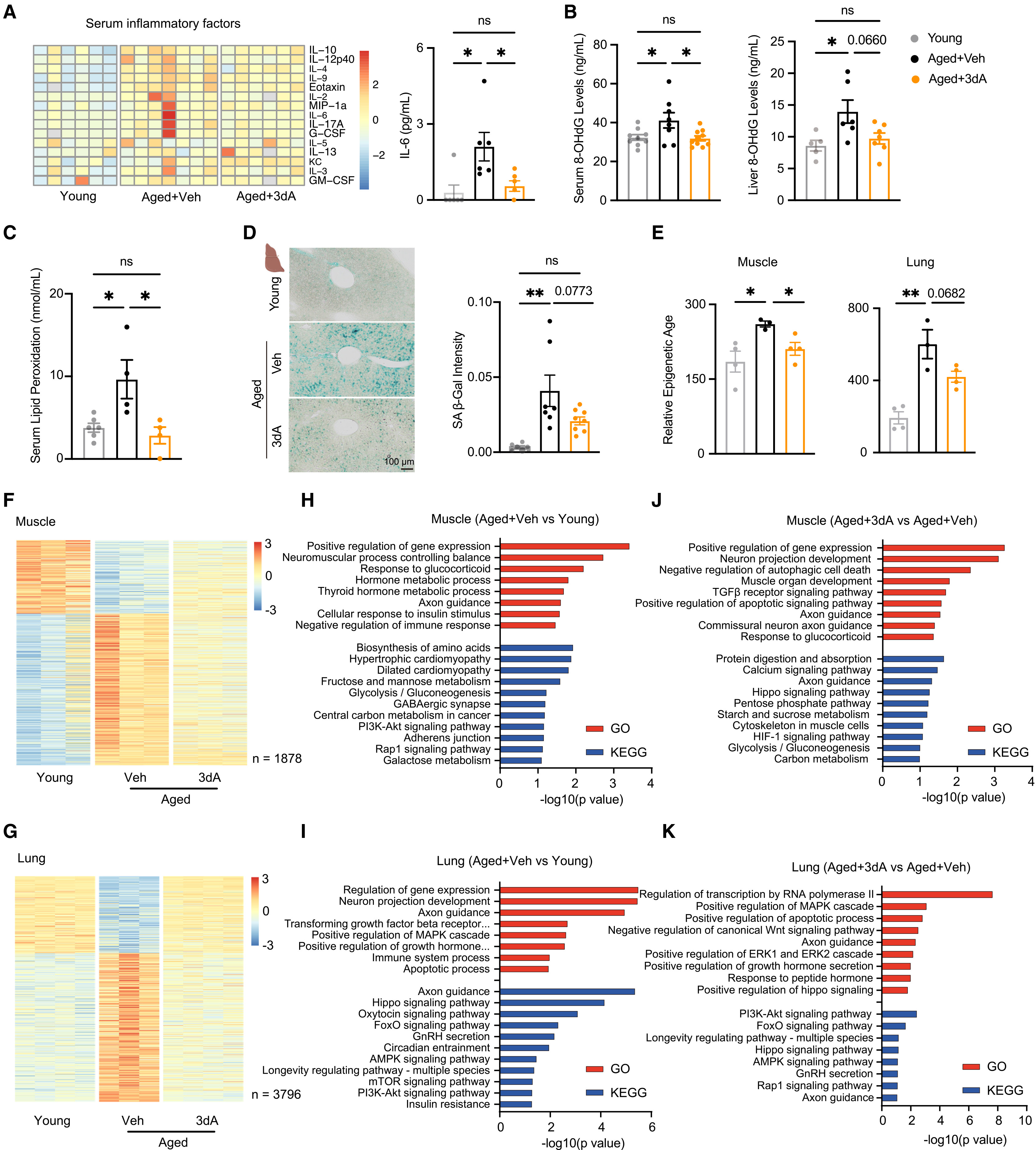
At the molecular and tissue level, 3dA treatment markedly reduced serum pro-inflammatory cytokines including IL-6, lowered serum and hepatic 8-OHdG and lipid peroxidation levels, and decreased hepatic senescence-associated beta-galactosidase (SA-beta-Gal) staining. Whole-genome bisulfite sequencing (WGBS) epigenetic clock analysis further demonstrated that 3dA significantly reversed age-associated DNA methylation drift, reducing epigenetic age in aged skeletal muscle and lung.
Complementary time-series hepatic RNA sequencing across six circadian time points (ZT0 to ZT20) and endocrine profiling (Figure 3) revealed that 3dA restored the oscillation amplitude and phase of age-dampened rhythmic transcripts and rejuvenated the blunted diurnal oscillations of serum corticosterone, testosterone, and liothyronine (T3) in aged mice, pointing toward an upstream central neuroendocrine mechanism.
Source: Haijiao Zhao et al. · Cell · © Elsevier Inc.
Original figure and English caption ↗
Brain-Wide Screening and In Vivo Recordings Pinpoint the Hypothalamic PVN Hub
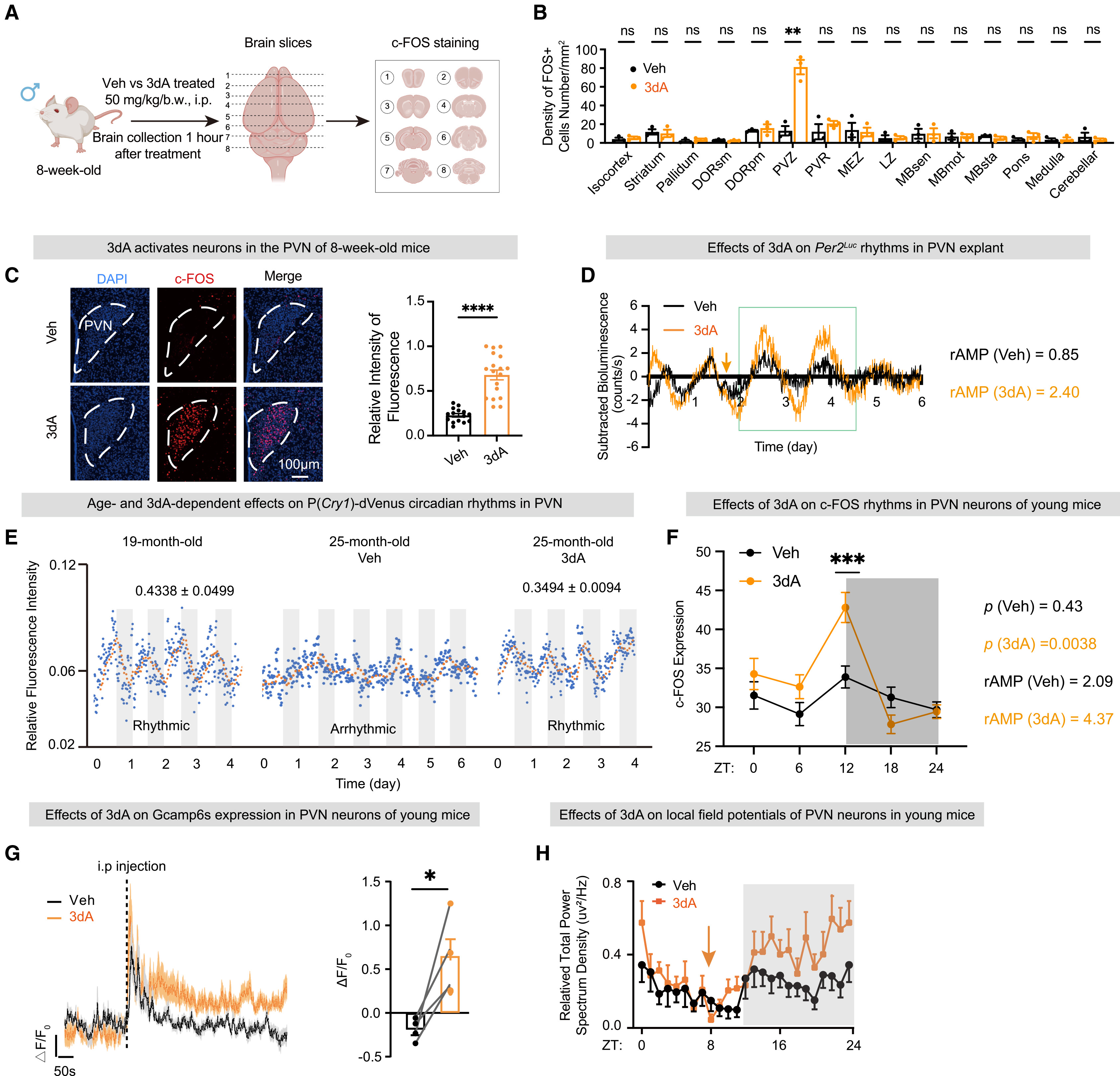
To identify the central target mediating 3dA's systemic effects, the authors quantified c-FOS induction across 14 brain regions following 3dA administration. Neuronal activation was most pronounced in the paraventricular zone, specifically within the hypothalamic paraventricular nucleus (PVN). Ex vivo bioluminescence recordings of Per2Luc PVN explants confirmed that 3dA boosted relative circadian oscillation amplitude from 0.85 to 2.40.
Longitudinal in vivo fiber photometry of the circadian reporter P(Cry1)-dVenus in the PVN of 25-month-old mice demonstrated that 3dA rescued age-abolished 24-hour fluorescence rhythmicity and doubled the diurnal amplitude of PVN c-FOS oscillation from 2.09 to 4.37. In vivo GCaMP6s calcium photometry and local field potential (LFP) recordings further confirmed that 3dA acutely elevates PVN neuronal calcium activity and restores dark-phase PVN network power.
Source: Haijiao Zhao et al. · Cell · © Elsevier Inc.
Original figure and English caption ↗
PVN-Specific Ruvbl2 Knockout Abolishes 3dA Systemic Anti-Aging Benefits
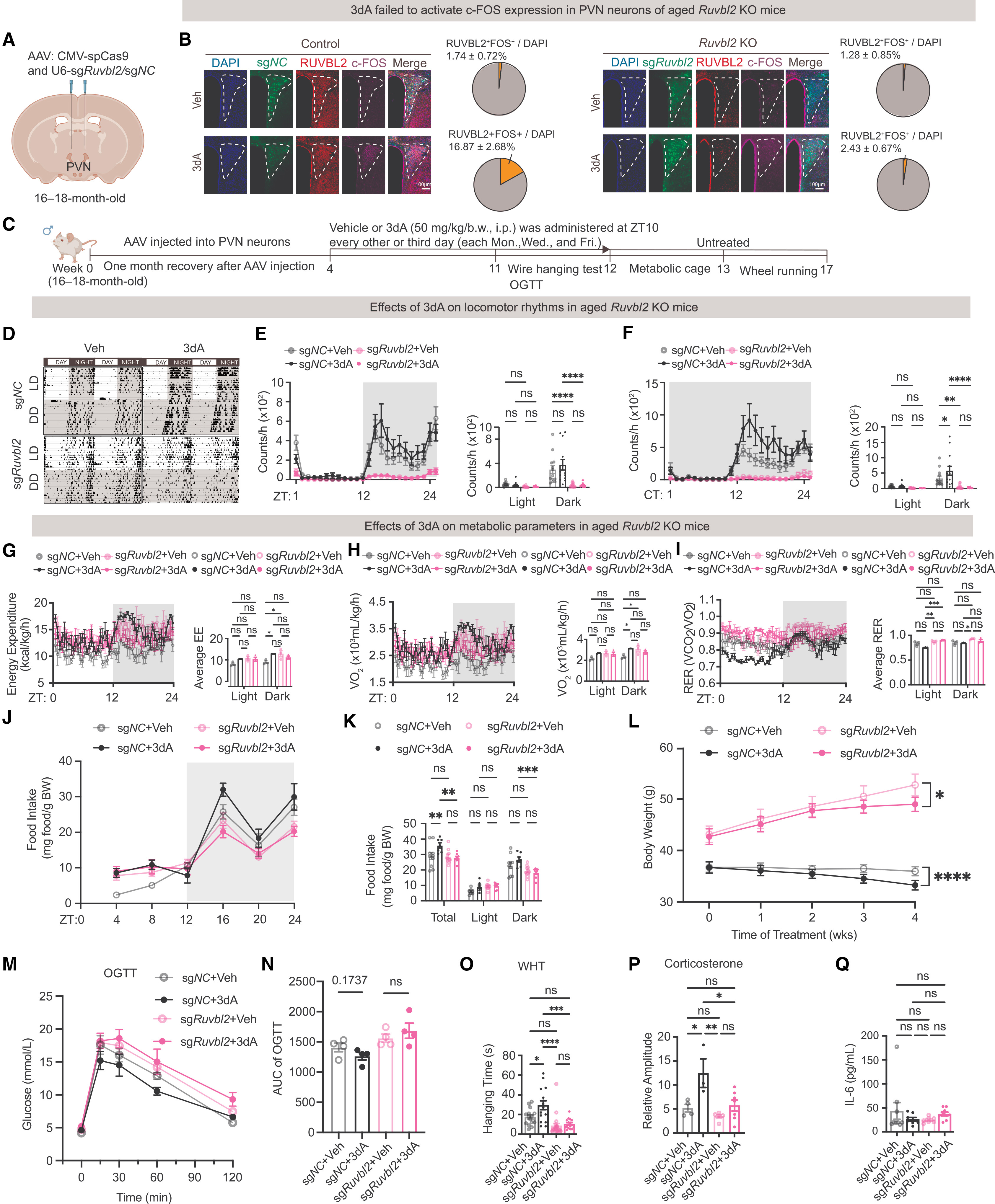
Because RUVBL2 is the direct molecular target through which 3dA modulates the core circadian clock complex, the authors tested whether PVN RUVBL2 is required for systemic rejuvenation by bilaterally injecting AAV-CMV-spCas9 and AAV-U6-sgRuvbl2 into the PVN of 16- to 18-month-old male mice. Whereas 3dA activated c-FOS in 16.87% of RUVBL2-positive PVN neurons in control mice, this response dropped to 2.43% in PVN Ruvbl2-knockout mice.
Crucially, PVN-specific CRISPR/Cas9 deletion of Ruvbl2 completely abolished 3dA-induced improvements in light-dark (LD) and constant-darkness (DD) locomotor rhythms, dark-phase energy expenditure and VO2, body weight control, oral glucose tolerance, wire-hanging strength, balance-beam coordination, serum corticosterone rhythmicity, and IL-6 suppression, proving that RUVBL2 signaling within the PVN is indispensable for systemic rejuvenation.
Source: Haijiao Zhao et al. · Cell · © Elsevier Inc.
Original figure and English caption ↗
Timed Chemogenetic Activation of PVN Neurons Phenocopies Systemic Rejuvenation
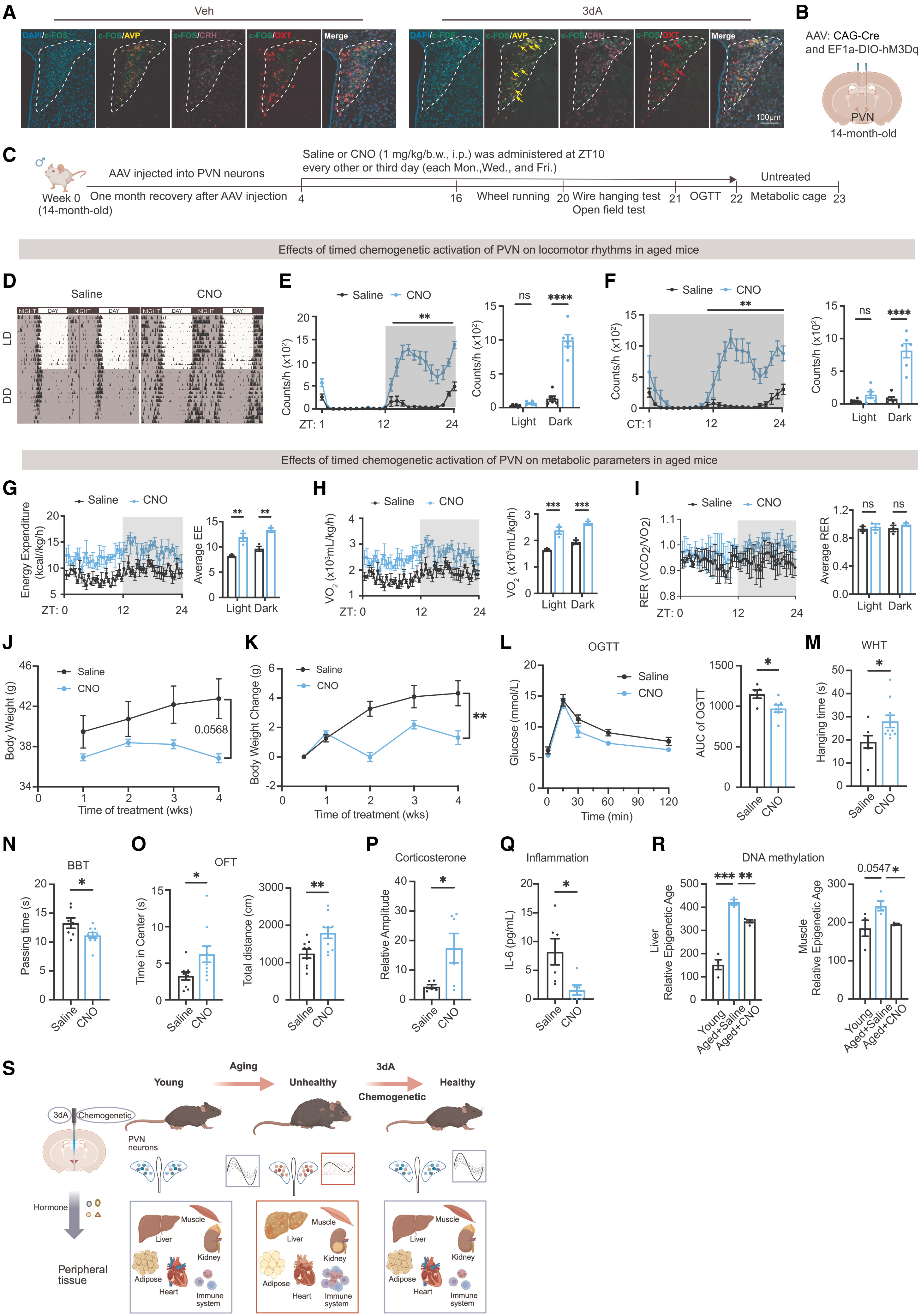
Double-labeling immunofluorescence revealed that 3dA-activated c-FOS-positive PVN neurons co-express the neuroendocrine peptides arginine vasopressin (AVP), corticotropin-releasing hormone (CRH), and oxytocin (OXT). To test whether directly boosting rhythmic PVN neuronal activity is sufficient to drive systemic rejuvenation, the authors expressed the excitatory DREADD hM3Dq bilaterally in the PVN of 14-month-old male mice and administered CNO on the same circadian schedule (Monday, Wednesday, and Friday at ZT10).
Timed chemogenetic activation of PVN neurons robustly restored diurnal and circadian wheel-running rhythms, elevated dark-phase energy expenditure and VO2, and improved glucose tolerance, wire-hanging strength, balance-beam performance, and open-field exploration. Moreover, rhythmic PVN activation restored serum corticosterone oscillation amplitude, reduced circulating IL-6, and significantly lowered DNA methylation epigenetic age in both liver and skeletal muscle, establishing PVN circadian restoration as a sufficiency hub for systemic anti-aging.
Source: Haijiao Zhao et al. · Cell · © Elsevier Inc.
Original figure and English caption ↗