Why does sleep loss impair the gut? Following a vagal pathway to intestinal stem cells
A mouse study links sleep deprivation to dorsal motor vagal output and enterochromaffin serotonin release, tracing how disrupted sleep affects intestinal stem cells.
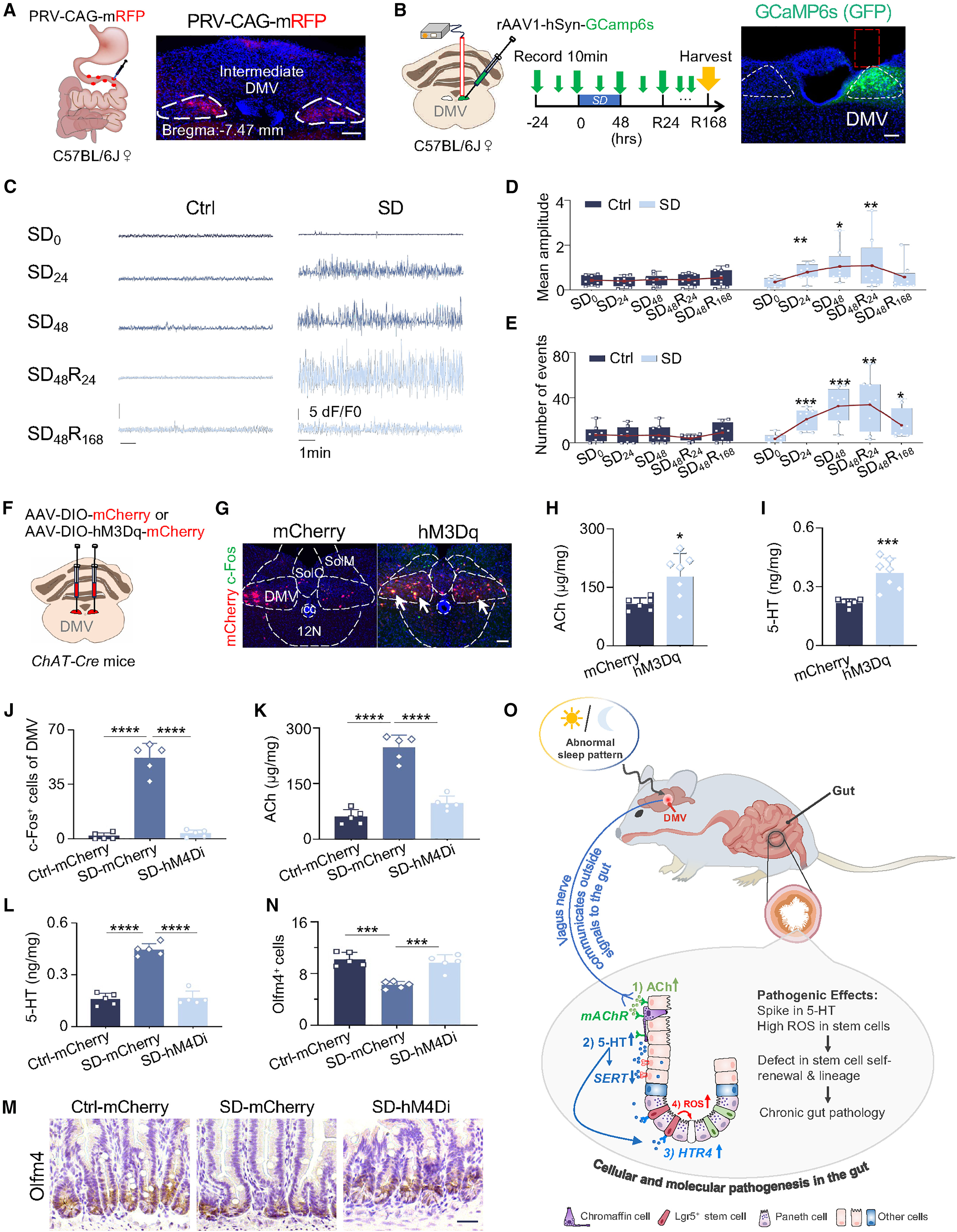
Sleep loss is a pervasive health challenge closely linked to gastrointestinal disorders such as inflammatory bowel disease (IBD). Rapid renewal of the intestinal epithelium every 3–5 days relies on Lgr5+/Olfm4+ intestinal stem cells (ISCs) residing at the crypt base, yet how sleep disturbance in the brain transmits long-range pathogenic signals to the gut to impair ISC self-renewal and mucosal regeneration has remained poorly understood. On February 1, 2026, Zhengquan Yu, Cong Lv, Feng Wang, Liping Wang, Maksim V. Plikus, and colleagues published a study titled "Sleep disturbance triggers aberrant activation of vagus circuitry and induces intestinal stem cell dysfunction" in Cell Stem Cell. Integrating retrograde viral tracing, in vivo fiber photometry, single-cell transcriptomics, and conditional genetics, the authors delineate a complete brain-to-gut cascade: sleep deprivation persistently hyperactivates cholinergic neurons in the dorsal motor nucleus of the vagus (DMV), driving vagal acetylcholine (ACh) release that acts on enterochromaffin CHRM3 receptors to trigger a gut serotonin (5-HT) surge, which in turn overactivates ISC HTR4 receptors to induce oxidative and endoplasmic reticulum stress.
Acute sleep deprivation triggers persistent hyperactivation of cholinergic neurons in the dorsal motor nucleus of the vagus (DMV), driving vagal acetylcholine release that stimulates enterochromaffin cells via CHRM3 to elevate gut serotonin (5-HT); excess 5-HT activates HTR4 receptors on intestinal stem cells, inducing ER stress and reactive oxygen species accumulation that impair stem cell renewal and regeneration.
Contents · 5 sections
Two-Day Acute Sleep Deprivation Impairs Intestinal Stem Cell Self-Renewal and Regeneration
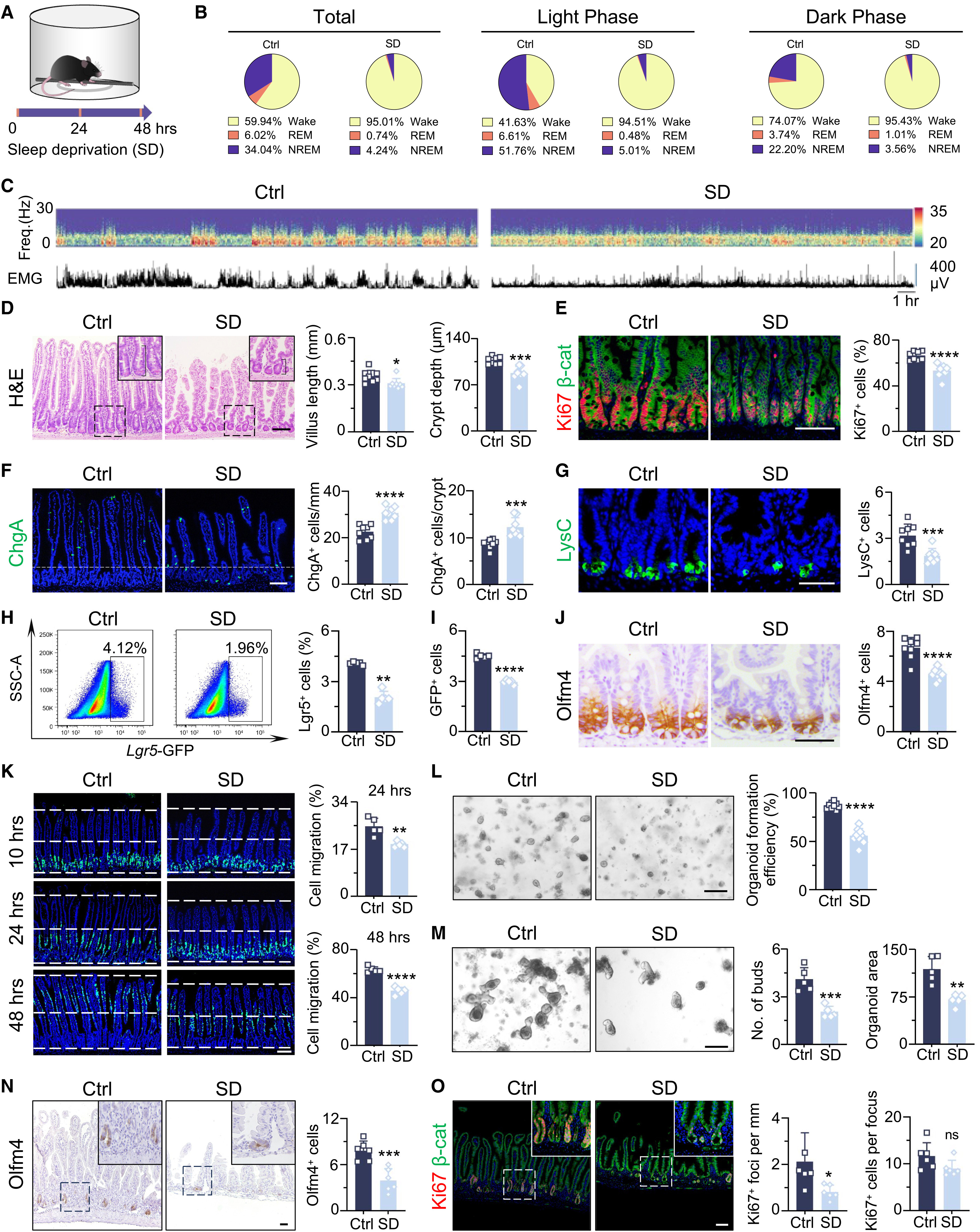
To capture early gut alterations without confounding systemic wasting, the authors subjected mice to 2 days (48 hours) of mechanical or multi-platform sleep deprivation (SD). Just 2 days of SD significantly shortened small intestinal villi and crypts, reduced Ki67-positive proliferating cells and lysozyme-positive Paneth cells, and expanded chromogranin A (ChgA)-positive enteroendocrine cells. In Lgr5-EGFP-IRES-CreERT2 reporter mice, SD reduced Lgr5-GFP+ and Olfm4+ intestinal stem cells (ISCs) by roughly half and slowed BrdU-labeled epithelial migration along the crypt-villus axis.
Ex vivo crypt culture assays confirmed that crypts isolated from SD mice exhibited markedly reduced organoid-forming efficiency, budding, and size, while in vivo mucosal regeneration following 12 Gy gamma-irradiation was severely compromised. Notably, depleting the gut microbiota with broad-spectrum antibiotics (ABX) failed to prevent SD-induced crypt hypoproliferation or ISC loss, indicating that acute sleep-loss-induced stem cell dysfunction operates independently of the microbiome.
Source: Mingxin Zhang et al. · Cell Stem Cell · © Elsevier Inc.
Original figure and English caption ↗
ER Stress, Integrated Stress Response, and Gut-Selective ROS Accumulation Drive ISC Injury
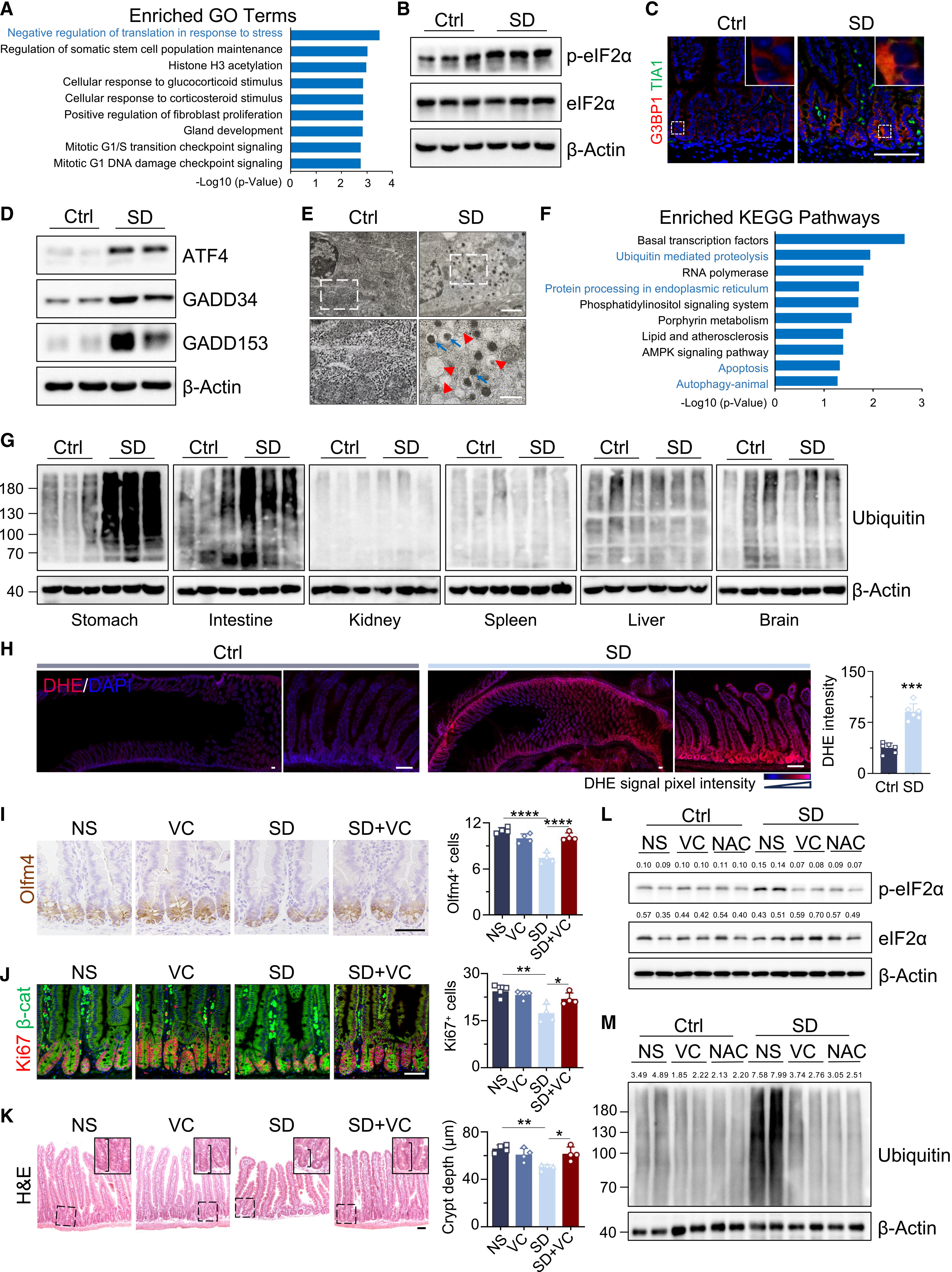
4D label-free quantitative proteomics of isolated intestinal crypts revealed that SD markedly downregulated Wnt/beta-catenin effectors (TCF1, LEF1, c-Myc) while strongly inducing endoplasmic reticulum (ER) stress and integrated stress response (ISR) pathways, evidenced by elevated p-eIF2alpha, ATF4, GADD34, and GADD153 (CHOP), widespread assembly of cytoplasmic G3BP1+/TIA1+ stress granules, K48-linked polyubiquitinated protein accumulation, and LC3B autophagy.
Dihydroethidium (DHE) staining revealed a striking organ-selective surge in reactive oxygen species (ROS) within the intestinal epithelium after 2-day SD—with no comparable ROS elevation in liver, lung, spleen, heart, kidney, or brain—accompanied by reduced Nrf2 expression and diminished antioxidant enzyme activities. Oral supplementation with the antioxidants vitamin C (VC) or N-acetylcysteine (NAC) abolished crypt ROS and stress granule formation and fully rescued Olfm4+ ISC numbers and epithelial proliferation.
Source: Mingxin Zhang et al. · Cell Stem Cell · © Elsevier Inc.
Original figure and English caption ↗
Enterochromaffin-Derived 5-HT Surge Acts on ISC HTR4 Receptors to Trigger Oxidative Stress
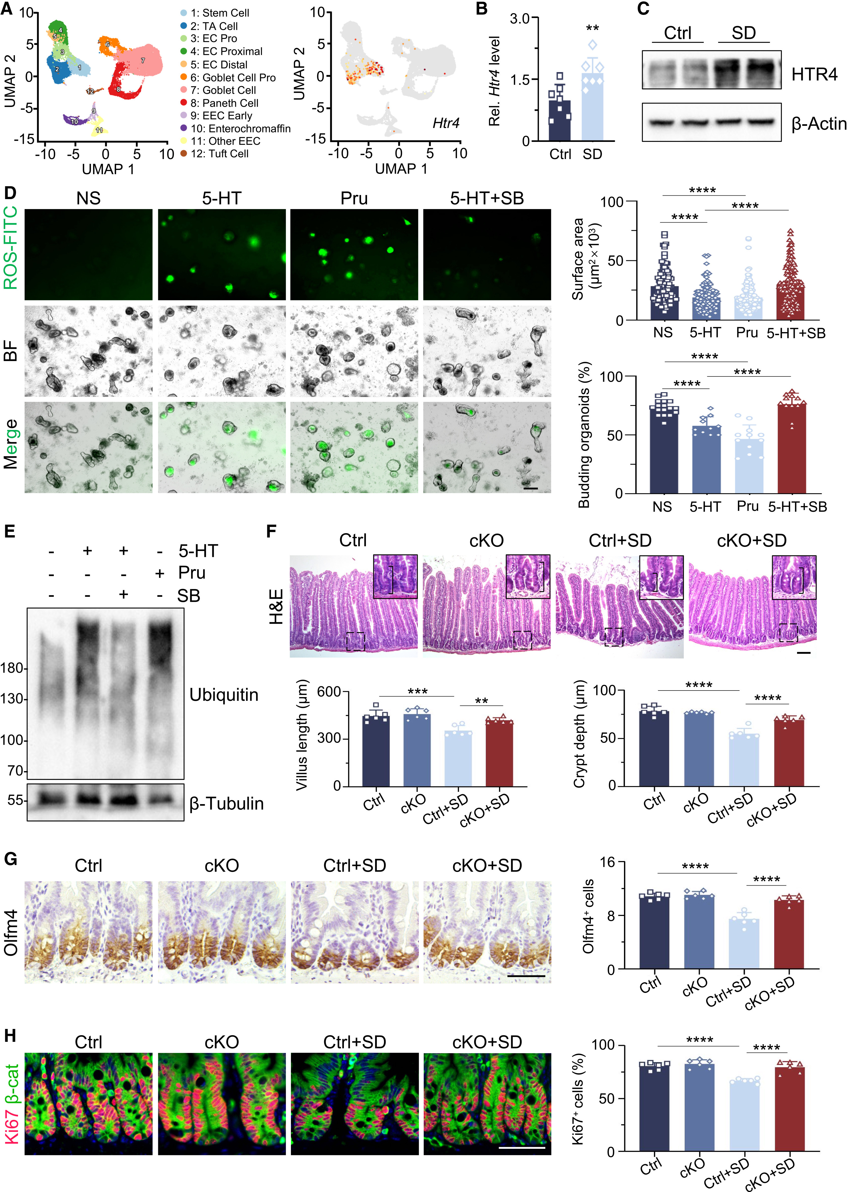
Targeted neurotransmitter metabolomics and ELISA identified serotonin (5-HT) as the most strongly upregulated neurotransmitter in the gut and serum after 2-day SD. This gut 5-HT spike resulted from both an expansion of Tph1-expressing enterochromaffin (EC) cells and promoter hypermethylation-driven downregulation of the serotonin reuptake transporter gene Slc6a4 (encoding SERT). In Tph1-/- mice lacking peripheral 5-HT synthesis, SD failed to induce gut ROS accumulation, stress granules, or ISC loss.
Single-cell RNA sequencing and in situ hybridization pinpointed Htr4 as the predominant 5-HT receptor enriched in ISCs and upregulated by SD. Administering the HTR4 agonist prucalopride phenocopied SD-induced crypt ROS accumulation and ISC depletion, whereas pharmacological HTR4 blockade (SB-203186 or GR113808) or conditional Htr4 deletion in intestinal epithelium (Villin-Cre;Htr4fl/fl) and Lgr5+ stem cells prevented cAMP/PKA/Ca2+ overload, ER stress, and ISC loss during SD.
Source: Mingxin Zhang et al. · Cell Stem Cell · © Elsevier Inc.
Original figure and English caption ↗
Subdiaphragmatic Vagotomy and Epithelial Chrm3 Deletion Block the Pathogenic Cascade
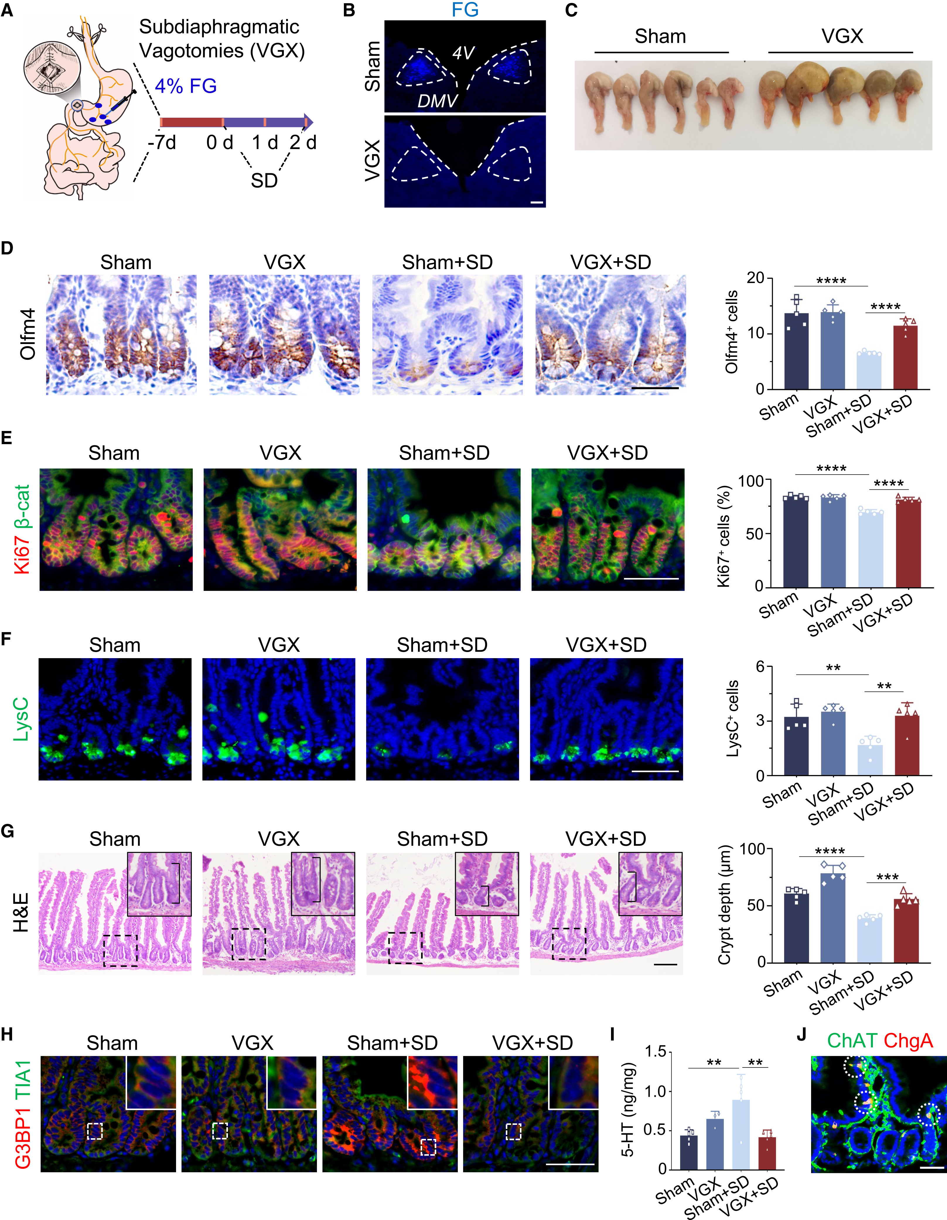
To determine how the sleep-deprived brain signals to the gut, the authors systematically tested three major brain-to-gut pathways. Neither blocking the HPA axis (via adrenalectomy or mifepristone) nor ablating sympathetic nerves with 6-hydroxydopamine (6-OHDA) rescued SD-induced ISC injury. By contrast, bilateral subdiaphragmatic vagotomy (VGX) completely prevented the SD-induced gut 5-HT surge, SERT downregulation, crypt stress granule assembly, and loss of Olfm4+ ISCs and Ki67+ proliferating cells.
Consistent with vagal hyperactivity, intestinal acetylcholine (ACh) levels rose markedly after SD in a vagotomy-sensitive manner. Exogenous ACh or carbachol directly stimulated EC cell 5-HT release and downregulated SERT, whereas blocking muscarinic receptors with atropine—unlike nicotinic blockade with hexamethonium bromide—or conditionally deleting the muscarinic M3 receptor in the intestinal epithelium (Villin-Cre;Chrm3fl/fl) abolished the SD-induced 5-HT surge and preserved ISC homeostasis.
Source: Mingxin Zhang et al. · Cell Stem Cell · © Elsevier Inc.
Original figure and English caption ↗
Persistent Hyperactivation of DMV Cholinergic Neurons Initiates the Brain-to-Gut Axis
To pinpoint the upstream central nucleus projecting to the small intestine during SD, the authors injected the retrograde trans-synaptic pseudorabies virus PRV-724 into the intestinal wall and performed brain-wide c-Fos mapping. Among gut-projecting brain regions, only the dorsal motor nucleus of the vagus (DMV) exhibited robust PRV+/c-Fos+ co-activation after SD, and PRV labeling in the DMV was completely abolished by subdiaphragmatic vagotomy.
Longitudinal in vivo GCaMP6s fiber photometry in the DMV revealed that both amplitude and frequency of spontaneous neuronal Ca2+ transients increased markedly at 24 and 48 hours of SD and remained persistently hyperactive for up to 7 days into recovery sleep before normalizing by day 14. In ChAT-Cre mice, chemogenetic activation (hM3Dq) of DMV cholinergic neurons was sufficient to elevate gut ACh and 5-HT and injure ISCs in well-rested mice, whereas chemogenetic inhibition (hM4Di) of DMV neurons during SD normalized gut ACh and 5-HT levels and preserved Olfm4+ intestinal stem cells.
Source: Mingxin Zhang et al. · Cell Stem Cell · © Elsevier Inc.
Original figure and English caption ↗