Why does feeding favor sleep? A vagal sensory route from the gut to hypothalamus
An adult male-mouse study follows upper-gut vagal inputs through NTS GABA cells toward hypothalamus, distinguishing feeding-related sleep from output-specific sleep structure.
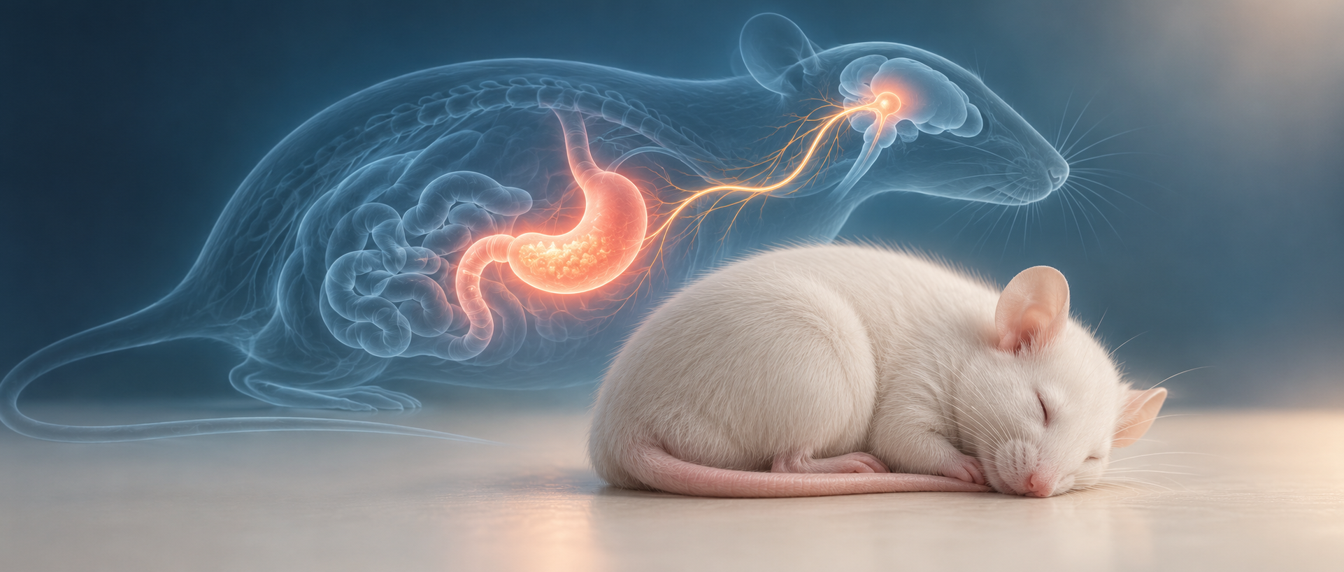
How does the body report digestive state to networks governing wake and sleep after a meal? Alongside nutrients and hormones, nerves can carry gut information directly to the brain. A December 2025 Nature Communications study follows stomach/duodenal vagal sensory inputs through brainstem nucleus of the solitary tract (NTS) and GABAergic outputs to hypothalamic PVH in adult male mice. Functional tests reveal an ascending route for feeding-related sleep and output-specific sleep organization.
Stomach/duodenal vagal sensory inputs promote NREM and contribute to the additional sleep after feeding. NTS GABA cells and their PVH output reduce wakefulness, with different effects on continuity from broad-cell and output-specific enhancement.
Contents · 6 sections
More sleep includes longer NREM bouts
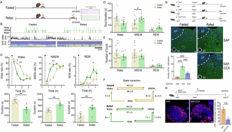
After overnight fasting and refeeding, mice showed less wakefulness and more NREM and REM. NREM bouts lengthened while bout counts and state-transition totals remained comparable. EEG and EMG identified sleep states, placing the effect in sleep structure rather than quiet behavior alone.
Feeding increased NTS activity labeling, which declined when upper-gut vagal sensory input was weakened. This connected gastrointestinal sensing with brainstem recruitment. Meals change nutrients, mechanics and hormones; the study followed a shared sensory route rather than assigning all effects to one stimulus.
Source: Chen K et al. · Nature communications · CC BY-NC-ND
Original figure and English caption ↗
Enhancing upper-gut signals increases NREM
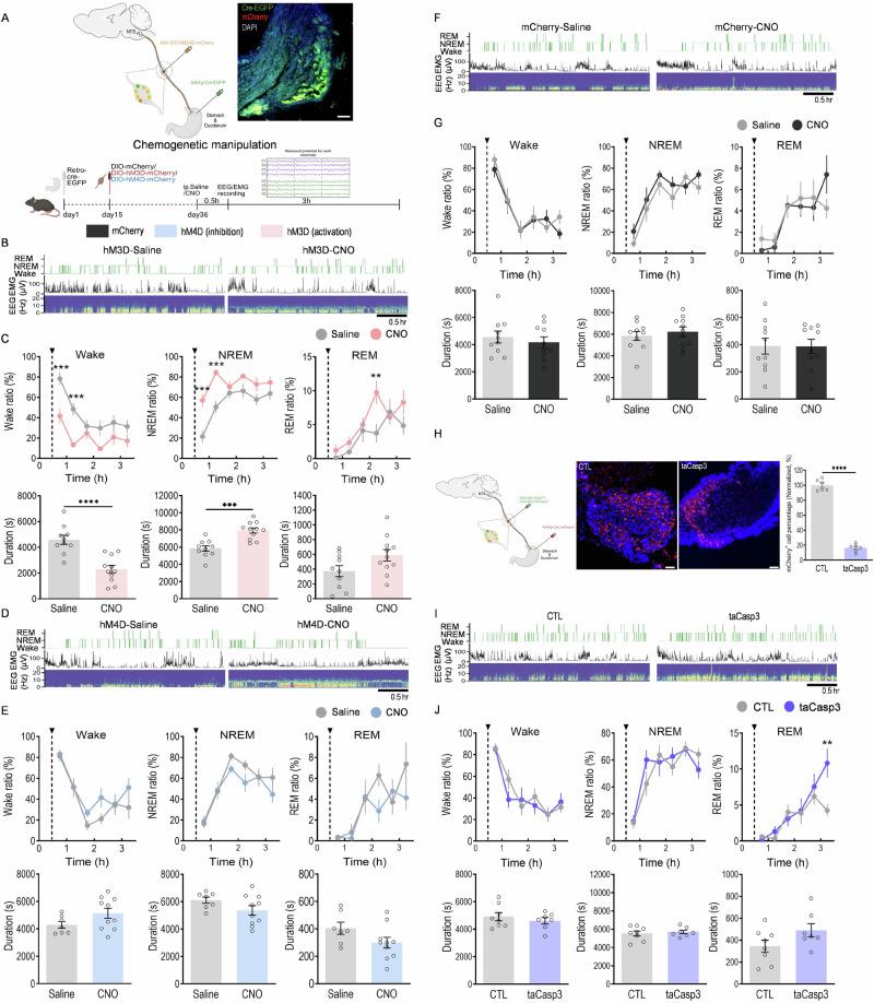
Enhancing stomach/duodenal vagal sensory cells reduced wakefulness and increased NREM totals and bout length, with REM totals broadly unchanged. Enhancement also promoted sleep during fasting. Similar manipulation of the studied small-intestinal sensory cells lacked matching effects, suggesting region-dependent input roles.
Suppressing or partially ablating these upper-gut cells did not clearly reduce general-state NREM totals, although structural measures such as wake bouts could change. The input thus responds to particular bodily states, while other systems also maintain ordinary sleep.
Source: Chen K et al. · Nature communications · CC BY-NC-ND
Original figure and English caption ↗
Suppression removes the clear feeding-related increase
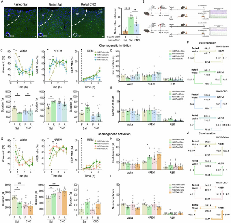
Controls showed less wakefulness and more NREM after refeeding; suppressing stomach/duodenal sensory inputs removed that clear feeding-related difference and weakened NTS recruitment. Enhancement and feeding-context suppression together established a functional contribution to postprandial sleep.
Together with general-state findings, the result distinguishes additional post-meal sleep from total sleep maintenance. Bodily state can add a feeding-related influence to existing regulatory systems rather than operate one independent sleep switch.
Source: Chen K et al. · Nature communications · CC BY-NC-ND
Original figure and English caption ↗
Feeding recruits excitatory and inhibitory groups
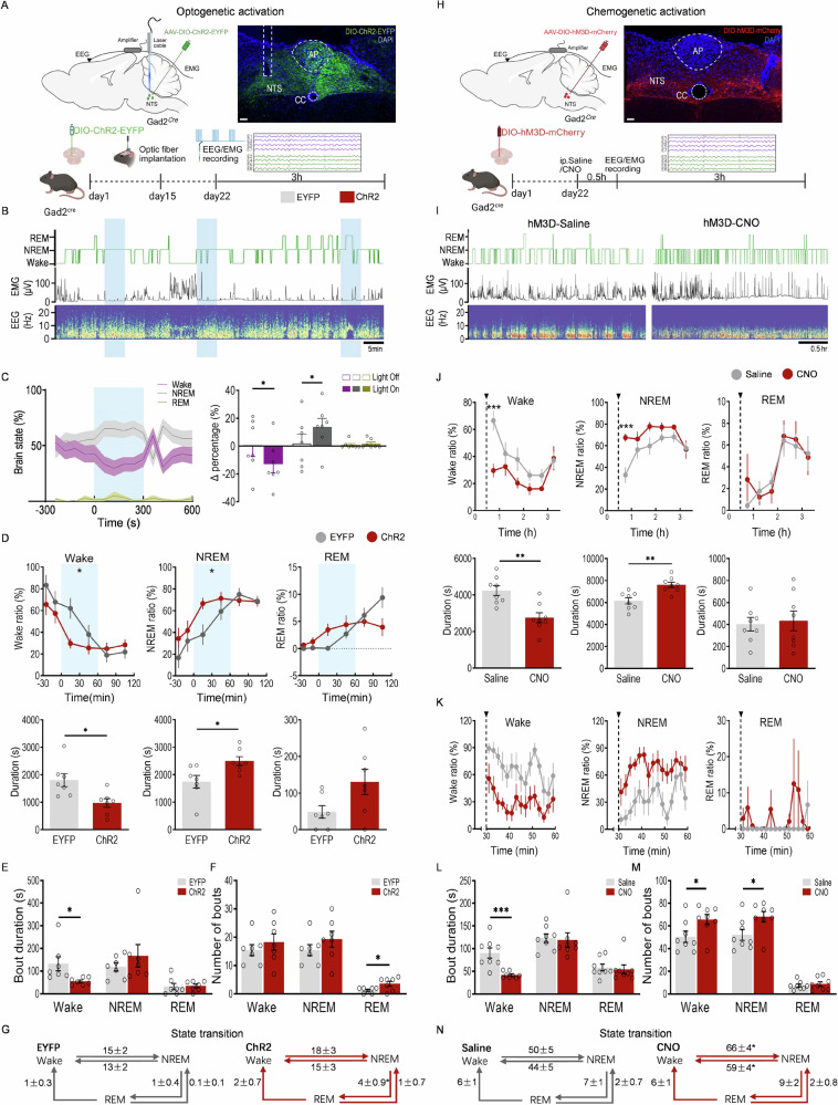
Feeding recruited glutamatergic and GABAergic NTS cells. Transsynaptic tracing and single-nucleus profiles identified several classes within upper-gut-input-associated labeled populations, with a substantial GABAergic fraction. Enhancing sensory input increased NTS GABA labeling, while suppression weakened its feeding response.
Enhancing NTS GABA cells reduced wakefulness and increased NREM, but sustained broad-population modulation also increased wake/NREM bout counts, producing fragmentation. More sleep did not automatically mean more continuous sleep. NTS also regulates autonomic functions, motivating output-specific analysis.
Source: Chen K et al. · Nature communications · CC BY-NC-ND
Original figure and English caption ↗
The PVH route produces a more continuous NREM pattern
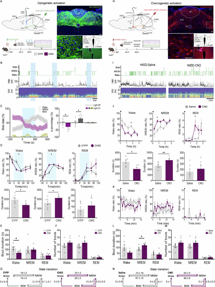
NTS GABA cells project to PVH and BNST among other sites. PVH-terminal enhancement reduced wakefulness and increased NREM; sustained local modulation lengthened NREM bouts without matching count or transition changes. This output did not produce the broad-population fragmentation pattern.
BNST output differed: optical stimulation did not clearly change sleep totals, while sustained chemogenetic modulation reduced wakefulness with a smaller NREM increase and comparable NREM bout length. Distinct outputs separated the brainstem population’s effects, with PVH particularly supporting wake suppression and NREM extension.
Source: Chen K et al. · Nature communications · CC BY-NC-ND
Original figure and English caption ↗
Bodily state enters sleep regulation through neural routes
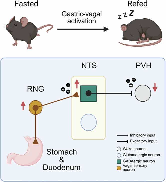
The proposed ascending model links stomach/duodenum, right nodose ganglion, NTS GABA cells and PVH. Several PVH classes support wakefulness, and inhibitory input may reduce their arousal drive. Recipient cells and obligatory steps remain to be resolved, while current evidence connects sensory, brainstem and hypothalamic components to feeding-related state changes.
The study concerns healthy adult male mice and short recordings, with sleep-disorder effects unresolved. It adds a neural account to postprandial somnolence: gut messages help organize subsequent wake and sleep alongside feeding, and their output route shapes continuity.
Source: Chen K et al. · Nature communications · CC BY-NC-ND
Original figure and English caption ↗