More alert, more connected? Locus coeruleus and an inverted-U relationship
Cross-species imaging and causal mouse experiments show that arousal shapes whole-brain functional connectivity, with changes depending partly on the starting brain state.
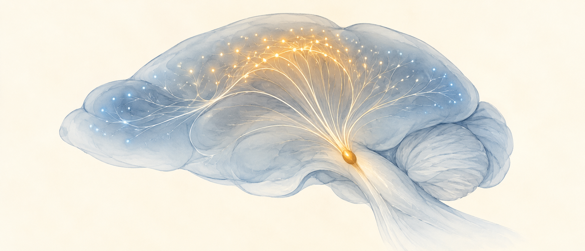
As the brain moves from drowsiness toward alertness, do connections between regions keep strengthening? A Nature Communications study finds a relationship with a turning point. Human task and resting observations, alongside several awake-mouse recordings, show higher global functional connectivity at intermediate arousal than at either end. Altering locus-coeruleus norepinephrine activity can drive corresponding changes. The starting brain state matters as well as the direction of the change.
Moving awake mice away from intermediate arousal in either direction, increasing or decreasing locus-coeruleus norepinephrine activity can lower global connectivity. Increasing activity under low-arousal anesthesia produces a different direction.
Contents · 6 sections
Connectivity here means signals fluctuating together
Functional connectivity describes correlations between regional signals over time, rather than newly formed axons or synapses. The study asks how these correlations change as a person or mouse naturally fluctuates in state during scanning. Anatomical routes can remain relatively stable while functional relationships vary over short periods.
Arousal is not simply a choice between waking and sleeping: wakefulness can range from drowsy to alert. EEG and pupil-related readouts track these fluctuations, allowing connectivity to be interpreted within the current state. This perspective can place apparently opposing findings within one broader relationship.
Intermediate arousal links connectivity with task performance
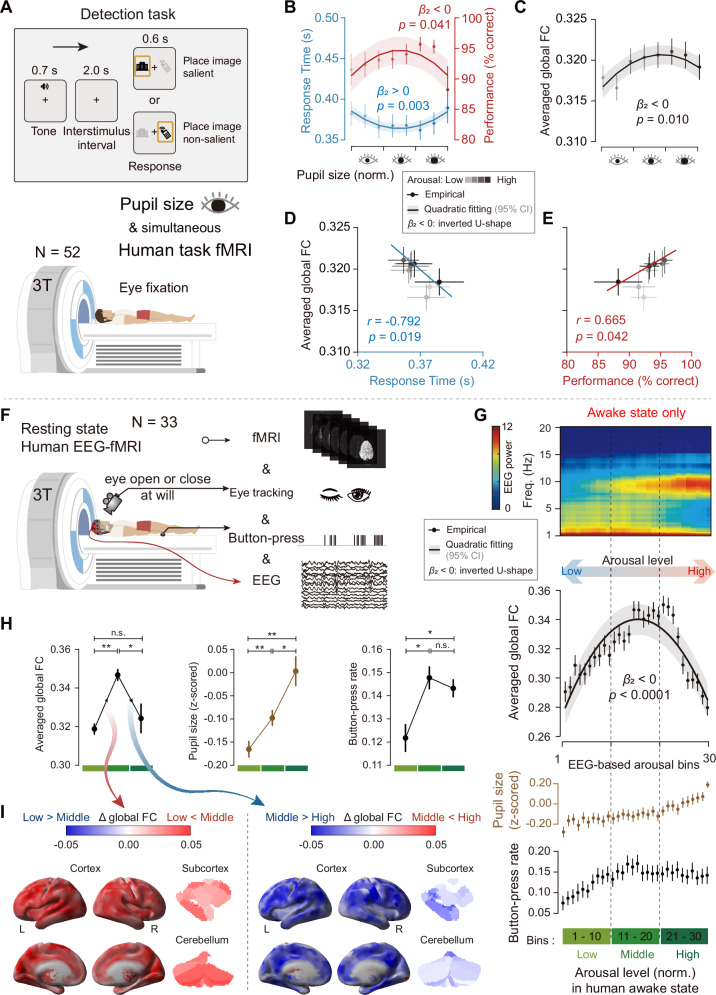
In a human visual-detection task, response time follows a U-shaped relationship with pupil-indexed arousal: responses are faster near the middle. Accuracy shows a weaker inverted-U pattern, as does global connectivity. Correlations between measures aggregated across arousal bins add a network-level clue to the association between state and task performance.
Resting observations yield a similar shape. Although recordings span sleep and wakefulness, the main connectivity analysis uses awake periods. Pupil size rises with EEG-indexed arousal, while connectivity first rises and then falls. Low and high arousal do not significantly differ in connectivity, so similar values can occur in distinct states.
Source: Tong C et al. · Nature communications · CC BY-NC-ND
Original figure and English caption ↗
Mouse networks show a similar turning point
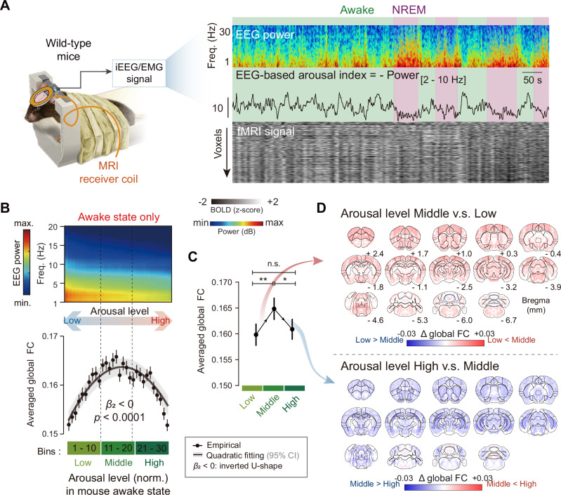
Combined EEG and functional imaging in awake mice also show higher global connectivity at intermediate arousal. Independent wide-field calcium analysis yields a similar relationship, extending the observation beyond blood-flow-related imaging. The readouts describe state-related activity from different perspectives and share related spatial patterns.
Regional sensitivity differs within the global average. Sensorimotor and attention-related networks, together with several brainstem regulatory nodes, respond strongly to arousal fluctuations. In mice, this distribution has a relatively strong spatial relationship with locus-coeruleus norepinephrine projections, bringing a small brainstem population into the explanation of whole-brain dynamics.
Source: Tong C et al. · Nature communications · CC BY-NC-ND
Original figure and English caption ↗
From the same starting point, either direction can lower connectivity
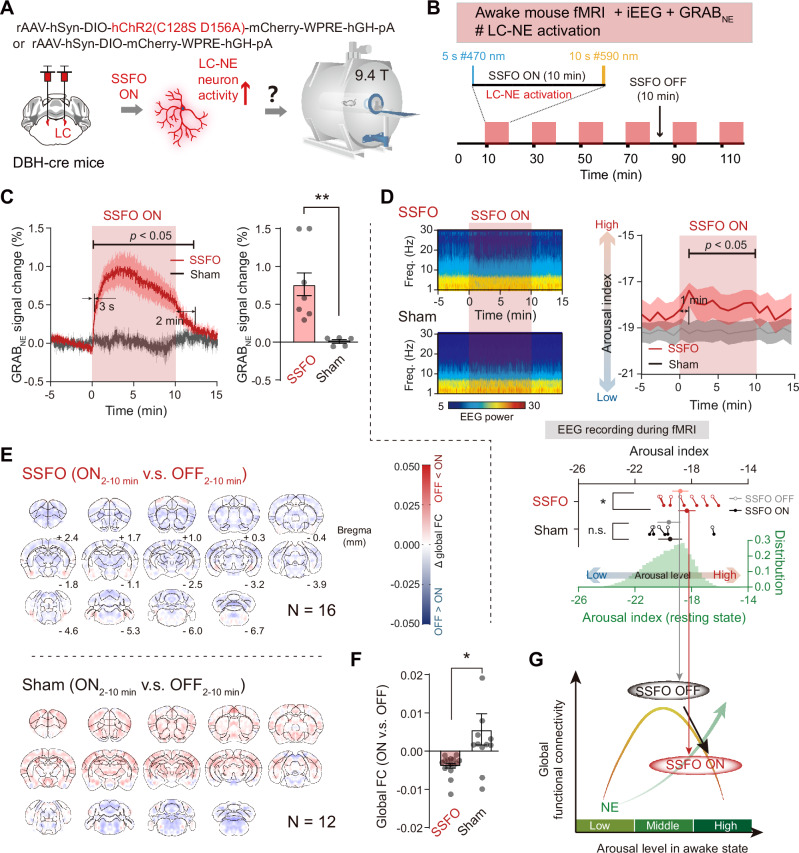
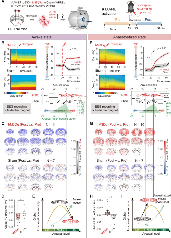
In simultaneous recordings, locus-coeruleus calcium changes precede cortical norepinephrine-associated signals and EEG arousal changes. Directly increasing locus-coeruleus activity in awake mice raises cortical transmitter signals and arousal, yet lowers global connectivity, matching the move from intermediate toward higher arousal.
Reducing activity lowers norepinephrine-associated signals and arousal, and also lowers connectivity, matching the move toward lower arousal. Together, the experiments show that the regulatory system can drive network changes without a simple more-transmitter–stronger-connectivity relationship. Moving away from the middle in either direction can leave the higher part of the curve.
Source: Tong C et al. · Nature communications · CC BY-NC-ND
Original figure and English caption ↗
The same increase has a different direction from low arousal
Another comparison examines wakefulness and isoflurane anesthesia. Increasing locus-coeruleus-associated activity raises arousal in both, but connectivity decreases in awake mice and increases under anesthesia. Different starting states allow the same direction of modulation to accompany different network outcomes, helping explain connectivity increases in earlier anesthetized studies.
Anesthesia changes neural and vascular baselines. The two sets of observations highlight how increasing locus-coeruleus activity can accompany different connectivity changes from different starting states. Bidirectional manipulation during wakefulness supplies causal evidence for this nonlinear relationship.
Source: Tong C et al. · Nature communications · CC BY-NC-ND
Original figure and English caption ↗
Arousal shapes the strength of regional relationships
The study joins human network associations with causal modulation in mice, showing that global connectivity varies nonlinearly with arousal. Human task performance and connectivity covary across arousal bins, while mouse experiments show how locus-coeruleus activity drives network changes. These complementary levels make state fluctuations during wakefulness part of the interpretation of imaging results.
Locus-coeruleus norepinephrine is one node capable of driving these dynamics, with other regulatory systems potentially contributing. Stronger connectivity has no context-free meaning: similar values can occur at opposite ends of the relationship, and the same direction of modulation can yield different outcomes from different starting states.