Why do two excitatory connections produce different anesthetic responses? Brainstem, thalamus and prefrontal cortex
A male-mouse propofol study follows righting reflex and EEG, finding different effects from brainstem–thalamic and thalamic–prefrontal activity changes.
Movement and EEG change during anesthetic induction and need not return together during recovery. A 2026 Drug Design, Development and Therapy study follows parabrachial nucleus, paraventricular thalamus and medial prefrontal cortex in male mice. Increasing brainstem–thalamic activity delays loss of righting and accelerates recovery; increasing thalamic–prefrontal activity shifts timing in the opposite direction.
Brainstem–thalamic activity has a relatively consistent bidirectional relationship with righting timing and cortical activity, while thalamic–prefrontal behavioral and spectral results diverge.
Contents · 6 sections
Righting and EEG offer different windows
Brainstem, thalamus and prefrontal cortex occupy different positions in state-regulating networks. Following connections during anesthesia asks how behavior and cortex change as a signal crosses a segment, beyond whether a region’s activity rises or falls. Comparing routes reveals structure and function separately.
Loss and recovery of righting provide behavioral landmarks. EEG describes cortical spectra and burst suppression, while deeper-anesthesia scores include breathing and limb activity. Together, the measures capture action, electrical activity and bodily responses.
When measures align or diverge is informative in itself. Recovery is more than one instant between stillness and movement: bodily responses can appear at different times, and cortical bands can change differently. Relating them to connections reveals the broader process.
Two connections bring three regions into one question
Tracing supports excitatory parabrachial (PB) input to paraventricular thalamus (PVT) and PVT output to medial prefrontal cortex (mPFC). The two segments were examined separately to ask whether they affect behavior and EEG similarly under propofol.
Regional connections do not assign the same task to every cell. PVT input recipients and cortical-projecting populations may contain different combinations. Studying segments separately reveals contributions beneath the overall structure and highlights source and target.
Brainstem-derived signals decline under anesthesia
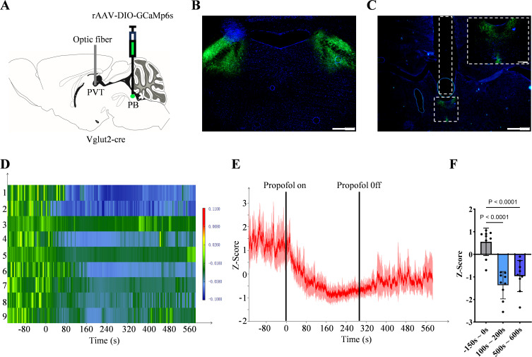
PB-derived terminal calcium signals in PVT decline during propofol and remain reduced for a period after withdrawal, linking the connection with the anesthetic trajectory.
The team then altered terminal activity while observing righting and EEG. Recording described when activity changed; interventions examined what changed with it, connecting induction, deeper anesthesia and recovery.
Source: Wang J et al. · Drug design, development and therapy · CC BY-NC
Original figure and English caption ↗
Brainstem–thalamic activity influences induction and recovery
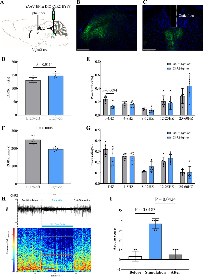
Increasing PB–PVT activity delays loss and accelerates recovery of righting, reducing delta power at loss. During deeper anesthesia, burst suppression declines and behavioral scores increase, chiefly through breathing and limb activity.
Reducing activity produces earlier loss and later recovery, with spectra shifting toward suppression. Recovery EEG comparisons are not all significant despite timing changes. The projection contributes at both ends of the transition.
Source: Wang J et al. · Drug design, development and therapy · CC BY-NC
Original figure and English caption ↗
Toward prefrontal cortex, behavior and EEG diverge
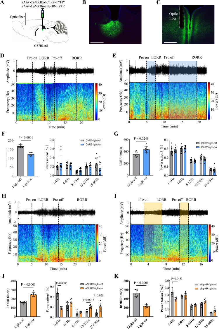
PVT–mPFC differs: activation advances loss and delays recovery without equivalently clear spectral changes. Inhibition delays loss and accelerates recovery with lower low-frequency and some higher-frequency activity.
The prefrontal output reveals dissociation between EEG and behavioral timing. Sources and targets shape outcomes as signals enter local cells and circuits, producing different overall responses.
The thalamus receives brainstem signals and sends activity toward cortex, yet those contributions do not simply add together. Activation of the two segments shifts righting timing in opposite directions, and the prefrontal segment’s behavioral changes need not coincide with clear spectral shifts. Different connections reveal different aspects of one anesthetic process: a signal’s position in the network matters alongside regional identity and transmitter type.
Source: Wang J et al. · Drug design, development and therapy · CC BY-NC
Original figure and English caption ↗
Similar connections can yield different network effects
The work situates anesthetic changes within brainstem, thalamic and cortical connections. Projections and measures differ, and behavioral timing together with EEG reveals network functions beneath similar transmitter identities.
The findings concern male mice under propofol. Excitatory identity alone does not determine a network’s final direction; recipients and state shape how signals become behavior and electrical activity.