How does thalamic reticular activity influence anesthesia and emergence?
A male-mouse study links anterior thalamic reticular GABAergic cells to isoflurane induction and recovery from two anesthetics. Activation and inhibition have asymmetric effects, while behavioral and EEG changes reveal different dimensions.
Entering anesthesia, remaining anesthetized and recovering responsiveness are more than a single switch. Drugs act on a brain whose existing networks also change state. A study published in CNS Neuroscience & Therapeutics on June 3, 2024 examines anterior thalamic reticular nucleus, an inhibitory region involved in thalamic processing and brain rhythms. In adult male mice, increasing relevant cell activity hastens loss of righting under isoflurane and delays righting recovery after propofol or isoflurane. Behavioral and EEG recordings reveal cellular contributions and distinct rhythmic changes during these transitions.
Activating anterior thalamic reticular GABAergic populations facilitates isoflurane induction and delays emergence. Inhibiting the broader GABAergic population mainly delays isoflurane induction without significantly changing recovery from either anesthetic. Activation strategies also produce different EEG spectral patterns.
Contents · 7 sections
An activity signature during anesthesia
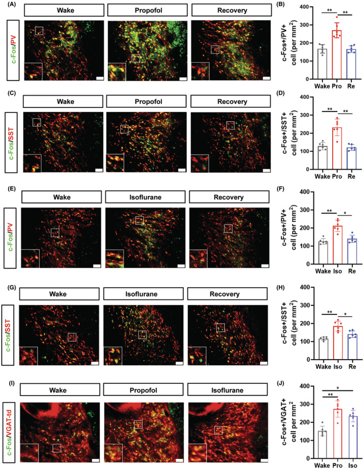
The study first compared the activity-related marker c-Fos across wakefulness, anesthesia and recovery. PV- and SST-labeled anterior reticular cells showed more c-Fos colabeling during propofol or isoflurane anesthesia, followed by a decline during recovery. Anesthesia therefore accompanied increased activity-related labeling in this inhibitory population.
Around two fifths of the examined cells expressed both PV and SST. The labels therefore identify overlapping rather than completely separate populations, providing cellular context for subsequent interventions.
Source: Yi R et al. · CNS neuroscience & therapeutics · CC BY
Original figure and English caption ↗
PV-cell activation delays recovery
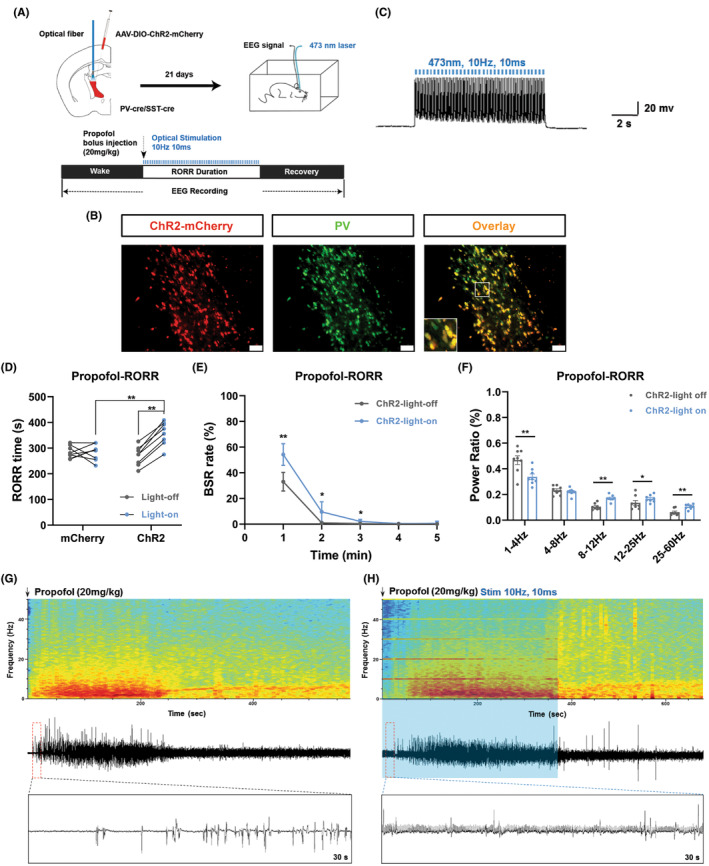
The propofol experiments focused on recovery of righting after injection. Activating anterior reticular PV cells prolonged recovery time and increased the EEG burst-suppression ratio. This measure describes alternation between activity bursts and relatively suppressed intervals, adding an electrical dimension to the behavioral change.
The findings link recovery timing to a defined population. Because righting was rapidly lost after propofol administration, this comparison addressed emergence. The isoflurane experiments then examined both induction and recovery.
Source: Yi R et al. · CNS neuroscience & therapeutics · CC BY
Original figure and English caption ↗
Faster induction and slower emergence
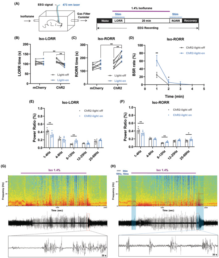
Under isoflurane, PV-cell activation shortened the time to loss of righting and prolonged recovery after anesthesia. The changes link this population to both entry and exit, with increased burst suppression during emergence.
Induction and emergence lie at opposite ends of an anesthetic episode but are distinct physiological transitions. Recording both exposes stage-dependent contributions. Here, activation consistently favored the onset or persistence of the state associated with lost righting.
Source: Yi R et al. · CNS neuroscience & therapeutics · CC BY
Original figure and English caption ↗
SST-labeled cells show related behavioral effects
SST-labeled cells showed related effects: delayed righting recovery after propofol, faster induction and slower recovery under isoflurane, with increased burst suppression. Findings across the labeling strategies provide additional evidence for an anterior reticular contribution.
PV and SST overlap substantially here, so the manipulations may recruit shared cells. The study does not separate the roles of shared and independently labeled populations, leaving a question about anterior reticular organization.
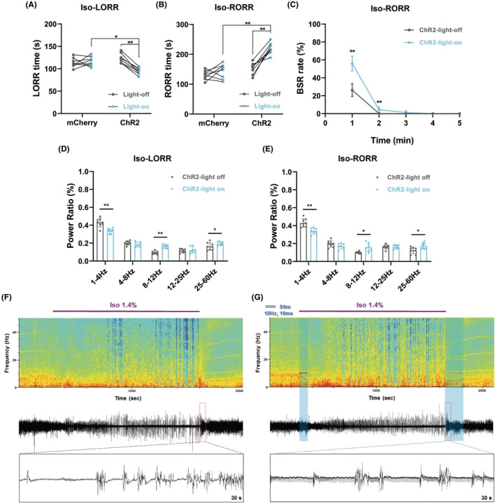
Source: Yi R et al. · CNS neuroscience & therapeutics · CC BY
Original figure and English caption ↗
Recovery slows with a different EEG pattern
Sustained PV-cell activation again delayed righting recovery after propofol and increased burst suppression. Its spectral pattern differed, however: relative delta power increased while relative gamma power decreased, unlike the preceding optogenetic findings.
Similar behavioral outcomes thus accompanied different EEG spectra. Because thalamic reticular cells contribute to brain rhythm generation, activation timing may influence the organization of cortical signals. The findings connect cellular activity, behavioral recovery and spectral composition while revealing their distinct dimensions.
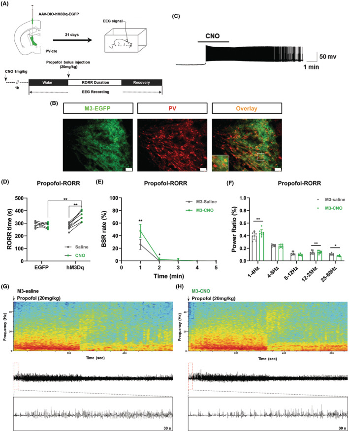
Source: Yi R et al. · CNS neuroscience & therapeutics · CC BY
Original figure and English caption ↗
Effects extend across isoflurane stages
Under isoflurane, sustained PV activation again hastened loss and delayed recovery of righting. Burst suppression increased during maintenance and emergence, while relative delta power increased across induction, maintenance and emergence. Behavioral timing and electrical changes provide complementary evidence for an anterior reticular contribution.
Different activation strategies slowed recovery without producing identical spectra. The authors propose that rhythmic activation may recruit EEG activity related to the stimulation rhythm and redistribute relative band power. Further recordings and interventions are needed to test that explanation and clarify the rhythmic contribution.
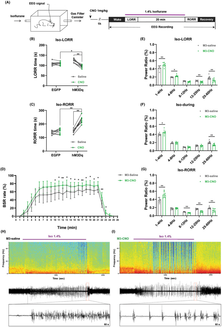
Source: Yi R et al. · CNS neuroscience & therapeutics · CC BY
Original figure and English caption ↗
Where does inhibition have its strongest effect?
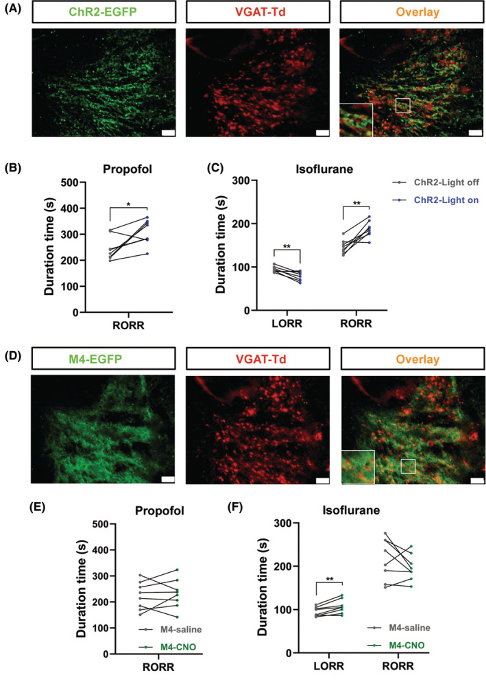
Activating the broader VGAT-labeled GABAergic population delayed propofol recovery and hastened induction while delaying emergence under isoflurane. Inhibition prolonged isoflurane induction but did not significantly change recovery from either anesthetic, revealing a stage-dependent asymmetry.
From activity-related labeling to intervention, this male-mouse study places anterior thalamic reticular cells within the network of anesthetic transitions. Entry and recovery are not a simple reversal, and one population can influence the stages differently. Separating overlapping populations and linking their rhythmic effects to downstream networks remain important questions.
Source: Yi R et al. · CNS neuroscience & therapeutics · CC BY
Original figure and English caption ↗