Still active under anesthesia: a sleep-related midbrain population
Sevoflurane does not silence every neuron. A sleep-related mouse midbrain population becomes more active, with GHSR providing a clue to its contribution to anesthesia.
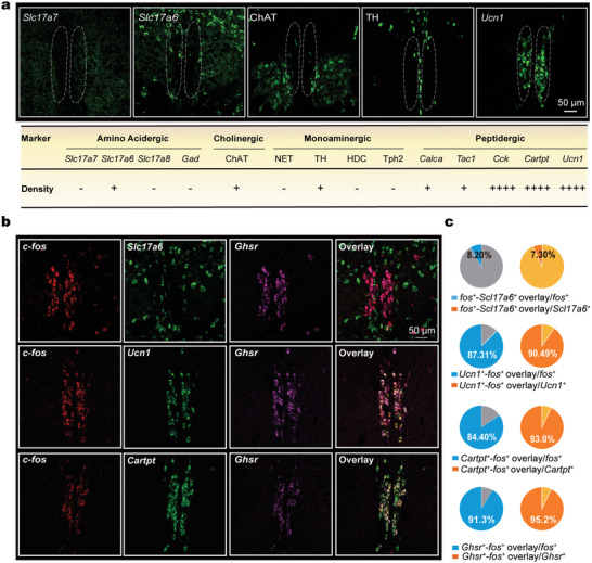
General anesthesia readily brings to mind a brain becoming quieter. Yet some cells may be working harder within that quieter state. A study published in Advanced Science on March 24, 2023 examined the centrally projecting Edinger–Westphal region, EWcp, in the mouse midbrain. One population became more active during sevoflurane anesthesia and was also more active during natural sleep. Following this clue linked an excitatory response involving the growth hormone secretagogue receptor, GHSR, to changes in anesthetic induction and emergence.
Sevoflurane-responsive cells largely carry UCN1 and CART markers and are enriched for GHSR. Suppressing the population or blocking local GHSR delayed induction and accelerated emergence in mice, implicating sleep-related excitation in sevoflurane action.
Contents · 6 sections
A population becomes busier as anesthesia begins
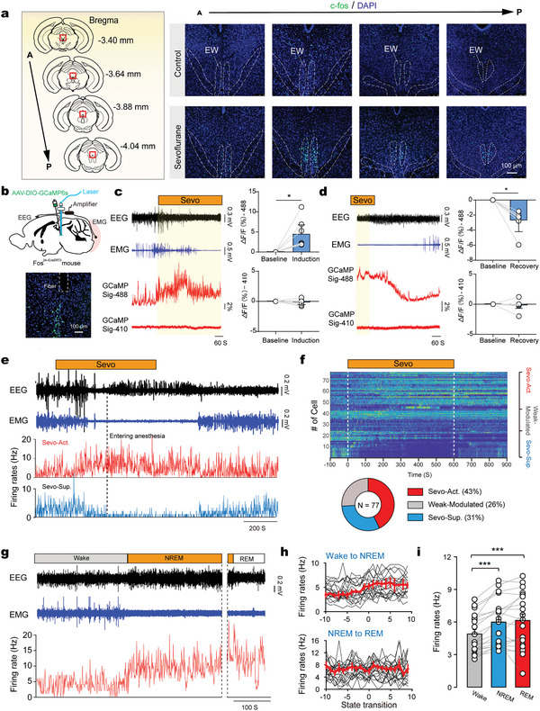
Activity mapping revealed a prominent cluster in EWcp. Calcium signals rose after sevoflurane began and declined during recovery after withdrawal. Electrical recordings also identified excited, suppressed and weakly modulated cells in the region, demonstrating heterogeneous local responses.
Among 77 recorded neurons, approximately 43% increased firing during sevoflurane anesthesia and 31% decreased it. Opposing responses across populations suggest that excitation of a selected group can contribute to a broader state transition.
Source: Yi T et al. · Advanced science (Weinheim, Baden-Wurttemberg, Germany) · CC BY
Original figure and English caption ↗
The same responsive cells are more active during natural sleep
Following 19 anesthesia-responsive neurons revealed higher average firing during both NREM and REM sleep than wakefulness. Activity rose before NREM onset, whereas no significant change appeared across the NREM–REM transition. These observations connect the population with sleep without erasing distinctions between its stages.
Shared cellular substrates are an appealing explanation for links between sleep and anesthesia. Yet sleep permits arousal by appropriate stimuli, whereas anesthesia involves sustained exogenous drug actions across additional molecular targets. A commonly active population offers a window onto their relationship, while its place in wider networks remains important.
Reducing activity changes entry into and recovery from anesthesia
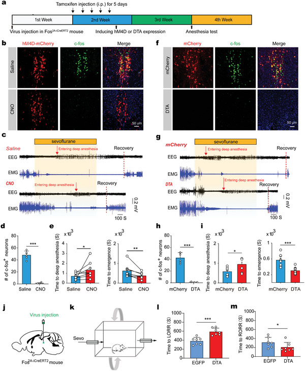
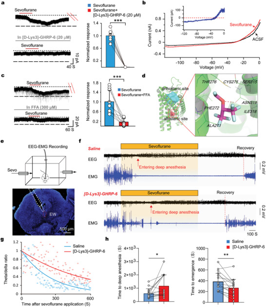
Suppressing the responsive population prolonged the time to an EEG-defined anesthetic state and accelerated recovery after sevoflurane withdrawal. Reducing the population produced changes in the same direction, with loss and recovery of righting providing complementary behavioral evidence.
EEG and righting behavior describe different aspects of the state. Together they support a contribution to anesthesia. Animals could still enter anesthesia, indicating altered induction and recovery dynamics alongside continuing actions at other targets.
Source: Yi T et al. · Advanced science (Weinheim, Baden-Wurttemberg, Germany) · CC BY
Original figure and English caption ↗
Two peptide markers point toward an enriched receptor
Most activated cells carried the peptide markers UCN1 and CART, with GHSR signals concentrated in this population. Other cell types were present nearby, but the responsive group had a distinct marker profile. Location, activity and molecular identity therefore converged on a receptor-level question.
GHSR is commonly associated with ghrelin signaling and bodily state. Its presence raises a specific question about sleep and anesthesia in these cells. Colocalization alone does not establish which peptide they ultimately release, making functional evidence essential.
Source: Yi T et al. · Advanced science (Weinheim, Baden-Wurttemberg, Germany) · CC BY
Original figure and English caption ↗
The receptor also influences time in wakefulness and NREM
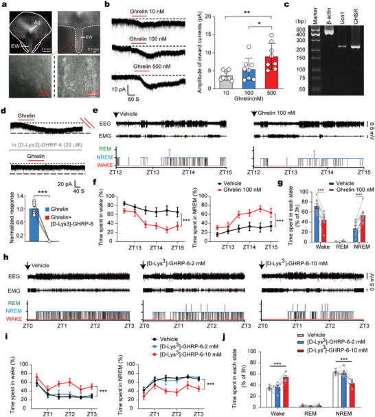
Ghrelin evoked excitatory electrical responses in relevant cells that were suppressed by GHSR blockade. In animals tested during the dark phase, local ghrelin increased NREM sleep, whereas a separate GHSR-blockade comparison during the light phase increased wakefulness and reduced NREM. Cellular responsiveness thus aligned with changes in behavioral state.
The effects were conditional: weaker conditions did not significantly alter sleep time, and REM did not change significantly alongside NREM. The receptor therefore contributes to state- and condition-dependent regulation rather than a uniform switch for sleep.
Source: Yi T et al. · Advanced science (Weinheim, Baden-Wurttemberg, Germany) · CC BY
Original figure and English caption ↗
How sevoflurane-associated excitation connects with GHSR
Sevoflurane also evoked an excitatory current in relevant cells. GHSR blockade markedly reduced it and nearly eliminated it under the corresponding condition. Local receptor blockade delayed induction and accelerated emergence, connecting receptor signaling, cellular excitation and whole-animal state.
Computational modeling proposed a possible GHSR pocket for sevoflurane, but the binding mode still requires structural and further functional evidence. This mouse-based study identifies a sleep-related contribution to anesthesia without demonstrating improved anesthetic safety or recovery quality from receptor modulation in humans.
Source: Yi T et al. · Advanced science (Weinheim, Baden-Wurttemberg, Germany) · CC BY
Original figure and English caption ↗