How can inhibitory cells support emergence? Orexin in the habenula
Orexin supports arousal, but how does it act in the lateral habenula? Male-mouse research links emergence with OxR2 in GAD2-marked cells while distinguishing induction, maintenance and recovery.
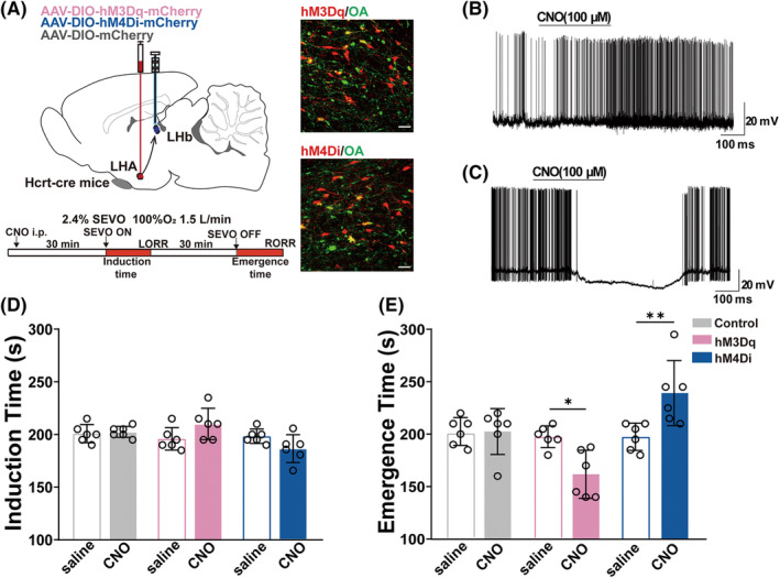
Inhibitory cells can contribute when a brain emerges from anesthesia, depending on their targets and local network. A study published in CNS Neuroscience & Therapeutics on February 5, 2023 followed an orexin-related arousal connection into the lateral habenula, LHb. In male mice under sevoflurane, enhancing relevant lateral hypothalamic input accelerated recovery of righting and altered maintenance-phase EEG. Further comparisons implicated orexin receptor OxR2 in GAD2-marked LHb cells, linking peptide signaling, cellular identity and local inhibition.
Enhancing orexin-related LHb input accelerated righting recovery, whereas suppressing it delayed recovery. Lowering OxR2 expression in GAD2 cells also delayed emergence without significantly changing induction. Local GABA-receptor findings support inhibitory involvement while leaving the full downstream organization unresolved.
Contents · 6 sections
Emergence changes without a significant induction change
The first comparisons modulated LHA orexin-related terminals in LHb. Enhancing activity accelerated recovery of righting after sevoflurane withdrawal, whereas suppression delayed it. Induction time did not change significantly in the same terminal comparisons.
Righting provides an observable behavioral state marker without directly measuring all subjective aspects of consciousness. Different sensitivities of induction and emergence indicate phase-dependent contributions rather than a uniform effect throughout anesthesia.
Source: Zhou F et al. · CNS neuroscience & therapeutics · CC BY
Original figure and English caption ↗
Input activity also changes suppression during deep anesthesia
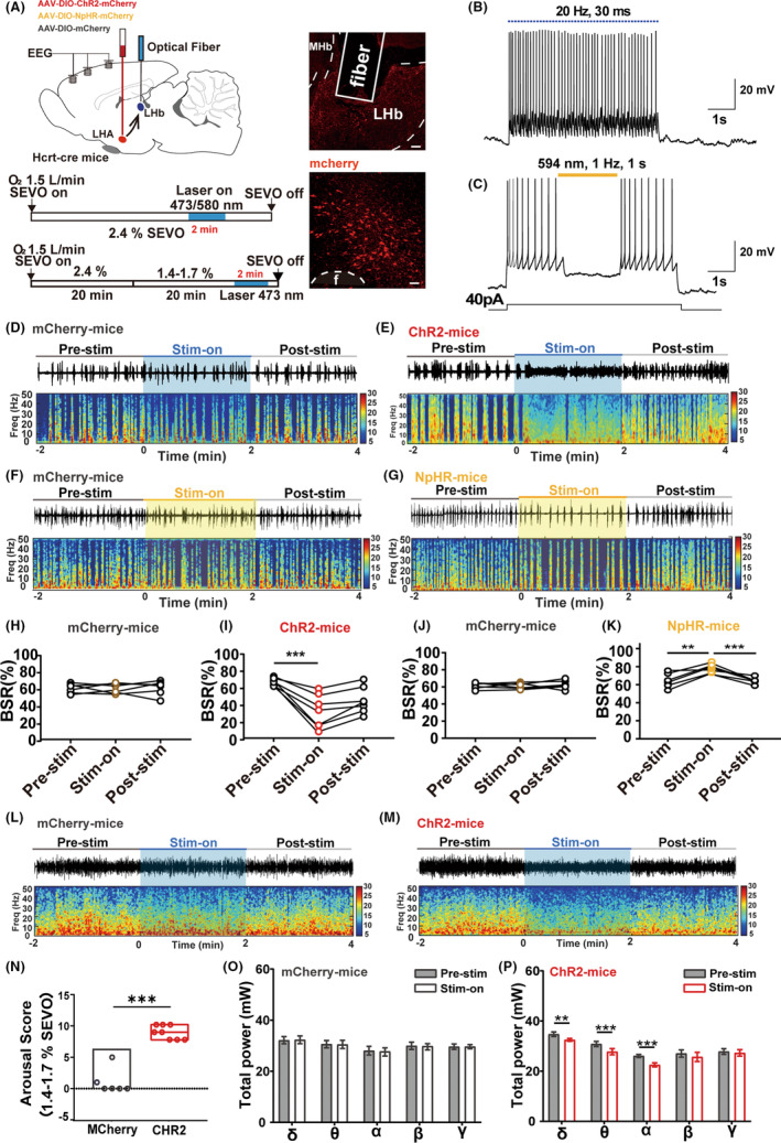
During deep anesthesia with stable EEG burst suppression, terminal activation reduced the burst-suppression ratio and suppression increased it. Measures returned after stimulation ended. EEG thus supplied evidence beyond recovery behavior that the input can influence maintenance-phase network state.
Under lighter anesthesia, activation accompanied movements and greater behavioral arousal, with lower delta-, theta- and alpha-related spectral power. Deep burst suppression and lighter-state behavior were examined in distinct states.
Source: Zhou F et al. · CNS neuroscience & therapeutics · CC BY
Original figure and English caption ↗
Local orexin comparisons highlight OxR2
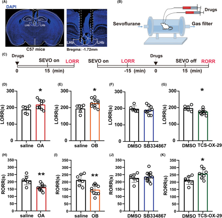
Locally in LHb, orexin A and orexin B prolonged induction and shortened emergence. OxR2 blockade produced shorter induction and slower emergence, whereas corresponding OxR1-blockade comparisons were not significant. The receptors therefore showed different evidence of involvement under these conditions.
These local pharmacological effects differed from terminal modulation: induction changed pharmacologically, whereas terminal effects centered on emergence. Exogenous local signaling and endogenous input differ in cellular coverage and duration, yielding different effects across anesthetic phases.
Source: Zhou F et al. · CNS neuroscience & therapeutics · CC BY
Original figure and English caption ↗
An emergence effect does not imply every EEG measure changes
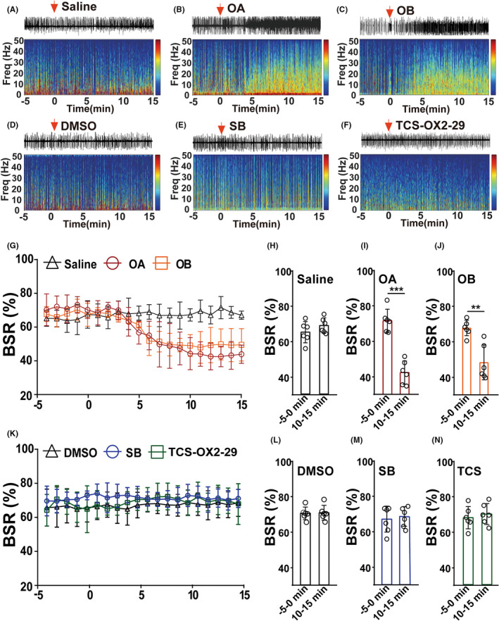
During deep maintenance, local orexin A or B reduced burst suppression, whereas OxR1 and OxR2 antagonism did not significantly change the corresponding ratio. This differs meaningfully from emergence-time findings, demonstrating outcome-dependent effects within the same signaling system.
Peptide-associated EEG changes remained evident over the measured subsequent window, whereas optical terminal effects returned more promptly. This distinguishes temporal scope without establishing a safer or better human anesthetic regimen.
Source: Zhou F et al. · CNS neuroscience & therapeutics · CC BY
Original figure and English caption ↗
Receptors in GAD2-marked cells link with emergence speed
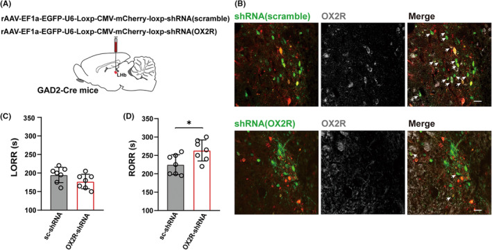
LHb is not a uniform population. Glutamatergic cells form a large component, while GAD2-marked inhibitory cells represent a different cellular contribution. Orexin acting on different cells can have different network consequences, making receptor location central to the question.
Lowering OxR2 expression in GAD2-marked LHb cells delayed righting recovery without significantly changing induction. Corresponding receptor reduction in a glutamatergic-marked population did not significantly change either transition, localizing the finding to a cellular context rather than total regional receptor abundance.
Recovery still occurred after receptor reduction. The result supports participation in emergence dynamics without establishing a unique determinant or showing that this receptor fully accounts for every orexin-cell effect.
Source: Zhou F et al. · CNS neuroscience & therapeutics · CC BY
Original figure and English caption ↗
Inhibitory-cell activity falls and rises across anesthesia
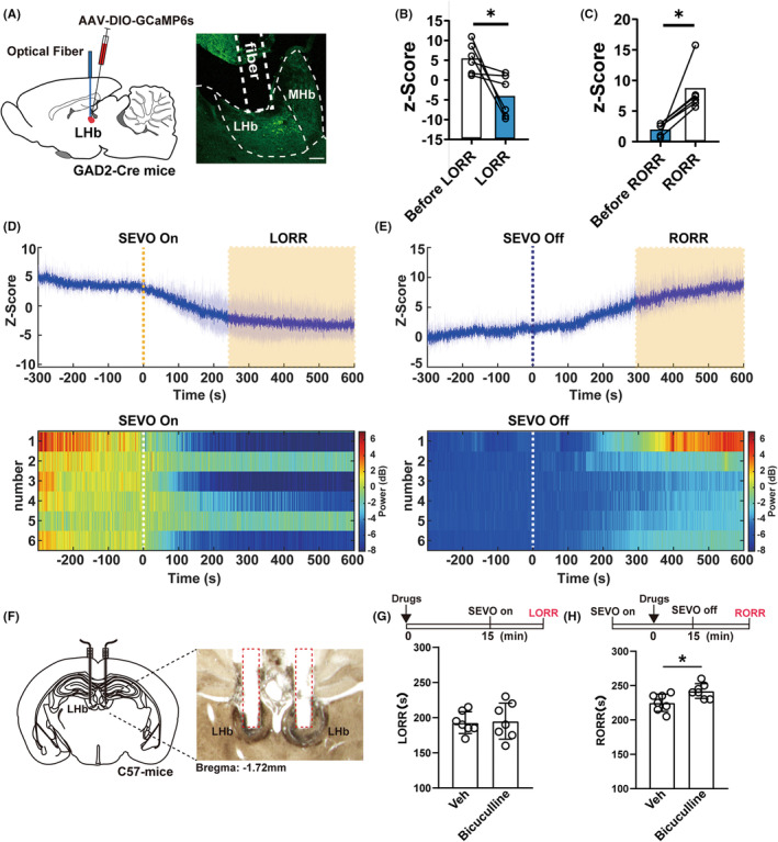
GAD2-cell calcium declined during anesthesia and rose during emergence. Local GABA-receptor blockade delayed recovery without significantly changing induction. These findings align with a possible local inhibitory contribution, in which inhibition of selected anesthesia-promoting activity could indirectly support emergence.
The network explanation remains incomplete. Terminal orexin release was not directly measured, and local versus distant outputs were not fully separated. These findings in adult male mice illuminate habenular cellular diversity while leaving other states and human relevance for further investigation.
Source: Zhou F et al. · CNS neuroscience & therapeutics · CC BY
Original figure and English caption ↗