Prefrontal PV cells share a beat within faster oscillations
Mouse prefrontal PV cells show different firing patterns across waking and sleep, yet all recorded cells favor a high-frequency oscillation phase during the tested active-wake and slow-wave-sleep states.
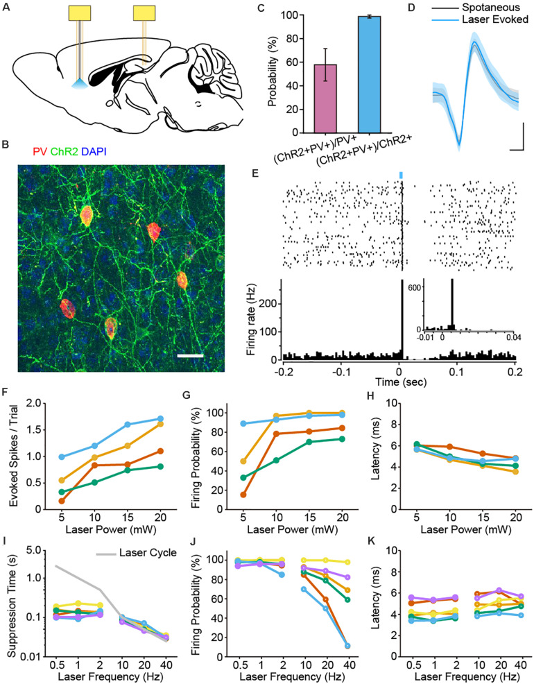
Do cells sharing the PV marker perform like a single coordinated orchestra? A mouse study published in Frontiers in Cellular Neuroscience on November 25, 2020 records prefrontal PV interneurons during natural waking and sleep. Their firing rates follow different state-dependent patterns, but spike timing reveals a shared feature: all recorded cells couple strongly to local high-frequency oscillations during active waking and slow-wave sleep. How often a cell fires and where its spikes fall within a rhythm are distinct questions.
The recorded PV cells form three state-related firing groups, without significant between-group differences in high-frequency coupling. Increasing PV activity strongly suppresses local pyramidal firing while changing field-potential power more modestly.
Contents · 6 sections
One molecular marker, three state-related patterns
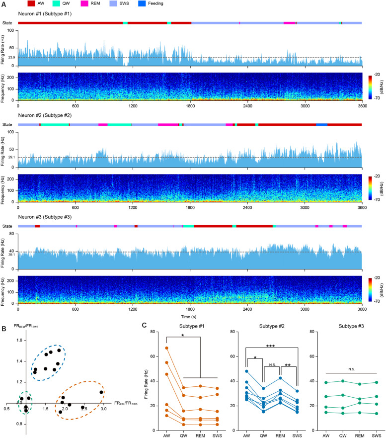
PV cells are an important component of cortical inhibitory networks and are often discussed together. The study follows eighteen identified PV cells from ten mice across active waking, quiet waking, REM sleep and slow-wave sleep, comparing state-related firing within each cell.
Six cells fire faster during active waking; eight show a pattern of higher activity during active waking and REM sleep; four show no significant firing-rate differences across states. A shared PV identity therefore contains different state-response profiles.
These groups describe activity profiles in the sampled cells. They expose diversity within the population: knowing a marker does not by itself reveal when a cell becomes more active.
Source: Yao Y et al. · Frontiers in cellular neuroscience · CC BY
Original figure and English caption ↗
Beyond rate, spike timing has a preferred phase
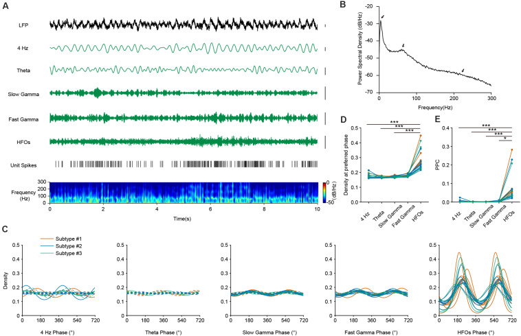
Local field potentials reflect electrical activity produced by the surrounding network and contain rhythms at different timescales. Phase coupling asks whether spikes tend to occur at a preferred point in a cycle. Ten claps can be scattered in time or repeatedly align with the same part of a beat.
During active waking, all eighteen recorded PV cells show significant phase coupling to high-frequency oscillations. Some also couple to slow and fast gamma, whereas most lack significant theta coupling. Coupling is strongest for high-frequency oscillations, rather than gamma coupling being entirely absent.
Source: Yao Y et al. · Frontiers in cellular neuroscience · CC BY
Original figure and English caption ↗
The common timing relationship persists in slow-wave sleep
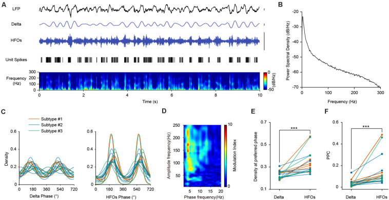
Slow rhythms become prominent during slow-wave sleep, with high-frequency activity embedded within them. Recorded PV cells couple to both delta and high-frequency oscillations, with stronger phase preference and consistency for the latter. The fast timing relationship remains clear in the changed sleep background.
In waking and slow-wave sleep, spikes favor phases near the high-frequency cycle’s trough. This does not require a spike on every cycle; it describes a timing preference when spikes occur. It links discrete cellular events with continuous local network activity.
Source: Yao Y et al. · Frontiers in cellular neuroscience · CC BY
Original figure and English caption ↗
Different rate profiles can share a timing relationship
The three rate-defined groups do not differ significantly in high-frequency phase consistency during either active waking or slow-wave sleep. A cell becoming busier or quieter in a state does not automatically determine how tightly its spikes align with the network rhythm.
The population thus has two organizational levels: state-dependent firing rates and a shared timing reference. Considering both reveals how heterogeneity and temporal coordination coexist, beyond a single population-average curve.
Single-cell firing and field potentials respond differently
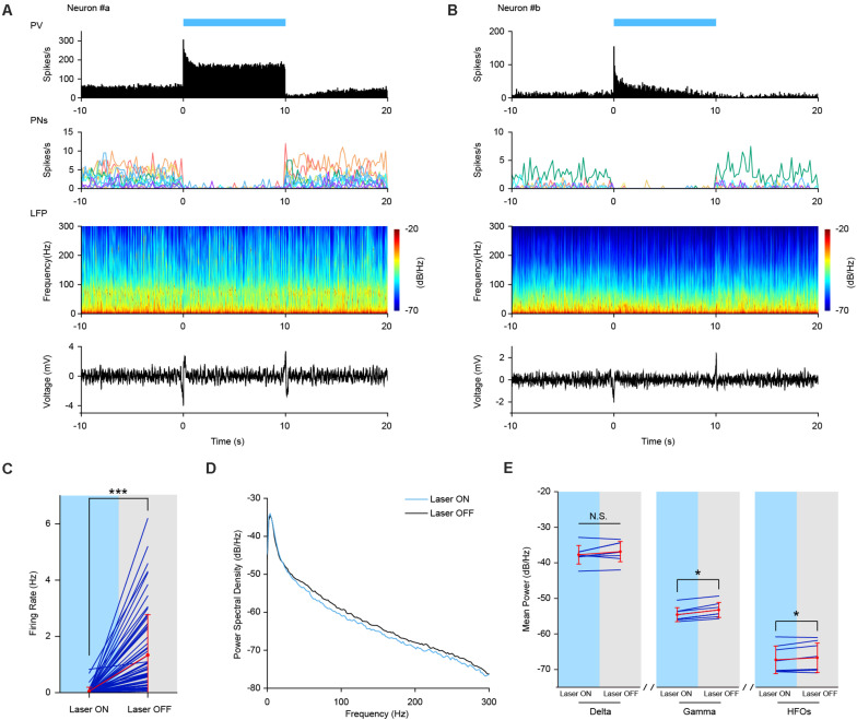
Increasing PV activity produces a pronounced initial firing increase, followed by elevated or gradually adapting responses. Nearby pyramidal-cell firing falls significantly, consistent with an inhibitory influence from the PV network.
Field-potential changes are more modest. During slow-wave sleep, gamma and high-frequency power decline slightly, without a significant delta change. Suppression of local pyramidal firing therefore does not produce a large reduction across all frequency bands.
Spikes and field potentials sample different scales. The extent of affected local cells and the combination of field-potential sources may contribute to their different effect sizes, making both cellular events and network context informative.
Source: Yao Y et al. · Frontiers in cellular neuroscience · CC BY
Original figure and English caption ↗
A timing clue awaiting its functional role
High-frequency activity invites comparison with hippocampal ripples, but similar timescales do not establish a shared generating mechanism. This study concerns local prefrontal spike–oscillation relationships, without directly measuring memory transfer across regions or testing these cells in attention or memory tasks.
The mouse findings provide a finer picture of inhibitory cells within network rhythms during natural waking and sleep. The source of the high-frequency rhythm, whether PV cells follow it, shape it or do both, and how the firing groups relate to finer cell identities remain open questions.