Orexin can stabilize REM sleep through coordinated pontine excitation
Rat and mouse findings link orexin input to SLD excitation and electrical coupling, helping REM episodes persist rather than acting solely as an arousal signal.
Orexin is commonly associated with maintaining wakefulness. REM sleep requires a different stability: active brain rhythms alongside low muscle tone. How can an excitatory signal help that state persist? A rat and mouse study published in Nature Communications on July 21, 2020 traces orexin signaling to pontine SLD. The signal changes coordination between cells as well as their activity, supporting more coherent network output.
SLD-associated orexin signaling enhances local excitation and electrical coupling and prolongs REM episodes. Reducing it shortens episodes and, over longer observation, affects REM amount and muscle-tone suppression.
Contents · 7 sections
A small projection population can differ from the whole
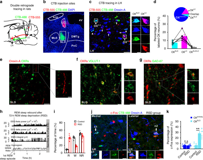
Hypothalamic orexin cells project widely, so whole-population measurements can combine cells with different destinations. During enriched rebound REM sleep, the SLD-projecting population shows increased activity labeling, while the entire orexin population does not change significantly.
These cells overlap little with orexin cells projecting to wake-promoting locus coeruleus, while orexin fibers and receptors are present in SLD. The same excitatory signal can take on different roles in different recipient networks.
Source: Feng H et al. · Nature communications · CC BY
Original figure and English caption ↗
Direct responses in some cells can spread more widely
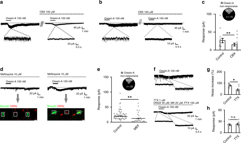
Rat SLD recordings show widespread excitation by orexin. When electrical coupling is disrupted, approximately one quarter of tested cells no longer respond directly, suggesting that the broad effect is not simply an identical direct action on every cell.
Gap junctions electrically connect neighboring cells, allowing sustained depolarization in directly responding cells to influence others. Excitation can therefore extend beyond its initial recipients. The approximate three-quarter estimate describes functional responses in the tested network, rather than a census of all pontine cells.
Source: Feng H et al. · Nature communications · CC BY
Original figure and English caption ↗
Orexin changes coupling as well as excitation
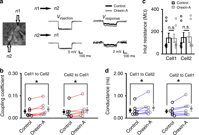
Paired recordings show stronger coupling in both directions and increased gap-junction conductance, without significant input-resistance changes. Orexin thus changes the route through which excitation spreads, as well as exciting individual cells.
Some cells receive the initiating signal while communication between cells becomes more effective. Together, these actions may help otherwise dispersed activity fluctuate in concert. The exact substrates exchanged through the junctions remain unresolved.
Source: Feng H et al. · Nature communications · CC BY
Original figure and English caption ↗
Activity becomes more coordinated, not just stronger
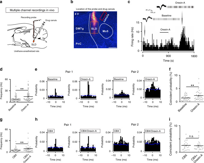
Recordings in anesthetized rats connect cellular coupling with network activity. Orexin increases firing in a subset of SLD units and raises near-coincident firing probability among pairs already showing significant interactions.
After electrical coupling is disrupted, coincident probability no longer significantly increases. The result supports a role in coordinating activity rather than uniform acceleration of every cell. These recordings concern the anesthetized network; effects during natural sleep require separate behavioral evidence.
Source: Feng H et al. · Nature communications · CC BY
Original figure and English caption ↗
Stabilizing REM differs from initiating it
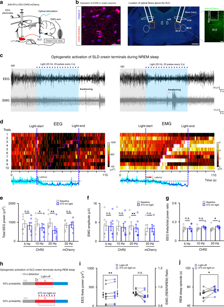
Increasing orexin-terminal activity in mouse SLD does not directly switch NREM into REM. During NREM it can increase the EEG theta component and reduce muscle activity, with occasional brief awakening, illustrating dependence on the current state.
During established REM, activation prolongs the episode and increases theta power. The muscle-activity ratio relative to preceding NREM trends downward without reaching significance. The clearest effect is persistence of an existing episode rather than easier initiation.
Source: Feng H et al. · Nature communications · CC BY
Original figure and English caption ↗
The signal contributes during natural REM
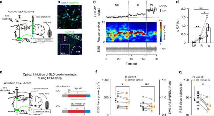
During natural sleep, calcium signals in SLD orexin terminals are higher in REM than preceding NREM, but lower than subsequent wakefulness. Local signaling therefore varies across states rather than following a simple awake-active, asleep-silent rule.
Briefly reducing terminal activity shortens REM episodes and lowers theta power, without a significant muscle-activity-ratio change. Activity measurements and intervention converge on episode maintenance, while calcium remains a population-activity measure rather than a direct readout of each peptide-release event.
Source: Feng H et al. · Nature communications · CC BY
Original figure and English caption ↗
With less of this input, REM becomes less stable
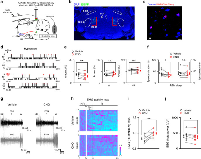
Over longer observation, reducing activity in SLD-projecting orexin cells lowers REM amount by about 17% relative to control. Episodes shorten without a significant change in episode number, wake amount or NREM amount. REM theta power falls and normal muscle-tone suppression is disrupted.
These findings extend orexin’s role from maintaining wakefulness to stabilizing a particular sleep state. Projection-defined cells may have other outputs, and coupling and behavior do not yet establish one exclusive pathway. Across rats and mice, recipient network and current state jointly shape the consequences of excitation.
Source: Feng H et al. · Nature communications · CC BY
Original figure and English caption ↗