Myelin damage affects anesthesia differently across brain regions
A male mouse study compares regional myelin changes: lateral-habenula damage hastens emergence, thalamic paraventricular damage hastens induction, while tested dorsal-raphe timings remain similar.
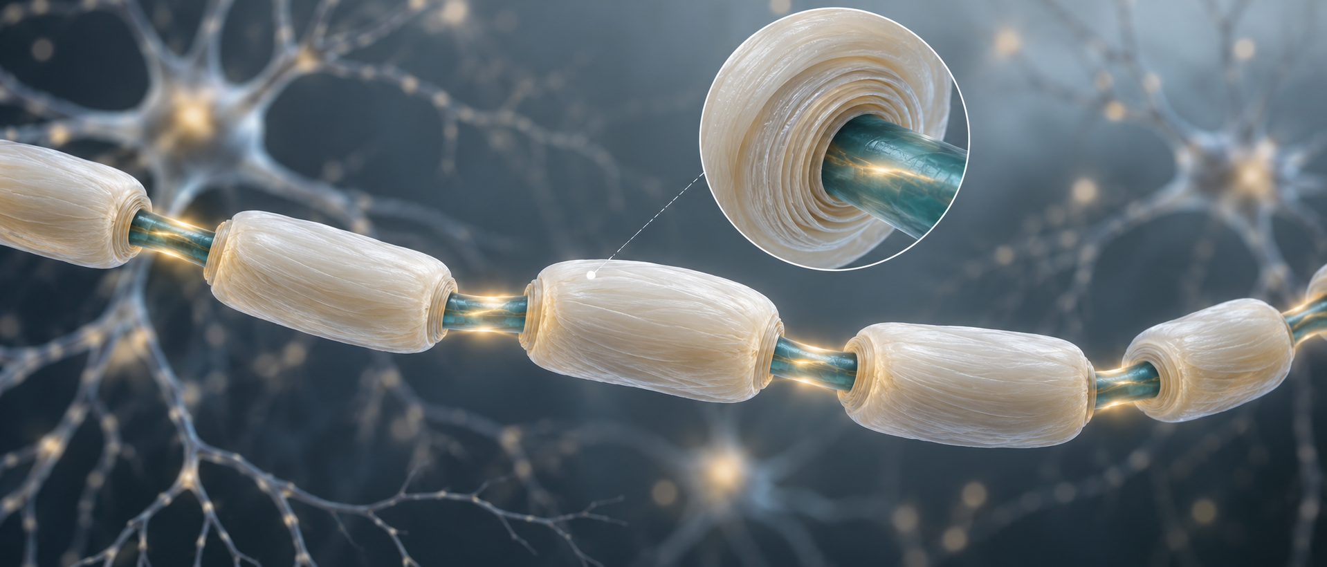
Anesthesia research often follows which neurons activate and which circuits support emergence. Myelin around axons also influences neural signaling. A study published in CNS Neuroscience & Therapeutics in 2024 examines this structure during isoflurane anesthesia: broader brain demyelination slows mouse emergence, whereas local damage produces different effects. Myelin location and the activity it supports give similar structural changes different behavioral consequences.
Myelin changes affect isoflurane induction and emergence differently across regions and accompany weaker population activity. Lateral-habenula and thalamic paraventricular activation effects weaken after demyelination.
Contents · 7 sections
Broader damage slows emergence
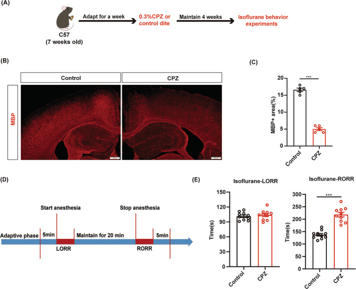
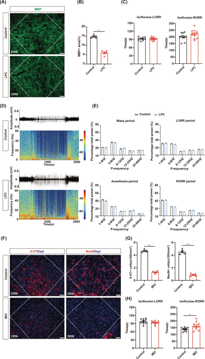
Oligodendrocytes form myelin around axons, supporting rapid, accurate signaling as well as protection and metabolic support. The study tracks structural changes using myelin basic protein, MBP, labeling, which decreases markedly in the broader demyelination model.
Under the same isoflurane condition, righting-reflex loss occurs at a similar time to controls, but its return after anesthetic cessation is later. Righting refers to recovering the normal body posture. Induction and emergence measure the two ends of the transition and need not change in the same direction. The effect concentrates on emergence, prompting examination of regional contributions behind the broader outcome.
Source: Wang X et al. · CNS neuroscience & therapeutics · CC BY
Original figure and English caption ↗
Lateral habenula: emergence occurs earlier
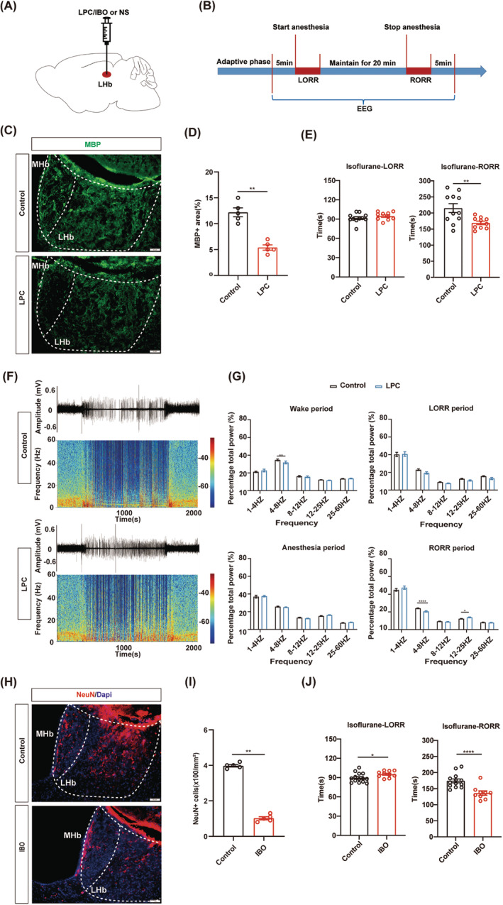
Lateral habenula, LHb, contributes to anesthetic-state regulation. Regional demyelination leaves induction time largely similar at the main tested condition but shortens emergence. EEG-band distributions during recovery also change alongside the behavioral effect.
Reducing LHb neurons likewise hastens emergence but delays induction. The interventions converge during emergence and differ during induction, showing that myelin changes and cell depletion do not have identical regional effects.
Source: Wang X et al. · CNS neuroscience & therapeutics · CC BY
Original figure and English caption ↗
Thalamic paraventricular nucleus: faster induction
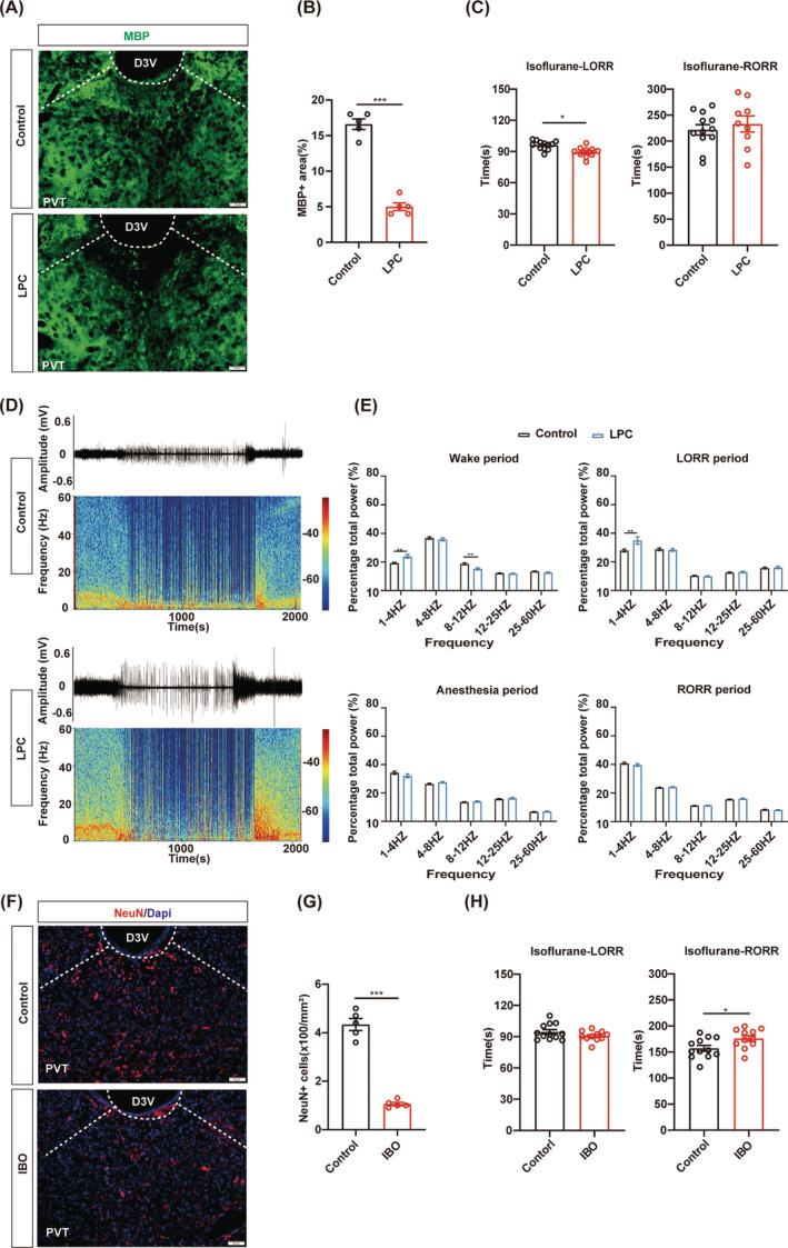
In thalamic paraventricular nucleus, PVT, demyelination primarily hastens righting-reflex loss, without a clear emergence-time difference. Before anesthesia, waking EEG shows more low-frequency power and less power in a faster band, indicating changes before anesthetic exposure.
PVT neuron depletion leaves induction largely similar while delaying emergence. At another, lower isoflurane condition, PVT demyelination also prolongs emergence. Region, intervention target and anesthetic condition influence which stage changes.
Source: Wang X et al. · CNS neuroscience & therapeutics · CC BY
Original figure and English caption ↗
Dorsal raphe: structural change without the same outcome
Dorsal raphe, DRN, contains diverse neurons. MBP labeling decreases there too, but induction and emergence remain largely similar at both main and lower isoflurane conditions. Tested EEG-band distributions likewise lack clear group differences.
DRN neuron depletion produces a different result: induction remains similar while emergence is delayed. Its neurons contribute to emergence, but the current regional demyelination condition does not clearly change behavioral timings despite reduced structural labeling.
Source: Wang X et al. · CNS neuroscience & therapeutics · CC BY
Original figure and English caption ↗
Population changes are more consistent than behavior
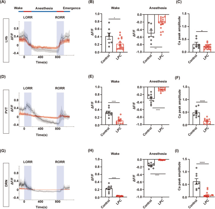
All three regions show fewer activity-labeled cells while awake after demyelination. Population calcium recordings also show smaller waking fluctuations and peaks. These shared changes do not automatically produce anesthetic behavior in the same direction.
During isoflurane exposure, control calcium signals decrease clearly, whereas changes are smaller after demyelination. Reduced population modulation across the transition offers an activity-level account of regional functional changes, with its relation to emergence depending on the region’s network role.
Source: Wang X et al. · CNS neuroscience & therapeutics · CC BY
Original figure and English caption ↗
Activation effects depend on the surrounding state
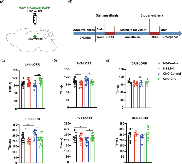
With relatively intact myelin, LHb activation prolongs emergence, whereas PVT activation hastens it. Demyelination weakens both of these oppositely directed activation effects. In DRN, induction and emergence do not clearly differ across groups under the same tested condition.
Myelin thus contributes beyond static wrapping: whether recruited cells generate their usual network effect also relates to surrounding structure. The same activation need not produce the same behavioral outcome in different myelin states.
Source: Wang X et al. · CNS neuroscience & therapeutics · CC BY
Original figure and English caption ↗
Anesthetic-state networks need structural support
These findings come from adult male mice under isoflurane. Regional myelin can surround local axons or incoming axons passing through, so its changes do not map solely to one population. The study connects anesthetic-state activity with structures supporting signaling.
Broader and regional damage produce different effects, and similar activity reductions can accompany different timing changes. Understanding regional contributions to anesthesia therefore involves both when cells activate and the structures supporting transmission through their networks.