After blood flow returns, how do mitochondria regain balance?
A mouse and cell study links early sevoflurane postconditioning protection to Shh–YAP1 and reveals compensatory interactions between two Drp1 modifications.
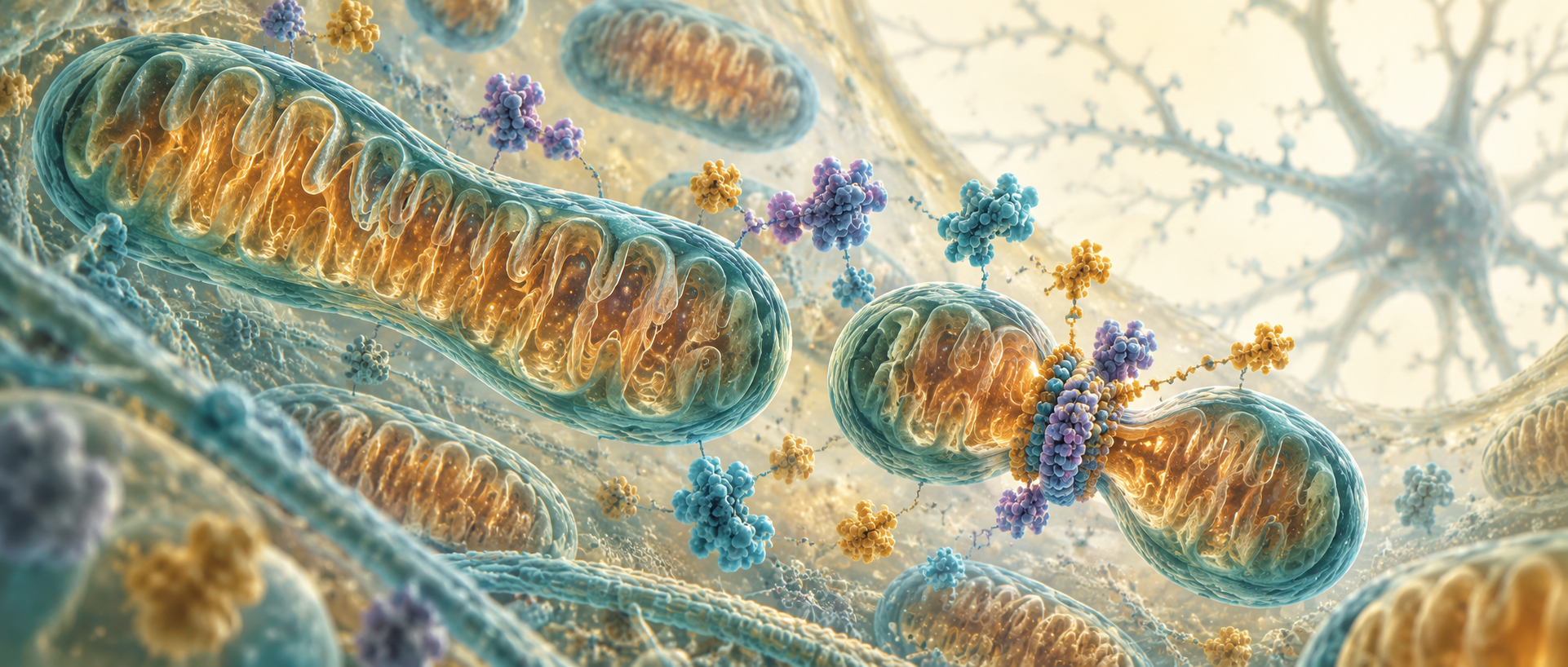
Returning blood flow does not immediately restore ischemic cells: mitochondrial shape, energy and membrane function are still changing. Researchers applied sevoflurane postconditioning at reperfusion and followed early protection through Shh–YAP1 to Drp1. Two modifications compensated for one another rather than acting as independent switches.
Postconditioning reduces early injury and improves some mitochondrial measures, weakened by Shh or YAP1 intervention. Drp1 Ser616 phosphorylation and SUMOylation show compensatory interactions.
Contents · 6 sections
Blood flow returns while organelles keep adjusting
Mitochondria are often called cellular powerhouses, but they are dynamic structures. Fission, fusion and permeability shape their function during ischemia and reoxygenation. The study combined vascular occlusion/reperfusion in mice with oxygen-glucose deprivation/reoxygenation in culture.
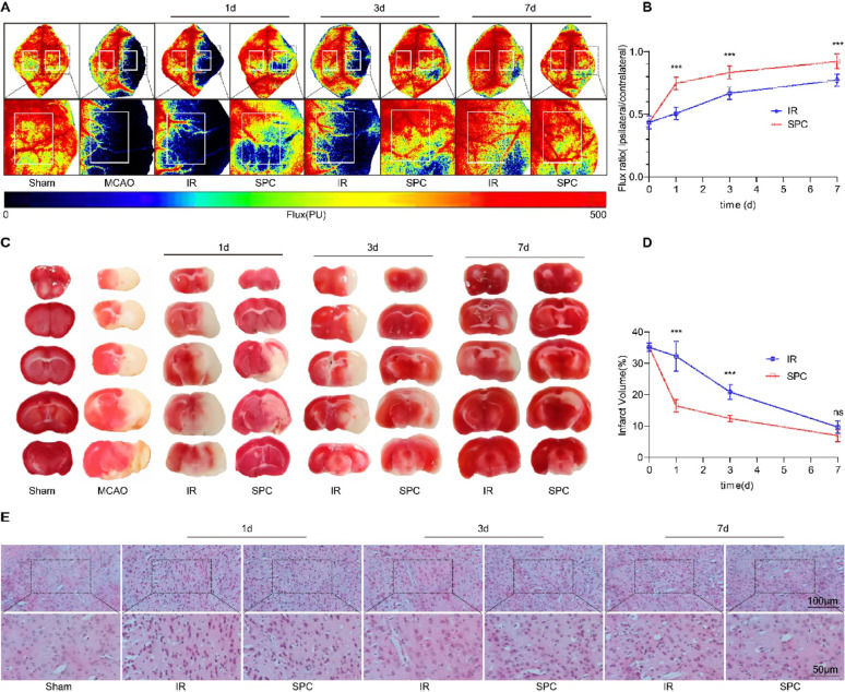
Mice received one hour of sevoflurane at reperfusion, and cells were treated after deprivation. Marked blood-flow and infarct changes occurred early, leading to a mainly twenty-four-hour mechanism assessment of this vulnerable transition.
The mouse model captures blood flow and tissue injury, while culture permits deprivation, reoxygenation and molecular manipulation under simpler conditions. The former locates early protection and the latter follows cellular signaling, moving from tissue phenomena to protein modifications.
Source: Peng L et al. · Molecular brain · CC BY
Original figure and English caption ↗
Cell survival improves and mice enter a new arm more often
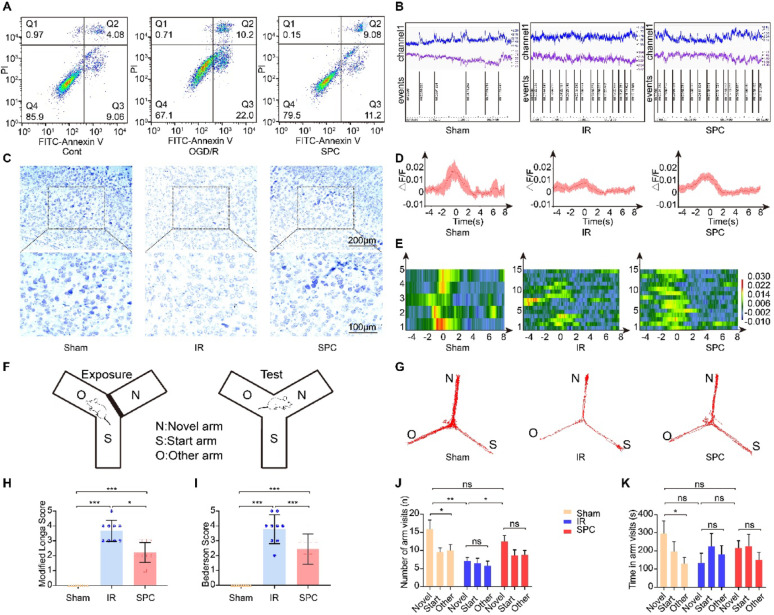
Postconditioning mainly reduced early apoptotic cells, with smaller effects on late apoptosis/necrosis. Mouse CA1 calcium activity, Nissl survival measures and neurological scores improved, revealing early protection with differences across readouts.
New-arm entries increased in the Y-maze, while dwell time showed only a nonsignificant trend. More frequent entry did not accompany the same dwell change. Transcription and protein measurements then directed attention to Shh and YAP1.
Source: Peng L et al. · Molecular brain · CC BY
Original figure and English caption ↗
Shh and YAP1 contribute to postconditioning responses
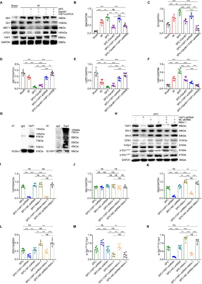
Transcription highlighted sonic hedgehog (Shh) and Hippo–YAP programs. Injury already recruited part of the response, and postconditioning further increased Shh, Gli1 and YAP1. Reducing YAP1 or blocking the process weakened some anti-apoptotic and structural improvements. Like a repair crew mobilizing after damage, increased signaling accompanied the cellular response to injury.
Shh blockade lowered YAP1, whereas YAP1 reduction did not markedly lower Shh. Gli1 still responded to YAP1 manipulation, suggesting feedback. Co-immunoprecipitation associated YAP1 with Drp1, connecting signaling with mitochondrial shape regulation.
Source: Peng L et al. · Molecular brain · CC BY
Original figure and English caption ↗
Shape, membrane function and ATP show mitochondrial changes
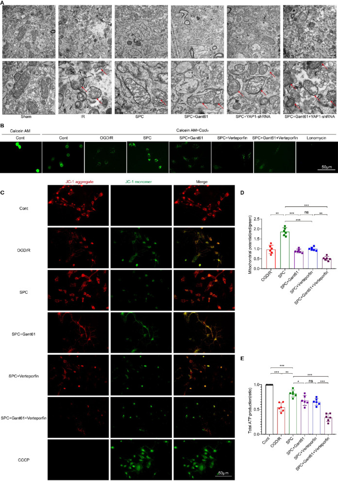
In mouse tissue, postconditioning reduced swelling, disordered cristae and fragmentation, weakened by Shh blockade or YAP1 reduction. Cultured neurons showed corresponding permeability-pore, membrane-potential and ATP changes, capturing structural and energetic facets.
Mfn2, OPA1 and Drp1 phosphorylation sites changed in different ways while Mfn1 remained comparable. The balance was not a uniform rise or fall, raising a question about whether one Drp1 modification compensates when another is limited.
Source: Peng L et al. · Molecular brain · CC BY
Original figure and English caption ↗
Restraining one modification brings another into play
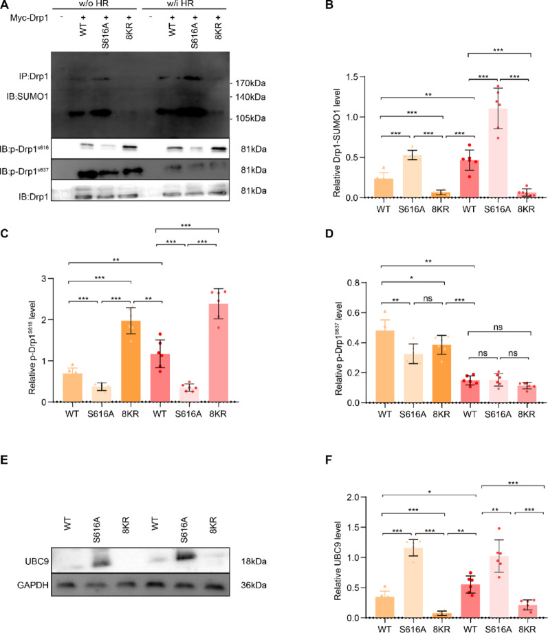
Postconditioning altered Drp1 phosphorylation and SUMO-related proteins. SUMO inhibition reduced the corresponding signal but increased Ser616 phosphorylation, without a similar Ser637 change, revealing site-specific compensation.
Mutations echoed the pattern: limiting Ser616 phosphorylation raised SUMOylation, while limiting SUMO sites increased Ser616 phosphorylation under deprivation/reoxygenation, with comparable Ser637. Drp1 behaved as a multi-regulated hub rather than a single-switch target.
Source: Peng L et al. · Molecular brain · CC BY
Original figure and English caption ↗
Cell protection involves a dynamic regulatory network
This young-male-mouse and cell study links early reperfusion protection with Shh–YAP1 and mitochondrial dynamics. Recovery involves shape, membranes and energy, with compensation between molecular modifications.
Early measurements provide a clear snapshot, while longer outcomes remain to be examined. Tracking how Drp1 compensation affects duration and mitochondrial state may reveal how intracellular balance continues rebuilding after flow returns.