An excitatory basal-forebrain–VTA connection in mouse anesthesia emergence
In male and female mice, optical activation of basal-forebrain glutamate cells or VTA terminals accelerates righting recovery after isoflurane, alongside changes in EEG, pupils, breathing and downstream calcium signals.
After anesthetic delivery stops, how does the brain return toward a state permitting action? Emergence involves reengagement of arousal networks as well as drug elimination. A study in The Journal of Neuroscience examines basal-forebrain, or BF, glutamate neurons and their ventral tegmental area, or VTA, output. Optical activation accelerates righting recovery in isoflurane-anesthetized mice, alongside cortical, pupil and respiratory changes.
BF-cell and VTA-terminal optical activation accelerate righting recovery without significantly changing induction time. Chemogenetic activation changes EEG but not recovery time. Terminal activation also increases VTA glutamate-population calcium signals.
Contents · 8 sections
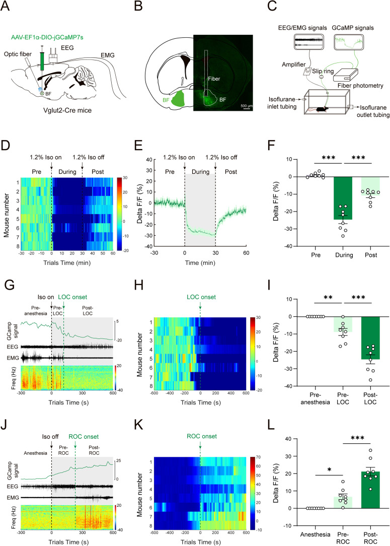
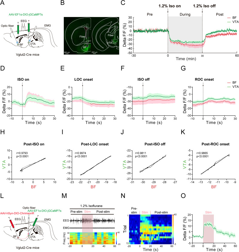
Signals decline during anesthesia and rise during recovery
BF glutamate-population calcium is recorded alongside EEG and muscle activity. Signals fall rapidly after isoflurane begins, remain lower during anesthesia and rise after delivery stops.
Aligned to EEG/EMG-defined transitions, the decline begins before loss of responsiveness and the rise before recovery. Continuous changes motivate direct testing of this arousal-associated population.
Source: Gong XT et al. · The Journal of neuroscience : the official journal of the Society for Neuroscience · CC BY
Original figure and English caption ↗
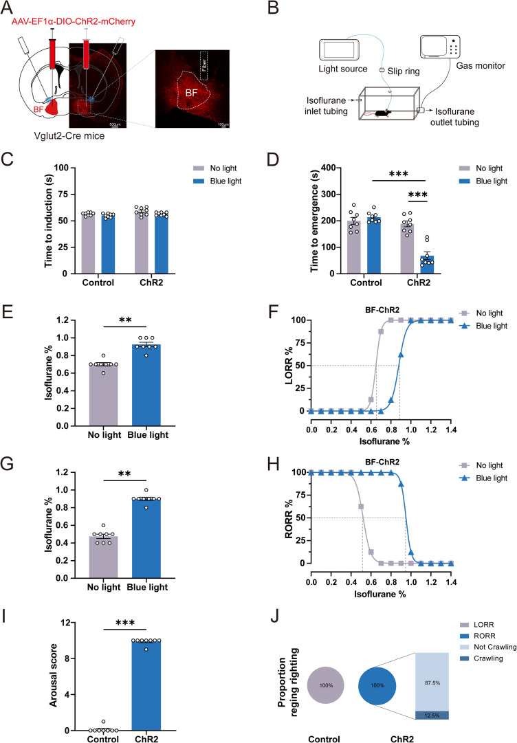
Optical activation accelerates righting recovery
BF glutamate optical activation shortens the interval from isoflurane cessation to righting recovery, without significantly changing time to righting loss during induction. Entry and exit respond differently to the intervention.
During concentration changes, activated mice lose righting at higher concentrations and regain it at higher concentrations. Stimulation during ongoing anesthesia also elicits movements, righting or crawling and raises behavioral arousal scores.
Source: Gong XT et al. · The Journal of neuroscience : the official journal of the Society for Neuroscience · CC BY
Original figure and English caption ↗
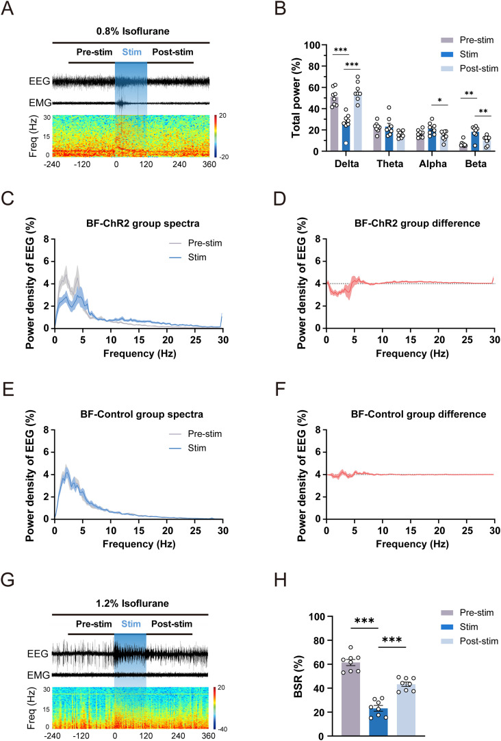
EEG shifts from slow waves toward faster activity
Under lighter anesthesia, BF activation decreases delta power and increases beta power. EEG shifts from higher-amplitude slow activity toward lower-amplitude faster activity, accompanying behavioral arousal.
Under deeper conditions, EEG alternates between bursts and suppressed periods. Activation lowers the burst-suppression ratio, which rises after stimulation ends, showing a temporally bounded cortical effect.
Source: Gong XT et al. · The Journal of neuroscience : the official journal of the Society for Neuroscience · CC BY
Original figure and English caption ↗
EEG changes need not shorten recovery time
Chemogenetic activation lowers delta power, raises theta power and reduces deeper-anesthesia burst suppression. Induction and righting-recovery times do not significantly change, distinguishing its behavioral effects from optical activation.
Chemogenetic inhibition does not significantly change induction or recovery times; optical inhibition leaves measured spectra and burst suppression unchanged. Inhibition does not necessarily reverse the effects produced by activation.
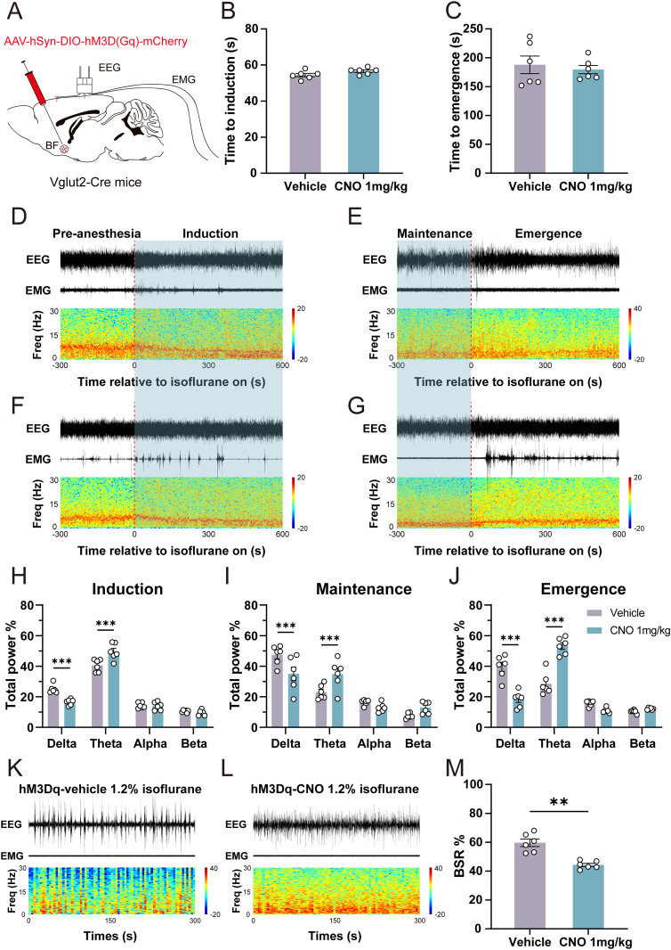
Source: Gong XT et al. · The Journal of neuroscience : the official journal of the Society for Neuroscience · CC BY
Original figure and English caption ↗
Pupils and breathing change during optical activation
BF optical activation dilates pupils and increases respiratory rate in anesthetized mice. Both return toward earlier levels after stimulation, adding physiological measures to cortical and behavioral outcomes.
Pupils and breathing have distinct time courses. Concurrent observation shows that one neural intervention affects multiple responses, motivating investigation of the BF outputs involved.
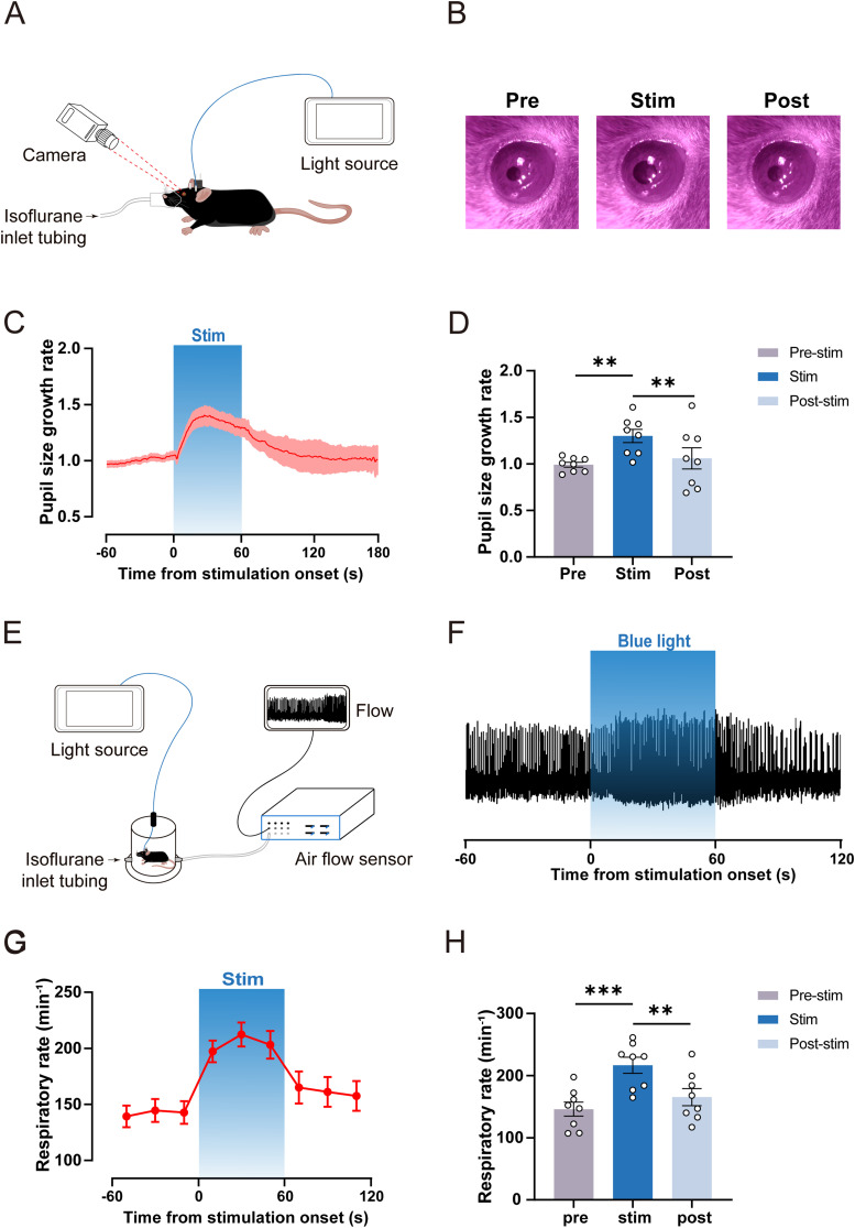
Source: Gong XT et al. · The Journal of neuroscience : the official journal of the Society for Neuroscience · CC BY
Original figure and English caption ↗
Activating VTA terminals also accelerates recovery
VTA is one of several BF glutamate targets. Stimulating BF-derived terminals there shortens righting-recovery time without significantly changing induction. Concentration responses and behavioral scores also shift toward greater arousability.
During ongoing anesthesia, terminal stimulation restores righting in some animals rather than all; other responses include movements or crawling. Defined terminal stimulation modulates behavior with variable response strength.
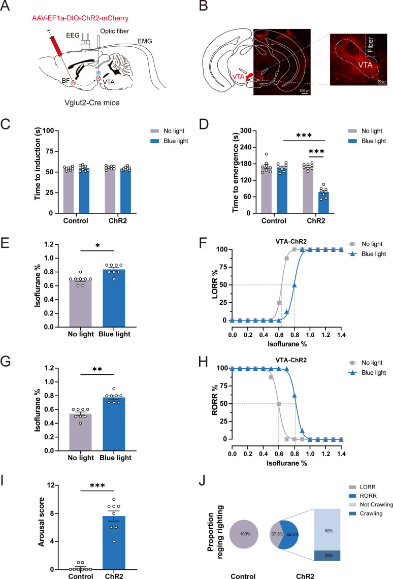
Source: Gong XT et al. · The Journal of neuroscience : the official journal of the Society for Neuroscience · CC BY
Original figure and English caption ↗
Terminal stimulation reproduces cortical and physiological responses
VTA terminal stimulation also decreases delta power, increases faster-band activity and reduces deep-anesthesia burst suppression. Pupils dilate and breathing accelerates before returning, paralleling BF-soma activation outcomes.
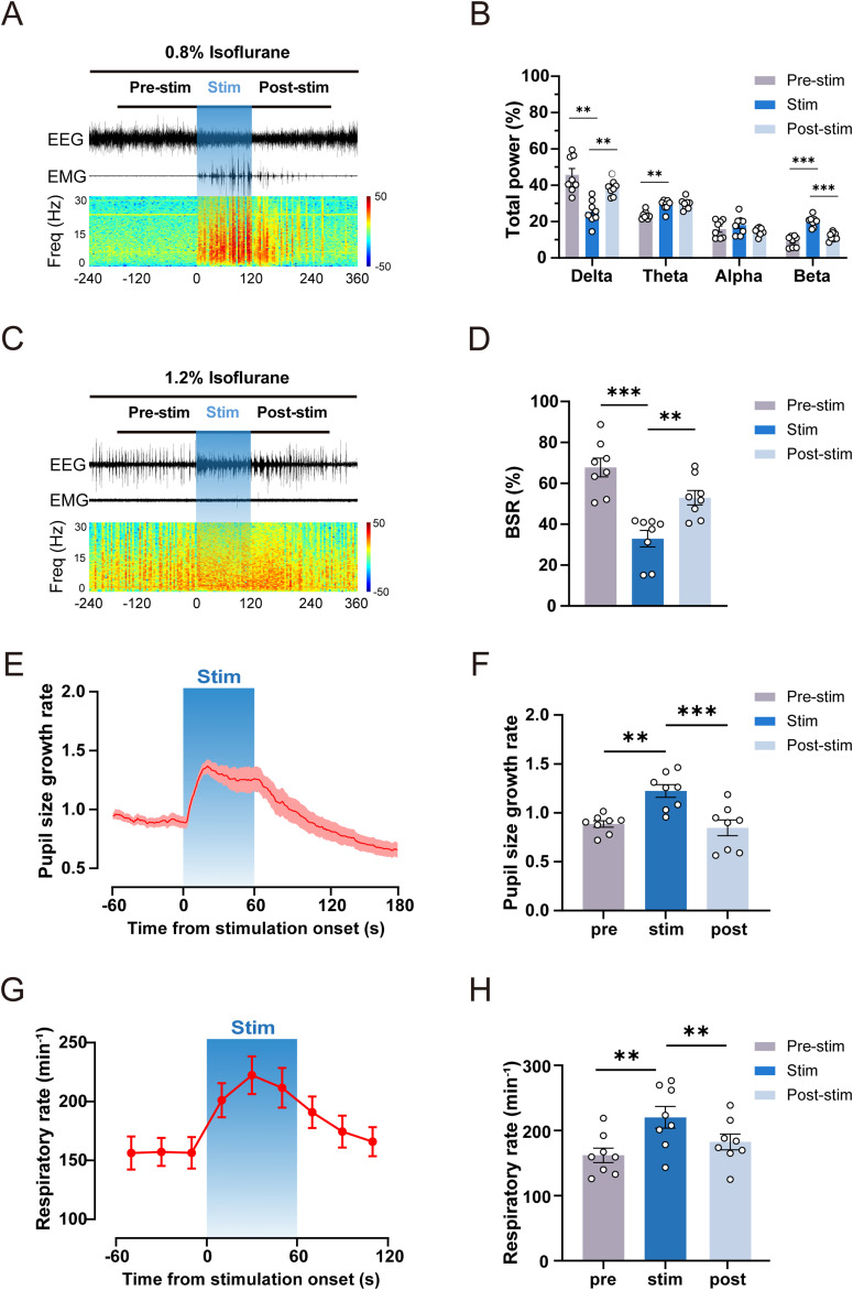
Source: Gong XT et al. · The Journal of neuroscience : the official journal of the Society for Neuroscience · CC BY
Original figure and English caption ↗
BF and VTA glutamate populations show linked dynamics
Dual-site recordings show linked BF and VTA glutamate calcium changes around anesthetic delivery, cessation and state transitions. BF-derived terminal stimulation increases VTA glutamate signals and shifts EEG toward faster activity.
This study including both sexes links post-isoflurane recovery with a recruitable excitatory output. Entry and exit respond asymmetrically, and EEG changes do not always accompany righting recovery, revealing more specific cellular and output contributions to emergence.
Source: Gong XT et al. · The Journal of neuroscience : the official journal of the Society for Neuroscience · CC BY
Original figure and English caption ↗