Midbrain inhibitory cells help mice emerge faster from anesthesia
A GABAergic population in the ventrolateral periaqueductal gray becomes less active during sevoflurane anesthesia. Activation accelerates emergence and changes EEG patterns; cortical input and output toward the ventral tegmental area also contribute.
Entering anesthesia and emerging from it may look like opposite processes, but emergence need not simply replay induction in reverse. A mouse study published in iScience on August 14, 2023 finds that activating GABAergic cells in the ventrolateral periaqueductal gray (vlPAG) accelerates return of the righting reflex without significantly changing its loss. Following cortical input and midbrain output, the study shows that inhibitory neurons can promote emergence in a particular circuit context.
Activating or inhibiting vlPAG GABAergic cells respectively accelerates or delays emergence from sevoflurane anesthesia, without significantly changing induction. Activating mPFC input to vlPAG or vlPAG terminals in VTA also promotes emergence, while local VTA GABA signals provide a dynamic link between the observations.
Contents · 6 sections
Signals decline during anesthesia and rise during emergence
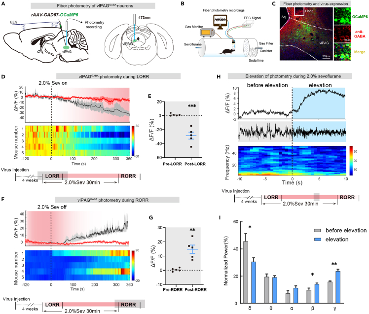
The vlPAG surrounds the midbrain aqueduct and contributes to sleep, defense and pain. The researchers first follow calcium signals in its GABAergic population. Signals gradually decrease after sevoflurane begins and rise again after inhalation stops, tracking the state transition.
Calcium elevations also occur during maintained anesthesia. Accompanying EEG shows less delta-band power and more beta and gamma power. These spontaneous co-occurrences raise a question: does the population merely follow state changes, or can it influence emergence?
Source: Guo Y et al. · iScience · CC BY-NC-ND
Original figure and English caption ↗
Emergence accelerates without a significant induction change
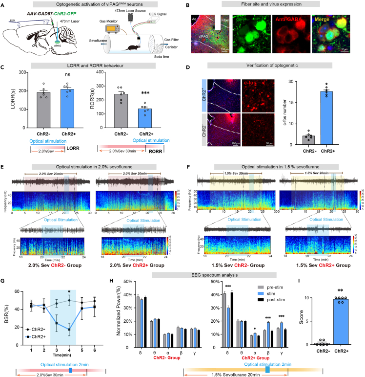
Activating the population makes mice regain the righting reflex sooner after sevoflurane stops. Activation during induction does not significantly change the time to lose the reflex. The different outcomes highlight a stronger effect on emergence under these conditions.
During deeper maintained anesthesia, activation also reduces the EEG burst-suppression ratio, the fraction of time spent in low-activity suppression intervals. Under lighter maintenance conditions, slow-wave power decreases, faster bands increase and body-movement scores rise. Both EEG and behavioral measures shift in an arousal-promoting direction.
Burst suppression is an EEG pattern alternating bursts of activity with low-activity intervals. The study examines both recovery after anesthetic cessation and whether the network can be shifted while anesthetic exposure continues. Maintenance EEG changes and earlier righting after cessation describe changes in network state and behavioral recovery, respectively.
Source: Guo Y et al. · iScience · CC BY-NC-ND
Original figure and English caption ↗
Inhibition prolongs emergence
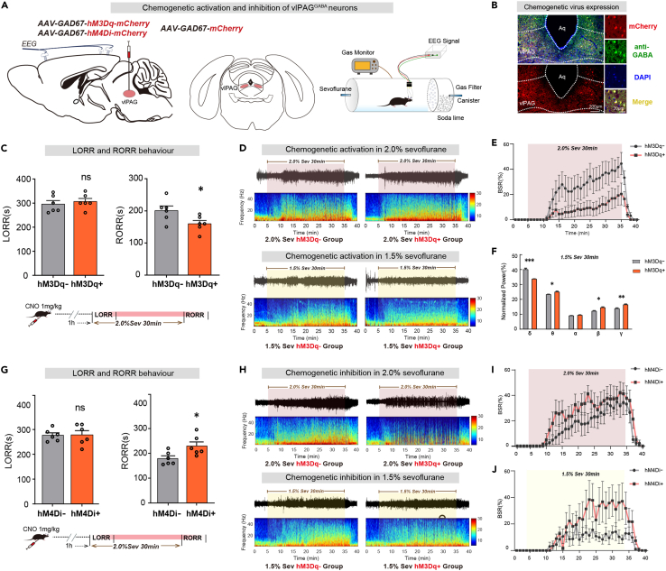
A second, more sustained activation approach produces similar results: faster righting-reflex recovery, less EEG burst suppression and no significant induction-time change. Converging approaches indicate that the finding is not restricted to a brief optical stimulus.
Inhibiting the population instead prolongs righting-reflex recovery and increases burst suppression during maintenance, again without significantly altering induction. Opposite behavioral effects of activation and inhibition connect the population to regulation of emergence.
Source: Guo Y et al. · iScience · CC BY-NC-ND
Original figure and English caption ↗
Terminals in VTA can also promote emergence
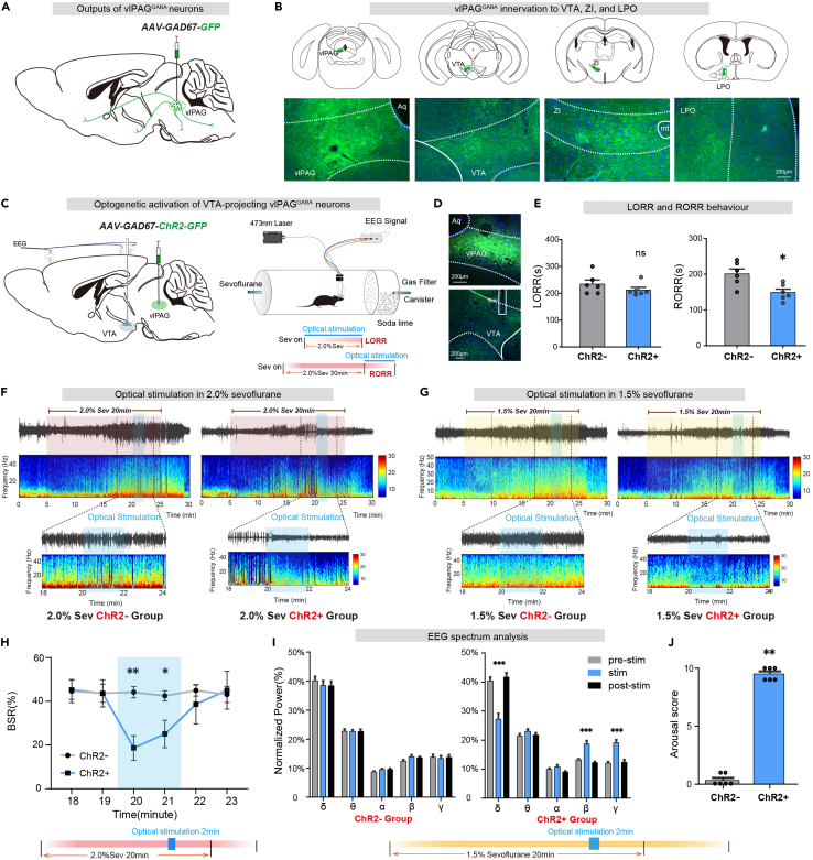
vlPAG GABAergic cells have multiple outputs. Tracing finds fibers in the ventral tegmental area (VTA), lateral preoptic area and zona incerta. In this study, activating terminals in the latter two regions produces no significant anesthesia effect, whereas VTA terminal activation shortens emergence.
Like activation of the broader population, VTA terminal activation leaves induction time without a significant change but lowers maintenance burst suppression. Under lighter conditions, slow waves decrease while faster bands and movement scores increase. These findings implicate the vlPAG output to VTA in promoting emergence.
VTA contains dopamine, GABAergic and glutamatergic cells. The network effect of an inhibitory input depends on its recipients. The authors propose that inhibiting VTA inhibitory cells could release other outputs from restraint. This is a model for the emergence-promoting effect; the specific functions of the recipients remain to be determined.
Source: Guo Y et al. · iScience · CC BY-NC-ND
Original figure and English caption ↗
The prefrontal cortex can influence the process upstream
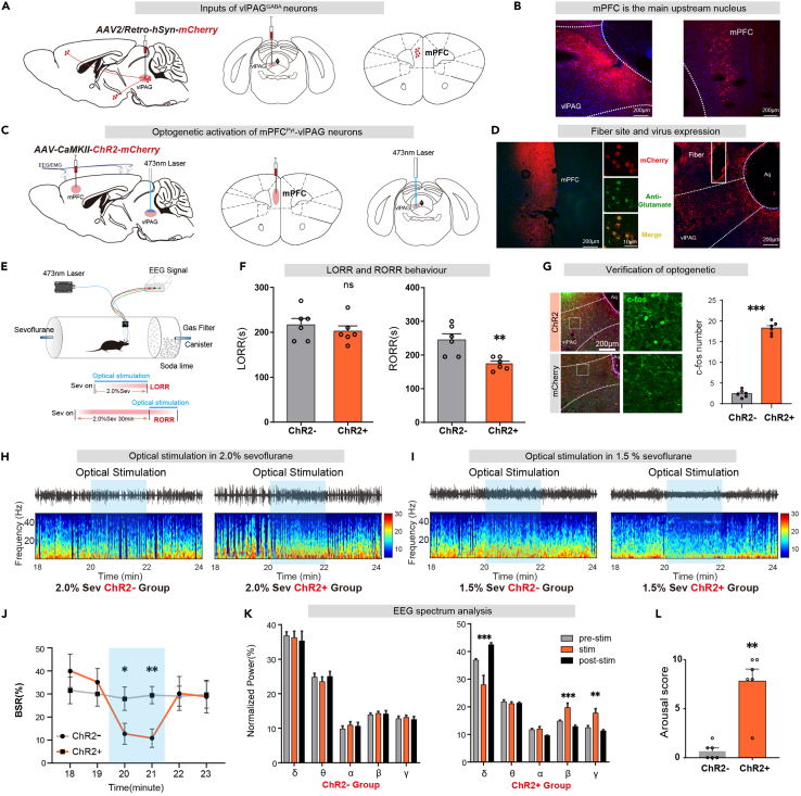
Upstream tracing identifies the medial prefrontal cortex (mPFC). Activating terminals of mPFC excitatory cells in vlPAG also accelerates return of the righting reflex without significantly changing induction time. The midbrain effect is thus linked to a cortical output.
EEG again shows reduced burst suppression and, under lighter anesthesia, less delta and more beta and gamma power. The findings support a cortical influence on midbrain regulation of anesthetic state, adding a descending contribution to ascending mechanisms of emergence.
Source: Guo Y et al. · iScience · CC BY-NC-ND
Original figure and English caption ↗
Local VTA GABA signals add another link
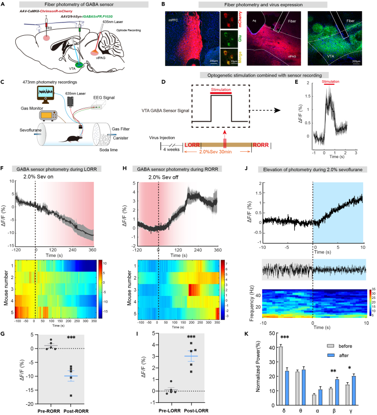
Activating mPFC input to vlPAG increases the GABA sensor signal in VTA. The sensor measures fluorescence changes related to local extracellular GABA. Its signal declines after sevoflurane begins, rises after cessation, and also co-varies with EEG during maintained anesthesia.
Together, the findings support a working model involving cortical input, vlPAG regulation and output to VTA. A local sensor alone does not identify the source of every GABA signal, and the crucial VTA recipients remain to be resolved. These adult-male-mouse findings illustrate how the behavioral meaning of an inhibitory transmitter depends on its network: here, GABAergic cells can promote emergence.
Source: Guo Y et al. · iScience · CC BY-NC-ND
Original figure and English caption ↗