Locus-coeruleus signaling helps mice recover after sedation
Midazolam transiently abolishes the righting reflex and reduces locus-coeruleus activity in mice. Enhancing related noradrenergic signaling shortens recovery, with VLPO α1 receptors contributing.

How do arousal systems re-engage after a sedative takes effect? A 2024 eLife study follows locus coeruleus and its connection with preoptic regions in midazolam-treated mice. Locus-coeruleus population activity decreases during loss of the righting reflex, while increasing the relevant cells’ activity shortens recovery. The team then examines how local receptors connect this effect with neuromodulatory signaling.
Locus-coeruleus noradrenergic activity influences righting-reflex recovery after midazolam. Local VLPO α1-receptor blockade counteracts recovery-promoting effects, and locus-coeruleus GABAA-receptor interventions also alter recovery.
Contents · 8 sections
Tracking state changes through the righting reflex
Mice normally right their bodies after being turned over. Midazolam temporarily abolishes this reflex before it returns. The study compares the interval between these events and uses calcium signals and EEG to examine neural changes. Recovery here specifically refers to the return of righting ability.
Midazolam enhances GABA-related inhibition, while the progression from molecular action to recovery also involves regulation between brain regions. Norepinephrine, a contributor to arousal regulation, provides a candidate signal to follow.
Brainstem signaling falls, while enhancement speeds recovery
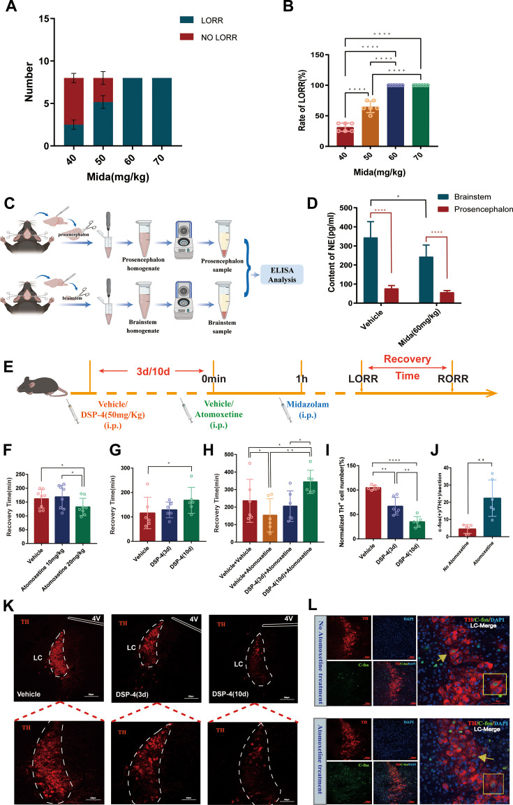
Norepinephrine content decreases in the sampled brainstem after drug treatment. An intervention enhancing central noradrenergic action hastens righting-reflex recovery, whereas damage to this system prolongs recovery and attenuates that enhancement effect. Tissue measurements and behavior point in a related direction.
These initial interventions act broadly. The study then focuses on locus coeruleus (LC), a major noradrenergic population connected with multiple regions and an important component of arousal regulation.
Source: Gu L et al. · eLife · CC BY
Original figure and English caption ↗
Population activity is low while righting is absent
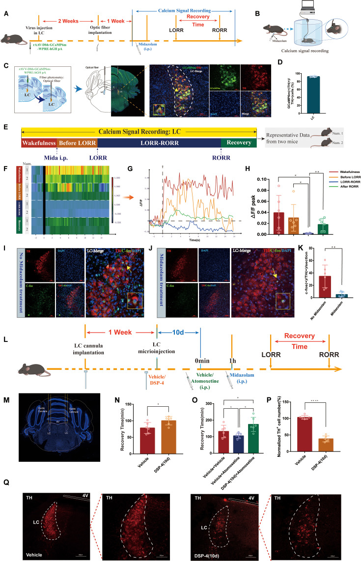
Calcium signals in the LC noradrenergic population decrease between righting-reflex loss and recovery and rise afterward. Activity-marker measurements also show reduced activity in the relevant cells after treatment. Both observations link the state change with the same region.
Reducing the relevant cells locally in LC prolongs recovery and attenuates the effect of enhancing noradrenergic action. The tissue-level clue thus narrows to a defined population that contributes to post-drug recovery.
Source: Gu L et al. · eLife · CC BY
Original figure and English caption ↗
Earlier recovery occurs in both males and females
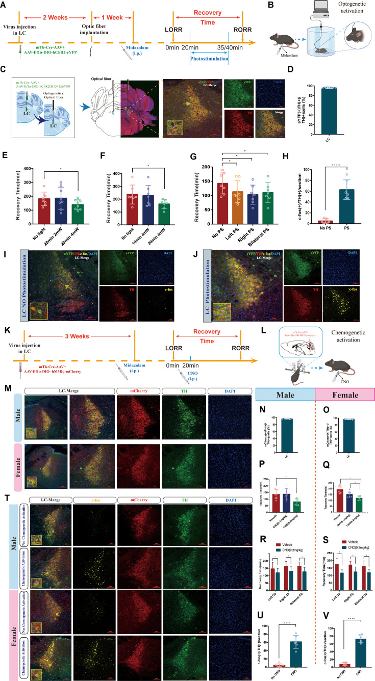
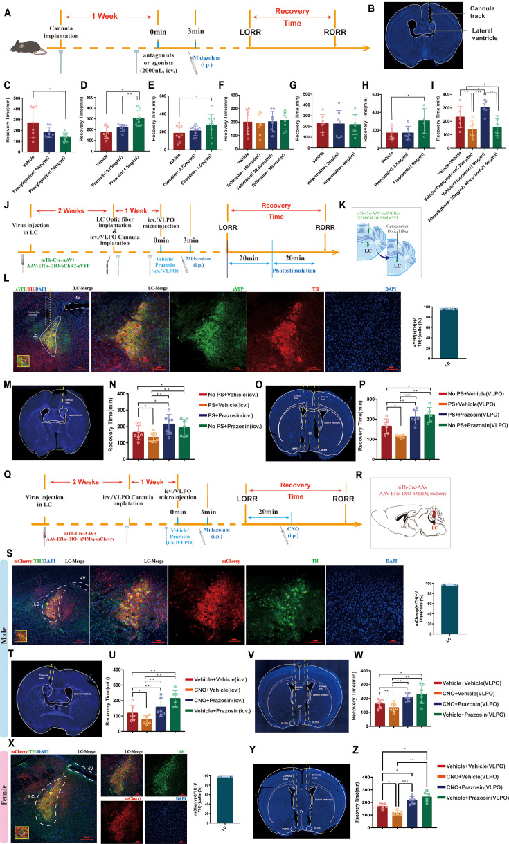
Increasing relevant LC-cell activity shortens recovery after midazolam. Unilateral and bilateral manipulations produce a similar direction under the tested conditions, and chemogenetic activation promotes recovery in both male and female mice.
EEG recordings also show altered activity patterns during drug action when LC is activated. Recovery is more than an endpoint: cellular activity and cortical signals change during the transition as well.
Source: Gu L et al. · eLife · CC BY
Original figure and English caption ↗
Recovery effects also appear at VLPO
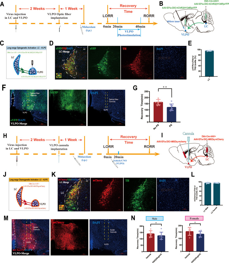
Ventrolateral preoptic nucleus (VLPO) contains diverse populations involved in sleep and arousal. LC activation increases related calcium signals in this region. Enhancing the labeled connection at VLPO also accompanies earlier righting-reflex recovery.
Local manipulation moves the question from whether LC changes recovery to where its signal acts. The experiments support an LC–VLPO contribution, while receptor comparisons further focus the inquiry on this target region.
Source: Gu L et al. · eLife · CC BY
Original figure and English caption ↗
α1-receptor blockade counteracts enhanced recovery
Pharmacological effects differ among adrenergic receptors. The tested α1 agonist shortens recovery and α1 blockade prolongs it. Some α2- and β-related interventions also affect recovery, while others show no significant effect, drawing attention to α1 signaling.
In particular, local VLPO α1-receptor blockade counteracts the faster recovery produced by LC activation, with corresponding chemogenetic results in both sexes. Increased cellular activity acts within a receptive local environment, where receptors provide an important link.
Source: Gu L et al. · eLife · CC BY
Original figure and English caption ↗
GABAA receptors connect with downstream signaling
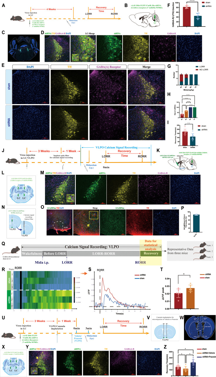
Local LC GABAA-receptor blockade also shortens recovery, and simultaneous VLPO α1 blockade counteracts this effect. Lowering GABAA-receptor expression in the relevant LC cells likewise produces shorter recovery; the mice still show righting-reflex loss under the tested drug conditions.
After receptor-expression reduction, related VLPO calcium signals are higher during recovery, and local α1 blockade counteracts the faster recovery. Drug-related inhibition and noradrenergic modulation converge in the behavioral and activity findings. Calcium traces aligned to righting-reflex recovery capture the ensuing brief signal changes.
Source: Gu L et al. · eLife · CC BY
Original figure and English caption ↗
Post-sedation transitions have a local regulatory logic
This adult-mouse study connects post-drug recovery with LC activity, preoptic signals and local receptors. The precise receiving VLPO cell types remain to be identified, while the findings already link the return of righting ability with activity in arousal-regulating systems.