Light alters sleep after stress through a visual-thalamic circuit
In male mice, chronic stress increases NREM sleep and light reduces that increase. Connections among visual thalamus, lateral habenula and RMTg contribute to the effect.

Stress changes sleep, but not always by reducing it. A study published in PLOS Biology on September 7, 2023 finds that repeated stress increases non-rapid-eye-movement, NREM, sleep and reduces waking in male mice. Light attenuates that change, with evidence linking visual thalamus to lateral habenula, LHb, and rostromedial tegmental nucleus, RMTg. The work examines how environmental light enters networks regulating stress and sleep, beyond its role in image formation.
Increased NREM after stress is linked to LHb–RMTg-related activity. Light reduces related cellular excitability and shifts sleep allocation. Suppressing the visual-thalamic input-related population or enhancing downstream NREM-promoting populations weakens the light effect.
Contents · 8 sections
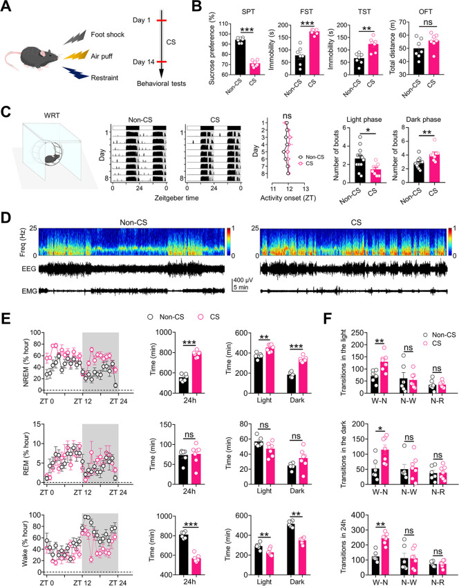
Stress increases NREM without a corresponding REM change
Chronic stress lowers sucrose preference and increases immobility in tail suspension and swimming without significantly changing total open-field distance. NREM increases and waking decreases during both light and dark phases, while REM does not change significantly.
Transitions also differ: wake-to-NREM events increase without parallel increases in the other two tested transition categories. Duration and transition counts provide distinct information beyond a general description of poor sleep.
Source: Huang L et al. · PLoS biology · CC BY
Original figure and English caption ↗
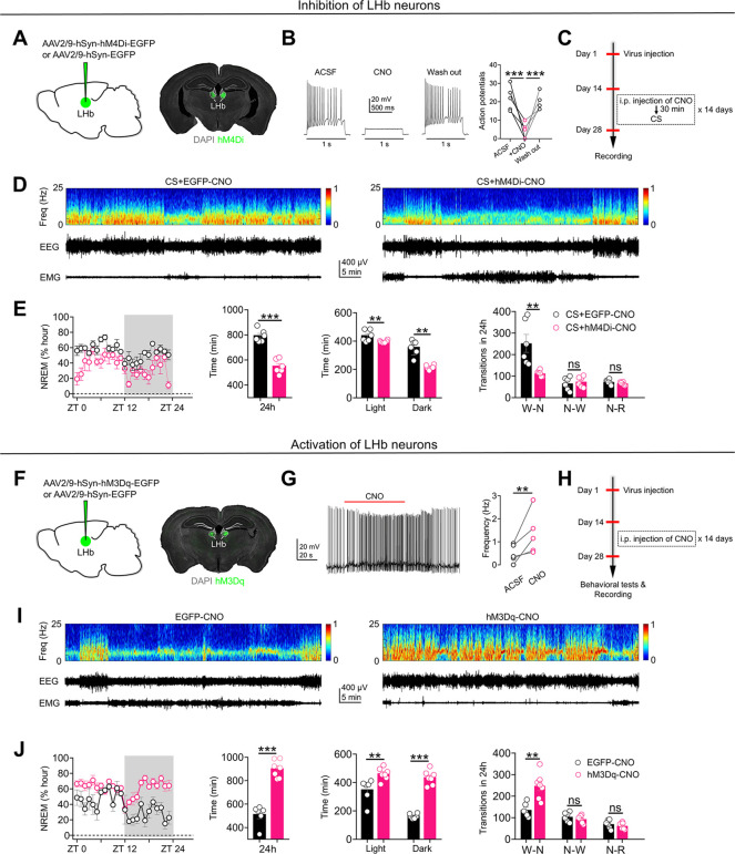
Changing LHb activity shifts sleep allocation
LHb processes stress-related information and sends outputs to several sleep-related regions. Suppressing LHb cells during stress attenuates the later NREM increase and waking reduction, with fewer wake-to-NREM transitions.
Sustained LHb activation without the same stress challenge also increases NREM, reduces waking and produces selected depressive-like measures. Functional intervention connects LHb activity with the sleep direction and motivates examination of its different outputs.
Source: Huang L et al. · PLoS biology · CC BY
Original figure and English caption ↗
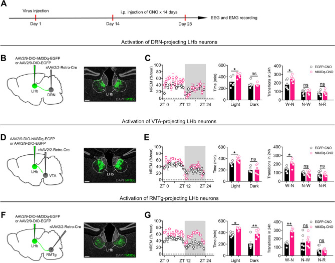
RMTg-projecting cells reproduce both-phase effects
Activating LHb populations projecting to dorsal raphe or VTA increases NREM during the light phase but not significantly during the dark phase. Activating RMTg-projecting cells increases NREM in both phases, more closely resembling the stress phenotype.
Cells sharing a source show different temporal effects according to output destination. These experiments manipulate projection-defined somata, which may have other branches; subsequent terminal and recipient-cell experiments examine the connection more closely.
Source: Huang L et al. · PLoS biology · CC BY
Original figure and English caption ↗
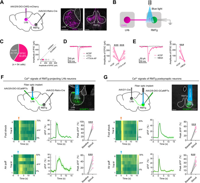
Excitatory LHb input recruits RMTg-related cells
Anatomical labeling identifies predominantly excitatory-related LHb cells and predominantly GABA-labeled RMTg recipients. Slice recordings support direct excitatory synaptic connections rather than proximity or coactivation alone.
Both recorded populations increase calcium during stress-related events. Population calcium describes event responses, while synaptic recordings characterize input to recipients. Together they connect stress-related information with this pathway.
Source: Huang L et al. · PLoS biology · CC BY
Original figure and English caption ↗
Activation promotes NREM and inhibition attenuates stress effects
Activating LHb terminals in RMTg increases NREM and reduces waking without significantly changing REM. Activating RMTg cells receiving LHb input also increases NREM duration and related transitions, linking the connection’s two sides through complementary experiments.
During stress, suppressing either RMTg-projecting LHb cells or LHb-recipient RMTg populations attenuates the NREM increase. The connection therefore contributes to the tested sleep alteration rather than merely accompanying it afterward.
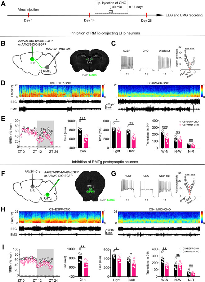
Source: Huang L et al. · PLoS biology · CC BY
Original figure and English caption ↗
Light attenuates increased NREM and measured excitability
The ventral lateral geniculate nucleus and intergeniculate leaflet, vLGN/IGL, provide an upstream visual-thalamic link. Tracing across two synaptic steps identifies predominantly GABA-labeled upstream cells, supporting a visual-thalamic route through LHb to RMTg.
With light treatment accompanying stress, the NREM increase is attenuated and waking increases, while REM totals remain nonsignificant. Relevant LHb and recipient RMTg cells show lower firing responses to matched current in slices, linking sleep outcomes with excitability changes.
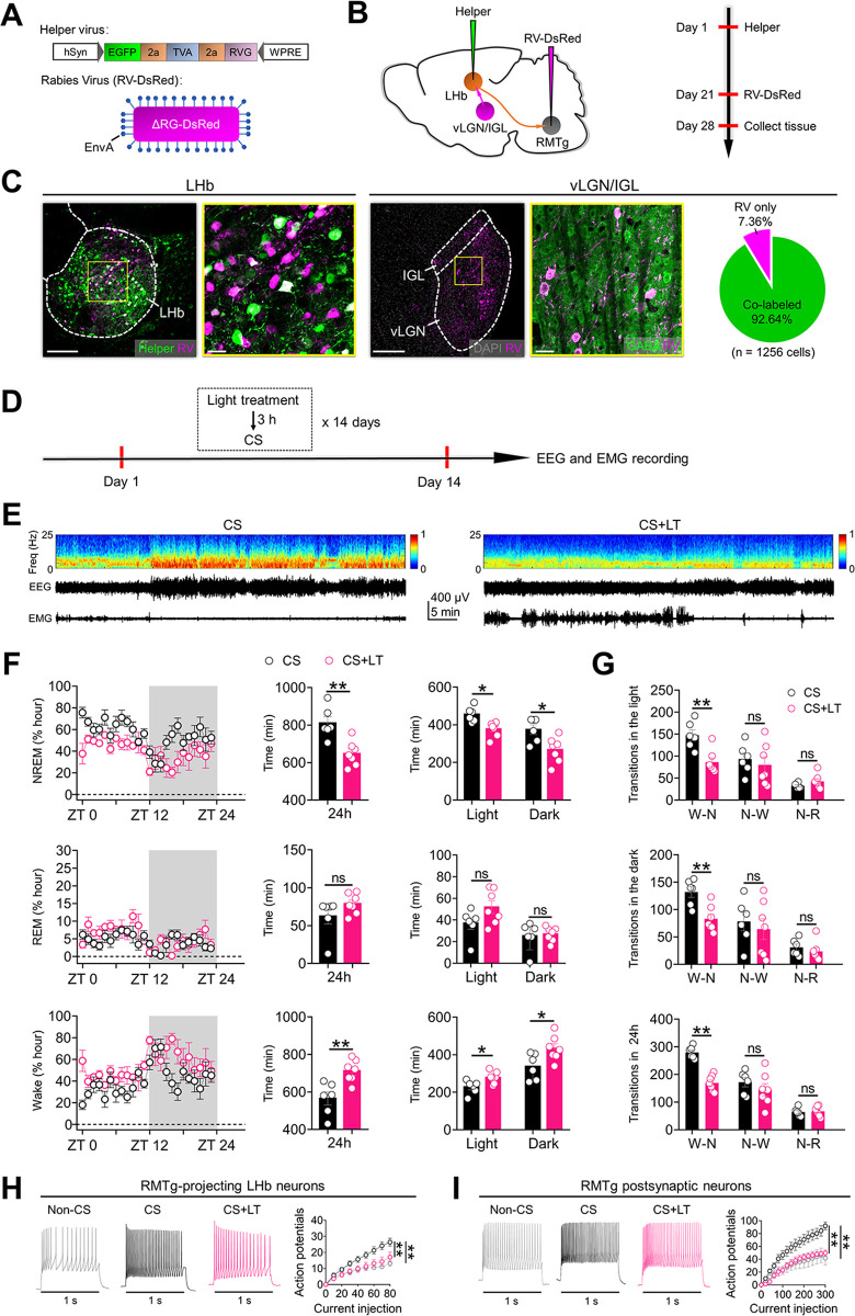
Source: Huang L et al. · PLoS biology · CC BY
Original figure and English caption ↗
Suppressing upstream or enhancing downstream activity weakens the light effect
Suppressing LHb-projecting visual-thalamic cells during light exposure weakens the NREM-reducing effect. Downstream tests act in the opposite direction: activating RMTg-projecting LHb cells or LHb-recipient RMTg populations likewise weakens the light effect.
The complementary directions support a model in which visual-thalamic recruitment permits reduced activity in downstream NREM-promoting populations. Visual information can reach sleep networks linked to stress, beyond pathways for image recognition.
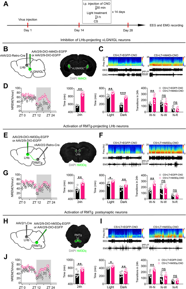
Source: Huang L et al. · PLoS biology · CC BY
Original figure and English caption ↗
The meaning of light depends on the physiological setting
The findings come from male mice aged six to eight weeks, with light accompanying stress. They concern an increased-NREM phenotype, not a human insomnia trial or a universal response to environmental light. Other light pathways, circadian states and prior experience remain relevant.
The study connects environmental input, aversive experience and sleep allocation. Sleep changes need not arise solely within sleep-regulating nuclei: specific visual-thalamic connections can alter how the stressed brain allocates waking and NREM.