How does the subthalamic nucleus affect emergence from propofol anesthesia?
STN CaMKIIα-expressing cells and their ventral-pallidal output bidirectionally alter EEG and righting-reflex recovery, without the same effect on induction.
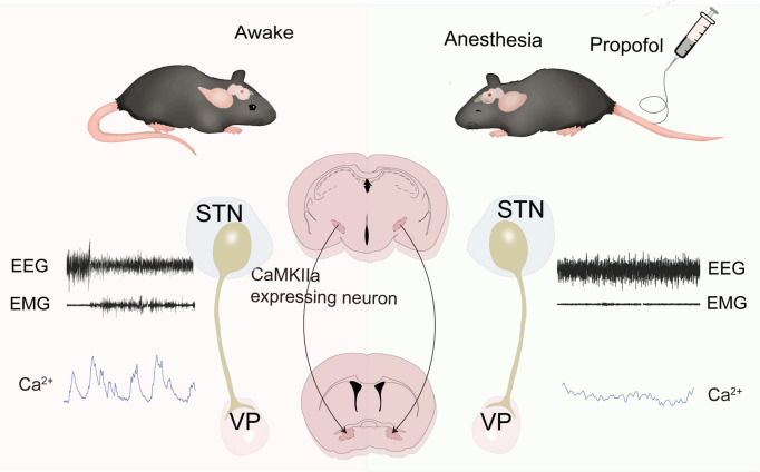
After anesthetic withdrawal, mice gradually emerge from slow-wave EEG and low activity. Is this simply waiting for drug levels to fall, or do circuits contribute? STN CaMKIIα-expressing cells decreased activity during propofol, recovered afterward and altered emergence timing when manipulated.
Activating STN cells or VP terminals promotes arousal-like EEG and faster righting recovery; inhibition has opposite effects. Chemogenetic manipulation leaves induction timing unchanged.
Contents · 6 sections
Entering and leaving anesthesia are different transitions
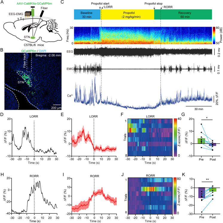
Induction, maintenance and emergence are different stages. STN, well known in motor regulation, also participates in brain states. Simultaneous cortical EEG, neck EMG and population calcium measurements aligned cellular activity, rhythms and behavioral events.
STN labeling and calcium signals declined during propofol and rose after withdrawal around righting recovery. Manipulations then tested whether the state-associated population could itself change emergence.
The transitions occur against different backgrounds: drug action suppresses the network during induction, while activity progressively recovers during emergence. Examining both stages reveals where a population’s effect is concentrated, and the subsequent bidirectional tests illustrate that distinction.
Source: Bai S et al. · Cell biology and toxicology · CC BY-NC-ND
Original figure and English caption ↗
Increasing STN activity accelerates recovery-related changes
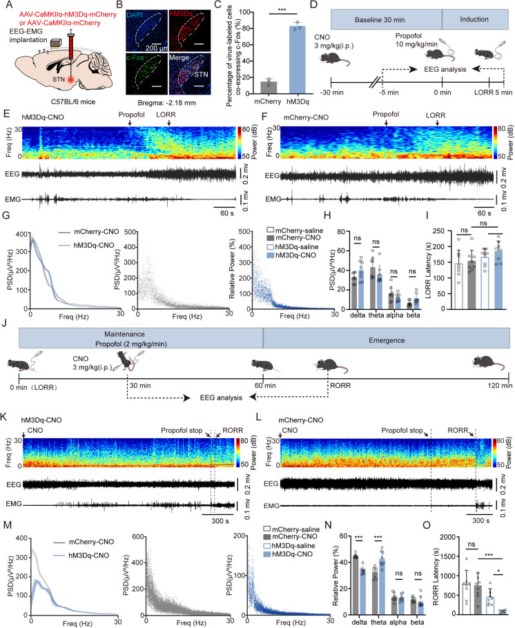
During maintenance, activation shifted strong slow waves toward lower-amplitude faster activity and increased EMG and behavior scores. After withdrawal it shortened righting recovery, with reduced delta and stage-dependent frequency changes absent from illuminated controls.
Chemogenetic activation also promoted emergence, but pre-induction activation did not significantly change righting-loss timing or relevant spectra. The population mattered more during emergence, with different induction and maintenance dosing conditions forming part of the context.
Source: Bai S et al. · Cell biology and toxicology · CC BY-NC-ND
Original figure and English caption ↗
Reducing activity reverses the recovery effect
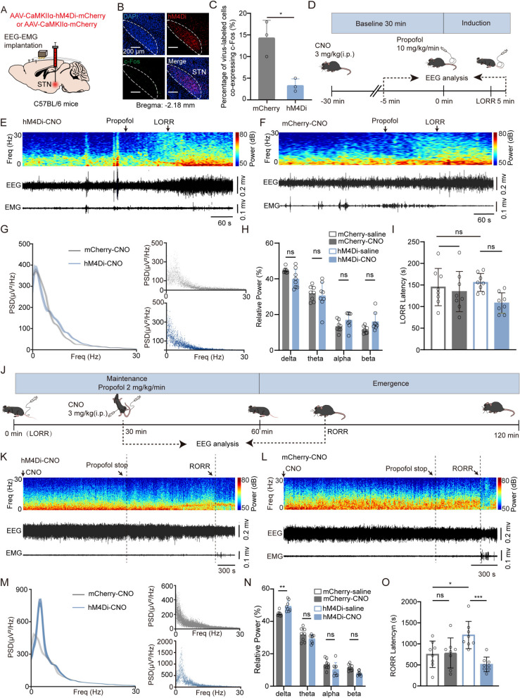
Optical inhibition favored slow waves and delayed EEG desynchronization and righting recovery. Some changes receded after light stopped. Chemogenetic inhibition increased emergence delta and prolonged recovery while some other bands remained comparable.
Inhibition also left induction timing unchanged. Opposite manipulations concentrated effects on recovery: greater STN activity sped it, suppression slowed it, showing a modifiable neural component alongside waning drug action.
Source: Bai S et al. · Cell biology and toxicology · CC BY-NC-ND
Original figure and English caption ↗
Ventral-pallidal output reproduces bidirectional effects
Tracing revealed several STN targets and retrograde labeling supported VP connectivity. VP-terminal activation produced arousal-like EEG, EMG and behavior and accelerated post-withdrawal recovery, retaining the direction of somatic activation.
Terminal inhibition increased delta and delayed recovery, matching somatic inhibition. VP contains multiple recipient types still to be identified; pathway experiments show that this output region can convey emergence-related modulation.
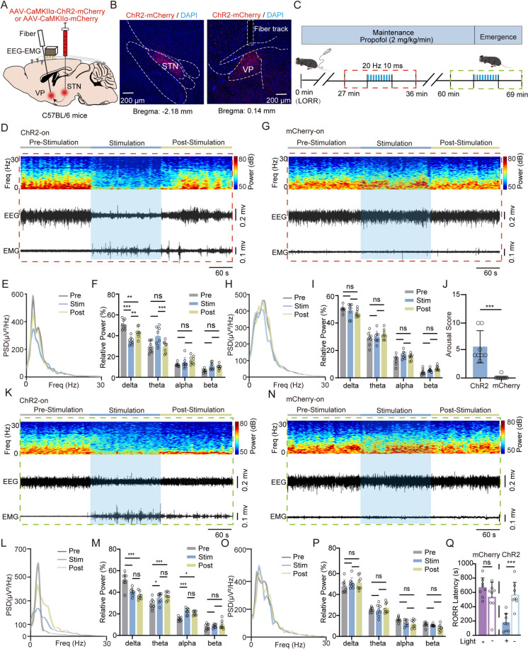
Source: Bai S et al. · Cell biology and toxicology · CC BY-NC-ND
Original figure and English caption ↗
EEG, EMG and reflexes describe different recovery facets
Slow-wave fractions describe cortical rhythms, neck EMG muscle activity, and righting reflects recovery of posture. Their corresponding changes reveal how state evolves around the recovery time rather than only a single number.
Because STN relates to movement and brain state, posture and EEG are informative together. Somatic and terminal interventions show both a local population effect and similar regulation through VP output under the mouse propofol conditions.
Emergence contains a modifiable circuit contribution
The adult-male experiments reveal a distinction between entering and leaving anesthesia. STN activity and VP output bidirectionally alter emergence, while corresponding manipulations leave induction timing unchanged.
After drug withdrawal, recovery involves more than a concentration clock: population activity and output pathways contribute to the transition. STN–VP demonstrates a neural component that can be accelerated or slowed in this mouse model.
Source: Bai S et al. · Cell biology and toxicology · CC BY-NC-ND
Original figure and English caption ↗