An arousal signal under anesthesia: from amygdala to basal forebrain
Activating central-amygdala GABA-marked cells delays induction and hastens emergence from isoflurane in mice. Their basal-forebrain terminals also promote behavioral and cortical activation.
The amygdala often brings fear and defense to mind, but its relationship with arousal may also affect emergence from anesthesia. A mouse study in Anesthesiology finds that central-amygdala GABA-marked cells reduce activity during isoflurane exposure and increase it during emergence. Activating them hastens the return of righting ability and changes cortical EEG. The team then follows their output toward basal forebrain.
Activating central-amygdala GABA-marked cells or their basal-forebrain terminals delays induction, hastens righting-reflex recovery and promotes cortical activation. Terminal activation produces a weaker behavioral effect.
Contents · 7 sections
Following cellular activity across a state transition
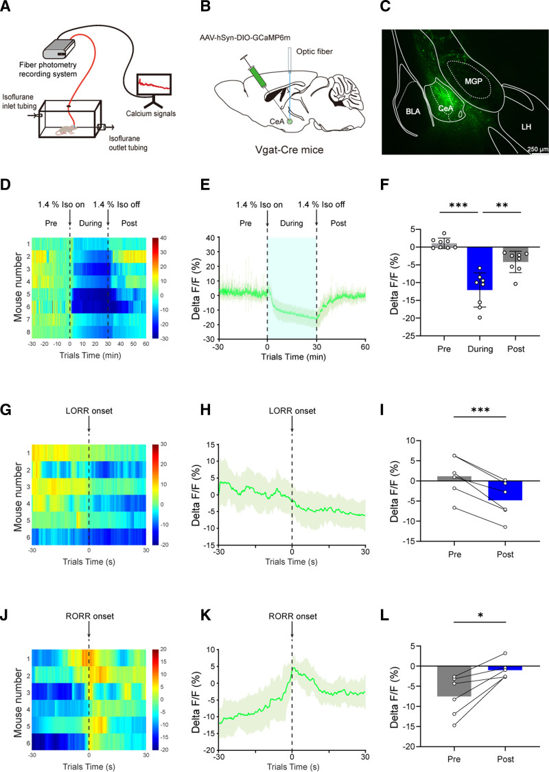
Central amygdala (CeA) connects with several arousal-related regions. Its GABA-marked cells have activity associations with wakefulness and arousal-related behavior, motivating examination across the onset, maintenance and cessation of isoflurane exposure.
Population calcium begins to fall before righting-reflex loss and rises before its return, peaking around recovery. This close relationship with behavioral timing makes CeA a candidate for causal intervention.
These recordings measure a pooled population signal. CeA contains cells with diverse molecular identities and connections, and defined subtypes in other studies can respond differently. The decrease describes the overall response of the labeled population under the tested condition and provides context for its subsequent activation.
Source: Zhang JS et al. · Anesthesiology · CC BY-NC-ND
Original figure and English caption ↗
Induction slows and post-anesthetic recovery accelerates
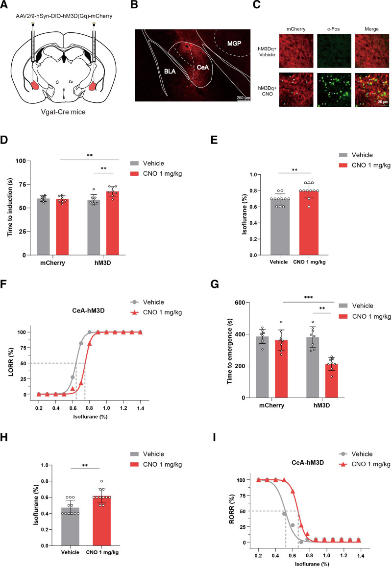
Chemogenetic activation prolongs the time to righting-reflex loss and shortens the time to regain it after anesthesia stops. Both induction and emergence shift toward maintaining or recovering behavioral activity.
With changing isoflurane concentrations, activated mice lose righting at a higher concentration and also regain it at a higher concentration. Controls lack the same significant changes. Altered cellular activity shifts the response to the anesthetic conditions.
Source: Zhang JS et al. · Anesthesiology · CC BY-NC-ND
Original figure and English caption ↗
Most mice right themselves, and some begin to crawl
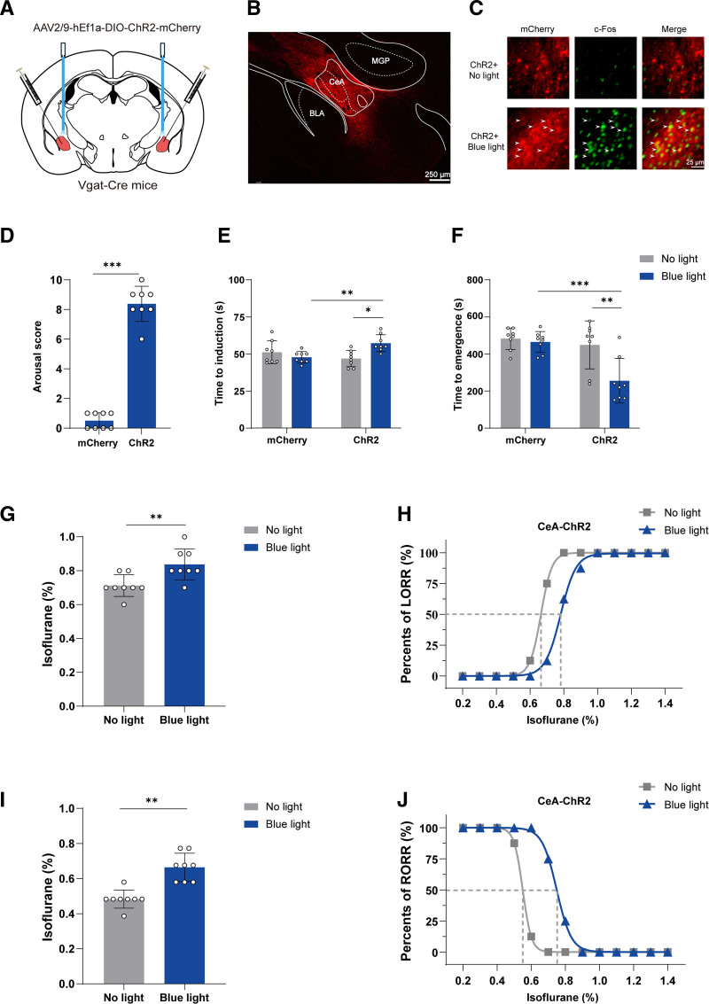
A faster activation approach also delays induction and hastens emergence. Under the tested anesthetic condition, activation produces limb, head and tail movements: seven of eight mice regain righting and four crawl, showing several levels of behavioral arousal.
Control stimulation does not produce the same changes. Naturally associated calcium activity and both slower and faster activation approaches converge on the population’s capacity to shift the tested anesthetic state.
Source: Zhang JS et al. · Anesthesiology · CC BY-NC-ND
Original figure and English caption ↗
EEG changes alongside behavior
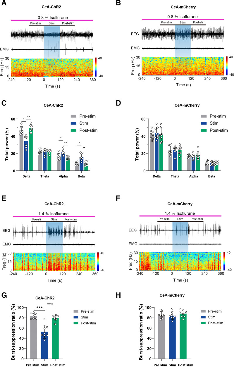
At the shallower tested anesthetic condition, CeA activation shifts EEG from high-amplitude slow activity toward lower-amplitude faster activity. Relative delta power decreases and alpha and beta power increase. EEG gradually returns toward its earlier pattern after activation stops.
At the deeper condition, EEG alternates between bursts and suppressed periods. Activation lowers the proportion of suppressed activity, which rises again afterward. Behavior and EEG reveal the state change at different levels.
Burst–suppression provides a particularly visible change: periods of electrical activity are separated by low-activity intervals, and activation reduces the proportion occupied by those intervals. Beyond observing movement, cortical recordings show that this population influences electrical patterns even at the deeper anesthetic condition.
Source: Zhang JS et al. · Anesthesiology · CC BY-NC-ND
Original figure and English caption ↗
Basal-forebrain terminals also affect emergence
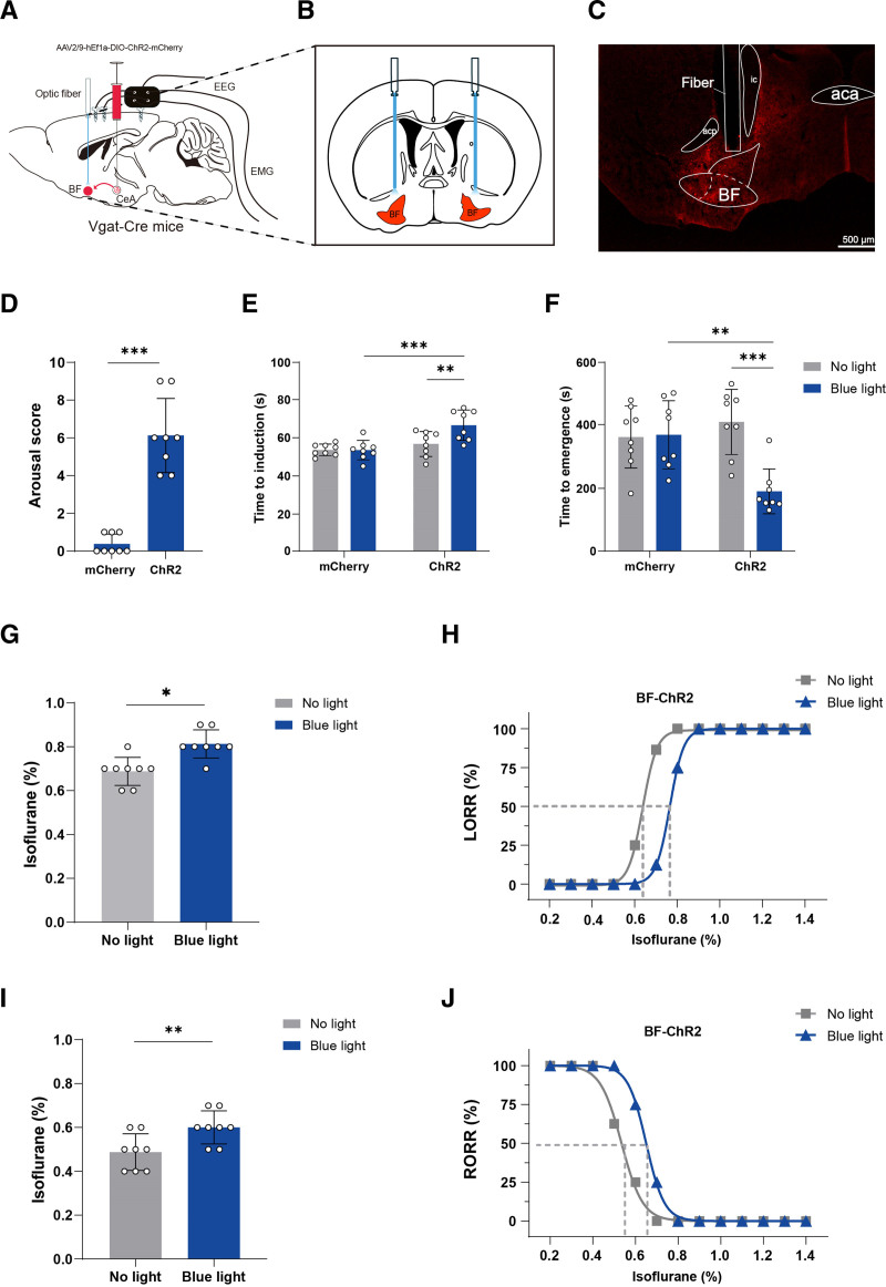
Basal forebrain (BF), an important arousal-regulating region, receives CeA output. Activating CeA terminals there likewise delays induction, hastens emergence after anesthesia stops and shifts concentration-related responses in the same direction.
The immediate behavioral response differs: all eight mice move, but only two regain righting and two crawl. This branch produces a weaker response than CeA-population activation. One output contributes to the overall effect, while other connections may also help drive the transition.
Existing anatomy also links CeA GABA cells with lateral hypothalamus and lateral habenula. BF is therefore one defined output branch. It reproduces the main induction and emergence effects while showing weaker immediate behavior, revealing a distinction between population-level and branch-level effects.
Source: Zhang JS et al. · Anesthesiology · CC BY-NC-ND
Original figure and English caption ↗
The branch also influences cortical activity
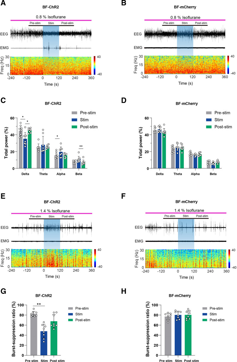
During terminal activation, relative delta power decreases and alpha power increases at the shallower condition, while burst–suppression ratio falls at the deeper condition. Controls lack the same significant changes. The BF branch thus influences both behavior and cortical activity, with its precise recipient cells still to be identified.
Source: Zhang JS et al. · Anesthesiology · CC BY-NC-ND
Original figure and English caption ↗
A network involving inhibitory cells can promote activity
This study includes adult male and female mice and links isoflurane-state transitions with CeA activity and its BF branch. GABA marking identifies a cellular signaling type, while a network’s behavioral effect also depends on its targets. Induction, emergence, movement and cortical activity together sharpen the picture of amygdalar involvement in arousal regulation.