How does a hypothalamic output influence anesthetic entry and recovery?
LHA glutamatergic neurons and their habenular projection contribute to state regulation during isoflurane anesthesia in mice. Righting-reflex and EEG effects are related but asymmetric and depend on anesthetic state.
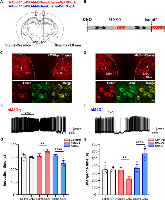
Even during anesthesia, the brain can respond differently to the same input. A mouse study published in Neuroscience Bulletin on April 13, 2021 examines how glutamatergic neurons in the lateral hypothalamic area (LHA) influence entry into, maintenance of and recovery from isoflurane anesthesia. Following their output to lateral habenula (LHb) links righting-reflex timing with EEG changes. The effects of increasing and decreasing activity are not always mirror images, particularly in deeper anesthesia.
Activating LHA glutamatergic neurons or their LHb terminals delays loss of righting, speeds recovery and reduces burst suppression in deeper anesthesia. Inhibition shifts reflex timing oppositely but does not significantly change deeper-anesthesia burst suppression.
Contents · 7 sections
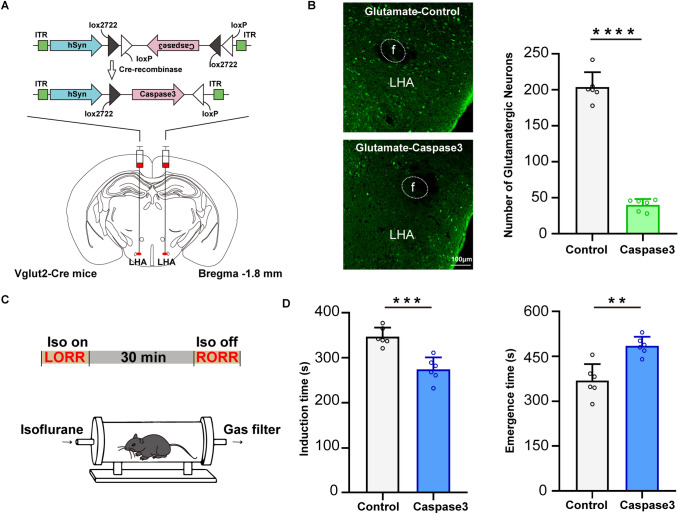
Reducing the population speeds induction and slows recovery
LHA contains several populations involved in waking, sleep and motivation, including a large glutamatergic population. Reducing these cells makes mice lose the righting reflex earlier and regain it later after anesthesia ends, supporting a contribution to opposing the anesthetic state and promoting recovery.
Isoflurane still produces anesthesia after cell numbers decline; the changed outcome is transition timing. This places the population in state regulation rather than making it an exclusive switch for anesthesia. Righting provides a concrete behavioral endpoint to compare with subsequent EEG findings.
Source: Zhao S et al. · Neuroscience bulletin · CC BY
Original figure and English caption ↗
Changing activity shifts transition timing in opposite directions
Activating the LHA glutamatergic population prolongs induction and shortens recovery; suppressing it produces earlier loss of righting and later recovery. Alongside population reduction, these activity manipulations provide a functional test at another level.
The population remains heterogeneous. LHA glutamatergic neurons can overlap with some orexin neurons and connect to several distant targets. The population-level result does not assign the entire effect to a single finer subtype. The discussion contrasts earlier orexin findings in which recovery changed without a significant induction effect. Targeting the broader glutamatergic population raises questions about functional divisions within LHA.
Source: Zhao S et al. · Neuroscience bulletin · CC BY
Original figure and English caption ↗
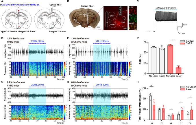
Activation reduces the proportion of EEG suppression
During deeper maintenance anesthesia, EEG alternates between bursts and suppression. Activating LHA glutamatergic cells markedly reduces the fraction occupied by suppression. This burst-suppression ratio, BSR, describes electrical organization rather than directly demonstrating restoration of all waking behaviors.
In lighter anesthesia, prominent burst suppression is absent. Activation instead lowers relative delta power and increases relative alpha, beta and gamma power. Different measures therefore capture effects of the same population under different states.
Source: Zhao S et al. · Neuroscience bulletin · CC BY
Original figure and English caption ↗
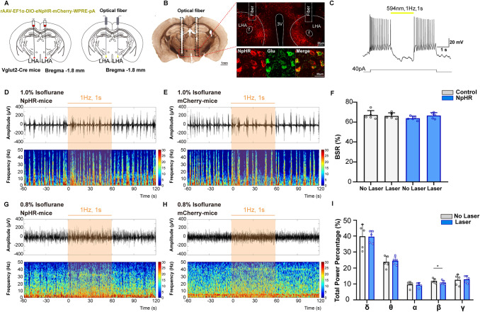
Further suppression does not produce a mirror-image EEG effect
Inhibiting the population does not significantly change BSR in deeper anesthesia. Under lighter anesthesia, the main detected effect is a small reduction in relative beta power, rather than a full reversal of every activation-associated spectral change.
The authors suggest that deeper anesthesia may already suppress the population enough to limit the effect of further inhibition. This remains an explanatory model; the study does not directly measure glutamate-release changes across that state.
Source: Zhao S et al. · Neuroscience bulletin · CC BY
Original figure and English caption ↗
Moving activation to habenular terminals retains key effects
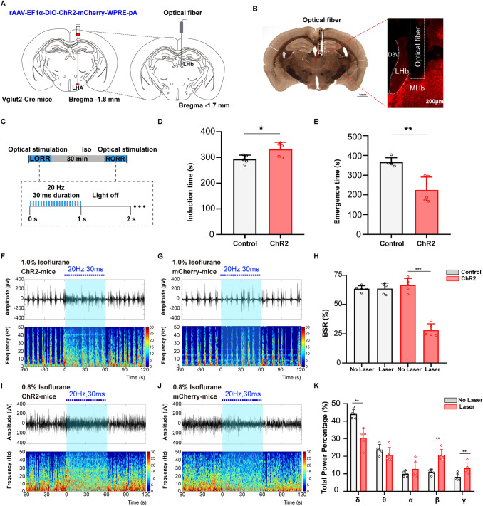
LHb is one of several targets of LHA neurons. Activating LHA glutamatergic terminals there likewise prolongs induction, shortens recovery and reduces BSR during deeper anesthesia. Terminal and somatic experiments provide evidence at two locations for this connection’s contribution.
In lighter anesthesia, terminal activation reduces relative delta power and increases relative beta and gamma power. These specific spectral effects do not assign a uniform arousal-promoting function to all LHb cells; the identities of the relevant recipients remain unresolved.
Source: Zhao S et al. · Neuroscience bulletin · CC BY
Original figure and English caption ↗
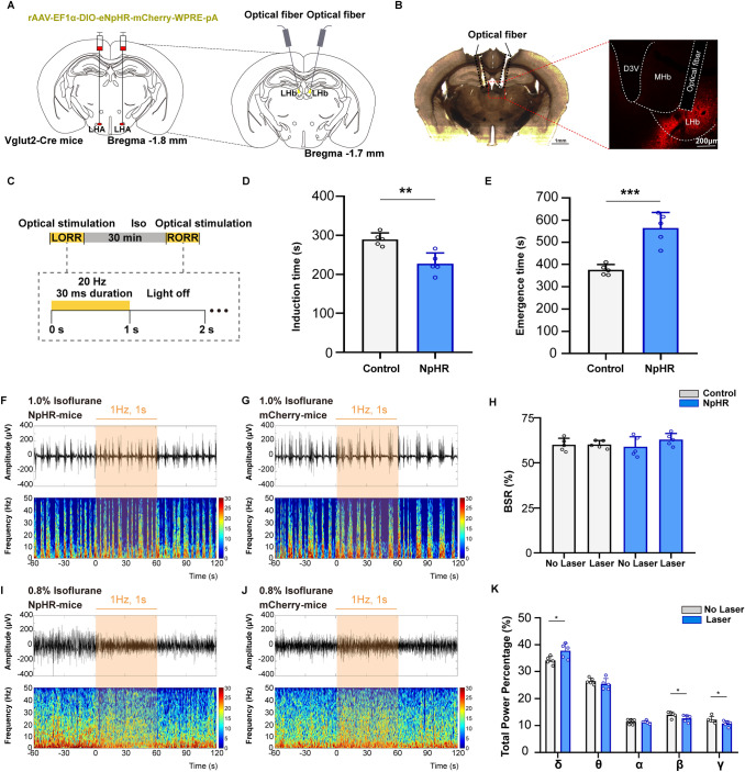
Terminal inhibition also depends on the state examined
Suppressing these terminals produces earlier loss of righting and later recovery. Deeper-maintenance BSR nevertheless does not change significantly; in lighter anesthesia, relative delta power increases while beta and gamma decrease. Transition timing and maintenance EEG thus provide related but distinct outcomes.
Source: Zhao S et al. · Neuroscience bulletin · CC BY
Original figure and English caption ↗
Arousal-related effects arise from particular cells and connections
This mouse study extends LHA glutamatergic contributions across recovery, induction and maintenance, with LHb providing one output. Other projections, local orexin-related connections and cotransmitters may contribute. Terminal stimulation can also influence other branches through axonal propagation, qualifying isolation of the connection.
A central remaining question is which LHb recipients produce the different outcomes across anesthetic states. The findings identify a state-dependent connection rather than a fixed map of arousal switches.