After stress, amygdala SST cells contribute to difficulty falling asleep
In mice, central-amygdala SST-labeled cells respond to acute stress, and activation delays sleep onset. Suppression shortens stress-associated sleep latency without significant changes in tested normal-sleep measures.
In an unfamiliar environment, sleep can fail to arrive when rest is due. How does stress interfere with the transition from waking to sleep? A study in Communications Biology follows somatostatin, or SST-labeled cells in the central amygdala of adult mice. They respond to several acute stressors, and increasing their activity delays sleep onset. The study also distinguishes difficulty initiating sleep from disruption continuing throughout sleep.
Increasing CeA SST-cell activity delays sleep onset, with increased waking mainly in the first hour. Suppression improves sleep latency in acute-stress models without significant effects on tested normal-sleep measures.
Contents · 7 sections
Different stressors evoke cellular responses
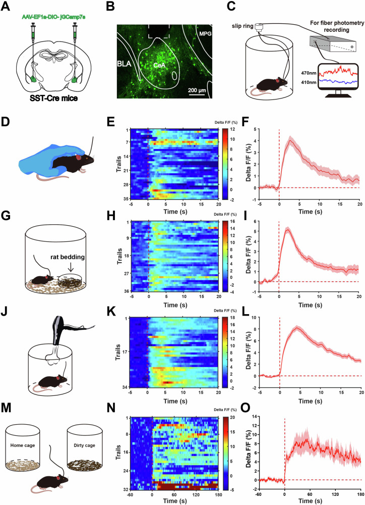
The study begins with acute stress. Brief restraint, rat-scented bedding, air puffs, and entry into a cage carrying other mice’s scents all raise calcium signals in the CeA SST population. Changes in the external context rapidly register in this population’s activity.
Increasing their activity also accompanies larger pupils, faster breathing, and less exploration of exposed spaces. Total distance in the open field and maze does not change significantly, so these exploratory changes are not accompanied by increased overall movement. Cellular activity and stress responses connect across several measures.
Source: Yao W et al. · Communications biology · CC BY-NC-ND
Original figure and English caption ↗
Activating the population makes waking faster
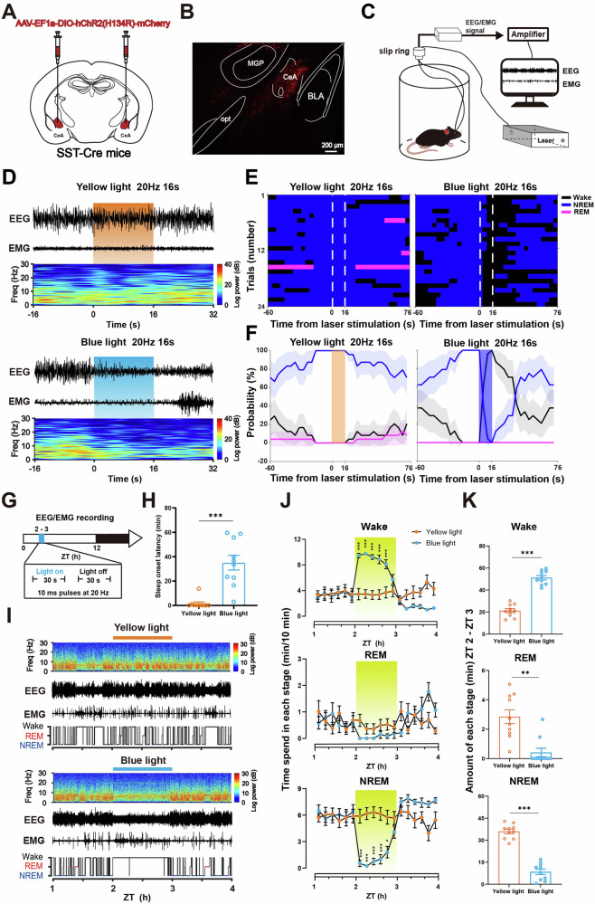
The team follows sleep states using simultaneous brain and muscle electrical recordings. After mice enter NREM sleep, activating CeA SST cells can rapidly promote waking. Across tested activation conditions, a faster activation rhythm also makes that transition more likely and faster.
A second question is whether this activity delays sleep in waking mice. During an hour of recruitment in the resting phase, sleep latency lengthens, waking increases, and REM and NREM sleep decrease. Activation affects both sleep onset and the distribution of states in this early interval.
Source: Yao W et al. · Communications biology · CC BY-NC-ND
Original figure and English caption ↗
Sleep is delayed without sustained waking
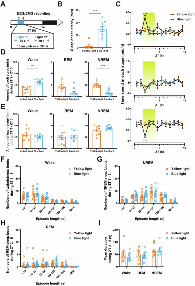
Extending observation to three hours shows increased waking mainly in the first hour, with no significant differences in subsequent sleep-state amounts. Numbers of episodes across length categories and average durations also remain similar. Longer activation does not turn the entire observation period into waking.
A different activation approach produces a similar pattern: delayed sleep onset, an early increase in waking, and no significant later changes in sleep or waking episodes. These temporal differences focus attention on sleep initiation rather than merging all sleep-related effects.
Source: Yao W et al. · Communications biology · CC BY-NC-ND
Original figure and English caption ↗
Spontaneous sleep transitions lack the same signal changes
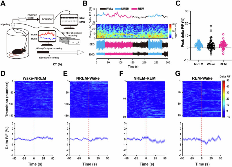
A population that can promote waking when activated need not show the same response at every spontaneous awakening. The study records calcium, brain activity, and muscle activity in freely moving mice, comparing normal waking, NREM, and REM sleep. Population signals do not differ significantly among these states.
Signals also show no significant changes across several sleep–wake transitions. This differs from their clear increases under stress. In these population recordings, the cells respond more prominently to stressful contexts than to spontaneous state transitions.
Source: Yao W et al. · Communications biology · CC BY-NC-ND
Original figure and English caption ↗
Reducing activity does not clearly alter normal sleep
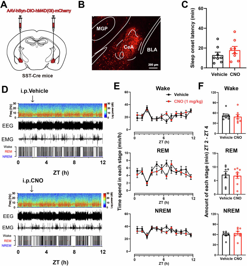
The team next suppresses CeA SST cells and follows normal sleep. Sleep latency does not change significantly, and waking, NREM, and REM amounts remain similar. The distributions of states in brain and muscle recordings show no clear alteration.
This places activation and suppression in different contexts: recruiting the population is sufficient to delay sleep, while reducing activity does not discernibly disturb sleep under the tested normal conditions. The next question is whether the activity becomes more consequential after stress.
Source: Yao W et al. · Communications biology · CC BY-NC-ND
Original figure and English caption ↗
Suppression advances sleep onset after cage change
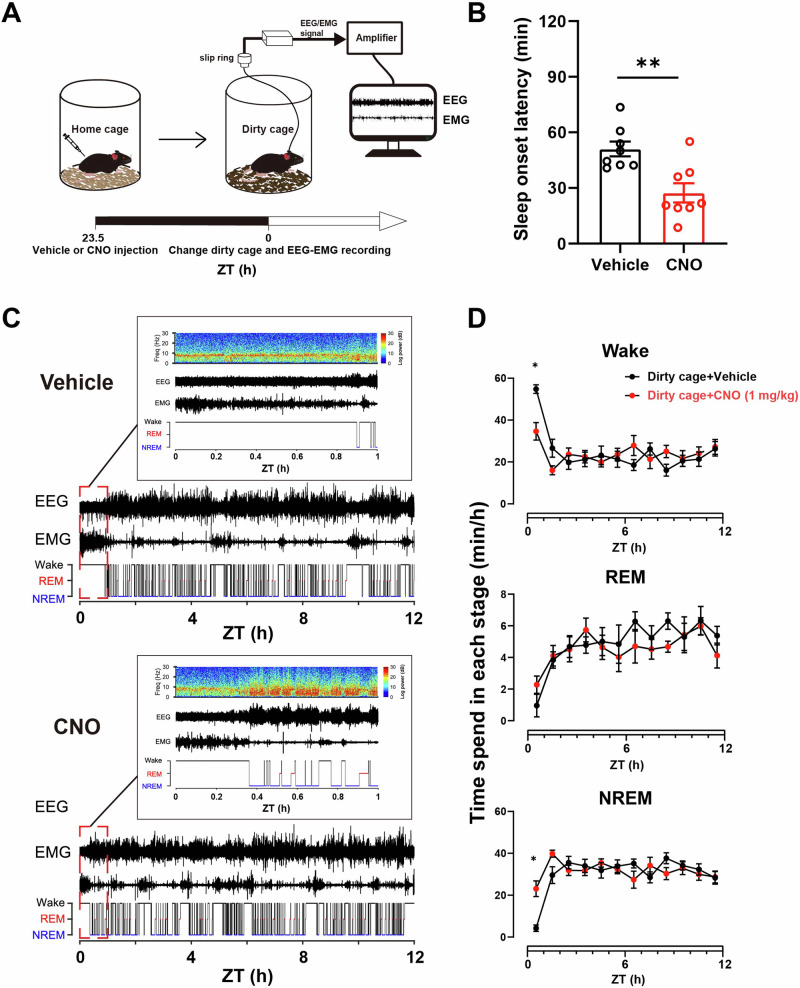
After entering a cage carrying unfamiliar mice’s scents, sleep onset is delayed and early waking increases. Suppressing CeA SST cells before cage change shortens latency, decreases first-hour waking, and increases NREM sleep. Improvement is concentrated near the beginning of the resting period.
A second acute-stress context involving restraint before rest also shows shorter sleep latency after suppression. The two contexts support the same interpretation: this population’s stress-associated activity contributes to waking that persists when sleep would otherwise begin.
Source: Yao W et al. · Communications biology · CC BY-NC-ND
Original figure and English caption ↗
Stress meets sleep at a particular transition
This study in adult mice of both sexes connects stress responses and sleep-onset timing with a labeled CeA population. Waking an animal and maintaining prolonged waking are distinct effects, just as stress-delayed sleep and ordinary sleep transitions can involve cells differently.
Across these acute-stress models and observation intervals, the SST population’s contribution centers on entering sleep. Examining a specific phase reveals how cells within one brain region link external stress with a sleep transition.