From tongue dragging to emergence:
Two neural circuits behind rescue-like behavior in mice
A mouse study follows helping behavior, tongue sensation and brain arousal to examine how social interaction changes a companion’s recovery from anesthesia.
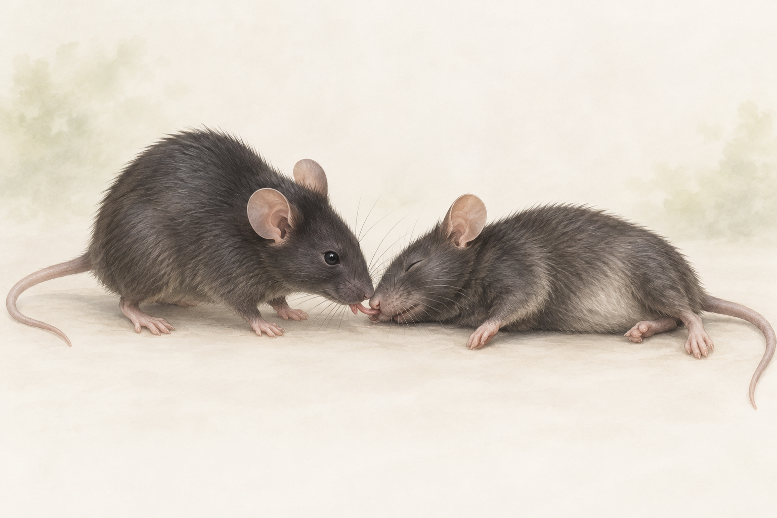
How might a mouse lend a hand when its companion lies unresponsive? Beyond sniffing, grooming and eye licking, researchers observed an unusual action: the awake mouse repeatedly pulled the anesthetized animal’s tongue outward. The recipient also emerged from anesthesia sooner. From tongue to brain, and from helper to recipient, a small interaction connected neural circuits in two animals.
The generation of helping and the recipient’s recovery involve circuits in two animals. Tongue sensory input links physical contact with brain arousal.
Contents · Seven sections
Helping has a receiving side
Interactions between social animals can influence one another's behavior and physiological state. Studies of helping naturally focus on the actor: does it detect an unusual state in its companion, and which neural activity accompanies approach or contact? A complete account also needs to follow changes in the recipient.
On January 22, 2025, researchers from the University of Science and Technology of China, Anhui Medical University and collaborating institutions published this study in Science Advances. They examined circuits in both animals: how helping behavior arises and how physical contact changes the recipient's arousal state.
An unusual action directed at an anesthetized companion
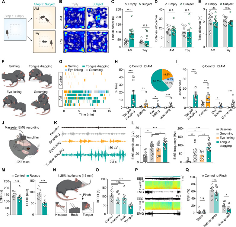
Bystanders sniffed, groomed and licked the eyes of anesthetized companions, and also grasped the tongue with their teeth and pulled it outward. The unexpected movement raised a question: was it a response to the companion’s condition or simply curiosity about an immobile object? Toy mice and other comparisons helped distinguish these possibilities.
A toy mouse provided a comparison. Bystanders spent longer near an anesthetized companion than near the toy, without a corresponding change in overall locomotion. The same tongue-dragging behavior was not observed toward dead mice, linking the interaction to the condition of a living anesthetized companion.
Masseter recordings helped distinguish tongue dragging from ordinary grooming. Muscle activity had greater amplitude and frequency during tongue dragging than during face grooming or eye licking. Female mice were less likely to drag the tongues of unfamiliar animals, whereas familiarity had no significant effect in males. Subsequent circuit experiments mainly used males, leaving sex and social familiarity as relevant factors for further study.
Does tongue dragging help the companion emerge sooner?
With an interacting bystander present, anesthetized mice regained their righting reflex sooner, while its loss during induction did not differ significantly. This ability to turn from the back into a normal posture served as the study’s behavioral measure of emergence. The interaction was linked to recovery, raising the next question: which form of contact contributed?
The team compared stimulation of the tongue, hindpaw and back during emergence from isoflurane anesthesia. Tongue stimulation significantly shortened righting-reflex recovery time; hindpaw and back stimulation did not show the same effect. The different responses brought the tongue into focus as a key sensory entry point.
EEG supplied complementary evidence. Tongue stimulation reduced the burst-suppression ratio during emergence, with no corresponding difference during wakefulness or anesthesia maintenance. Burst suppression alternates periods of greater activity with low-activity intervals. Its change supports an effect on the recovery process beyond a local movement response.
Source: Cao et al., Science Advances, 2025 · CC BY 4.0 · Original image unchanged
Original figure and English caption ↗
How does a tongue signal reach the arousal circuit?
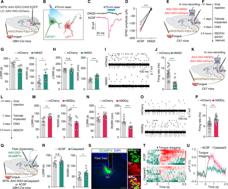
The next step connected peripheral tongue stimulation with brain activity. Tracing identified tongue-connected neurons in the mesencephalic trigeminal nucleus, or MTN, which contains primary afferent neurons involved in oral sensory processing. Further tracing showed projections from glutamatergic MTN neurons to the locus coeruleus, or LC.
Noradrenergic LC neurons are closely associated with arousal regulation. Recruiting them offers a possible route through which a local mechanical stimulus could influence a broader brain state. Recordings supported this idea: tongue stimulation produced stronger and faster responses in the identified LC neurons than hindpaw or back stimulation in the same comparison.
The team then observed the connection at the cellular level. Stimulating MTN projection terminals evoked excitatory postsynaptic responses in noradrenergic LC neurons, and AMPA receptor blockade suppressed them. The findings outlined a glutamatergic pathway: tongue sensory input → MTN → LC, linking a point of physical contact to a brain node involved in arousal.
Changing the circuit changes emergence
Intervention tested whether the pathway participates in recovery. Inhibiting tongue-connected MTN neurons delayed emergence and reduced LC firing. Activating these neurons instead accelerated emergence and increased LC activity. Opposing manipulations therefore produced corresponding changes in recovery speed.
Ablating the tongue-connected neuron population prolonged recovery from isoflurane anesthesia and weakened the LC calcium response to rescue-like interaction. Together with circuit inhibition, these results connect the behavior, anatomical pathway, neural activity and recovery outcome through causal evidence.
Source: Cao et al., Science Advances, 2025 · CC BY 4.0 · Original image unchanged
Original figure and English caption ↗
The helper’s brain has a circuit of its own
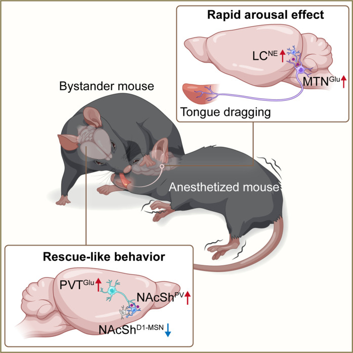
The researchers then turned to the bystander's brain. Activity changes in the paraventricular thalamic nucleus, or PVT, and nucleus accumbens shell, or NAcSh, suggested participation in behavior regulation. Glutamatergic PVT neurons provided input to NAcSh; inhibiting this projection reduced tongue dragging, whereas activating it increased its frequency and duration.
NAcSh does not behave as one uniformly changing population. Single-cell calcium measurements showed that some cells became more active during tongue dragging while others were inhibited. Parvalbumin-expressing PV neurons contributed to initiation: inhibiting them increased the latency before tongue dragging began, and activating them shortened it, without a notable effect on locomotor ability.
D1-type medium spiny neurons showed another direction of control: inhibiting them increased tongue dragging, while activating them reduced it. D2-cell manipulations had no significant effect. Combining these findings with earlier connectivity evidence, the authors proposed that PV cells may influence D1 cells through local inhibition. Distinct populations thus contribute to initiation and behavioral expression, with their local coordination remaining an interesting question.
Source: Cao et al., Science Advances, 2025 · CC BY 4.0 · Original image unchanged
Original figure and English caption ↗
Physical contact connects the brains of two animals
The study links two sides of the interaction: the helper's circuit influences rescue-like behavior, while tongue contact enters the recipient's sensory pathway and recruits arousal-related LC neurons. An interaction between two animals can thus be followed into a change in the receiving animal's brain state.
Rescue-like behavior is intriguing because it combines actions directed at an anesthetized companion with measurable recovery benefits. How do mice detect the companion’s state? Does the interaction occur in nature, or in other unresponsive conditions? These questions extend the finding into social behavior more broadly, while the helper’s subjective experience remains a further question.
By placing the generation of helping and the mechanism of benefit in one framework, the work offers a route for further experimental analysis and comparison in social neuroscience: study both why the actor acts and how the recipient is affected.