Brief theta waves recruit spatial experience again during sleep
After mice learn to find food in a maze, brief sleep theta waves accompany reactivation of learning-related ensembles. Suppressing thalamic input to entorhinal cortex weakens reactivation and subsequent spatial-task performance.

What happens in the brain after an animal learns a route and falls asleep? Memories continue to change after sleep begins. Ensembles associated with recent experience become active again, but what recruits them? A 2024 Nature Communications mouse study examines the connection between medial entorhinal cortex, MEC, and thalamic nucleus reuniens, RE. During non-rapid eye movement sleep, theta episodes lasting roughly three hundred milliseconds occur at a particular phase of slower activity and accompany reactivation of learning-related ensembles.
Closed-loop suppression of RE-to-MEC input reduces brief theta activity and ensemble reactivation during NREM sleep; mice subsequently take longer routes to food. Wake reactivation and measured sleep architecture show no significant changes.
Contents · 7 sections
Maze experience returns in sleep
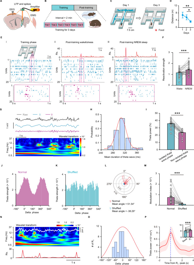
Mice search for food at a fixed location in a six-arm maze and take more direct routes with training. MEC ensembles show distinct coordinated firing patterns at different positions: some relate to the starting arm, others to turns or areas near the goal. Spatial experience is reflected in relationships among cellular activities.
Patterns from training provide a measure of how strongly similar ensemble activity recurs afterward. Reactivation is stronger during subsequently recorded NREM sleep than during wakefulness. This measure describes similarity in population firing patterns, allowing researchers to follow the recurrence of experience-related activity during sleep.
Source: Xiao Q et al. · Nature communications · CC BY-NC-ND
Original figure and English caption ↗
A short theta episode within slower activity
NREM sleep typically features slow delta activity, whereas theta often brings waking exploration to mind. MEC recordings reveal another form of theta: brief isolated episodes rather than sustained oscillation, nested in the more active phase of delta waves.
Peaks of learning-related ensemble reactivation coincide with these theta episodes. The overall slow rhythm of sleep remains, while brief changes may help recruit particular patterns of activity. The next question is which input supplies this transient signal to MEC.
Reuniens connects to superficial entorhinal cortex
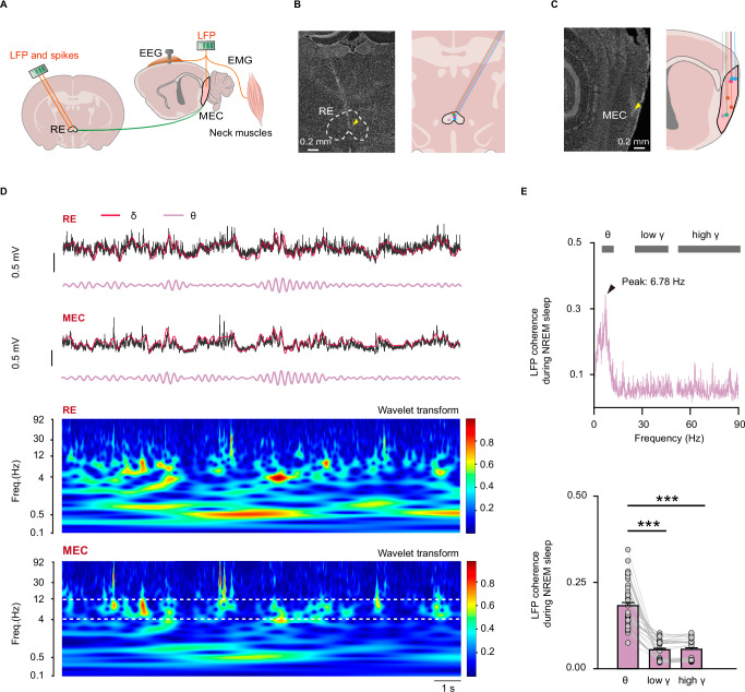
Tracing identifies inputs to MEC from several regions, with RE axons concentrated in superficial MEC. Theta activity in the two regions is strongly synchronized during NREM sleep, and time-series analysis indicates that RE activity predicts subsequent MEC changes. These observations focus attention on the connection.
Slice recordings show short-latency excitatory responses to RE input in MEC excitatory cells and interneurons. Receptor blockade eliminates these responses, supporting glutamatergic transmission. The regions share not only synchronized rhythms but also excitatory connections capable of directly influencing recipient cells.
Source: Xiao Q et al. · Nature communications · CC BY-NC-ND
Original figure and English caption ↗
The same input meets different cellular states
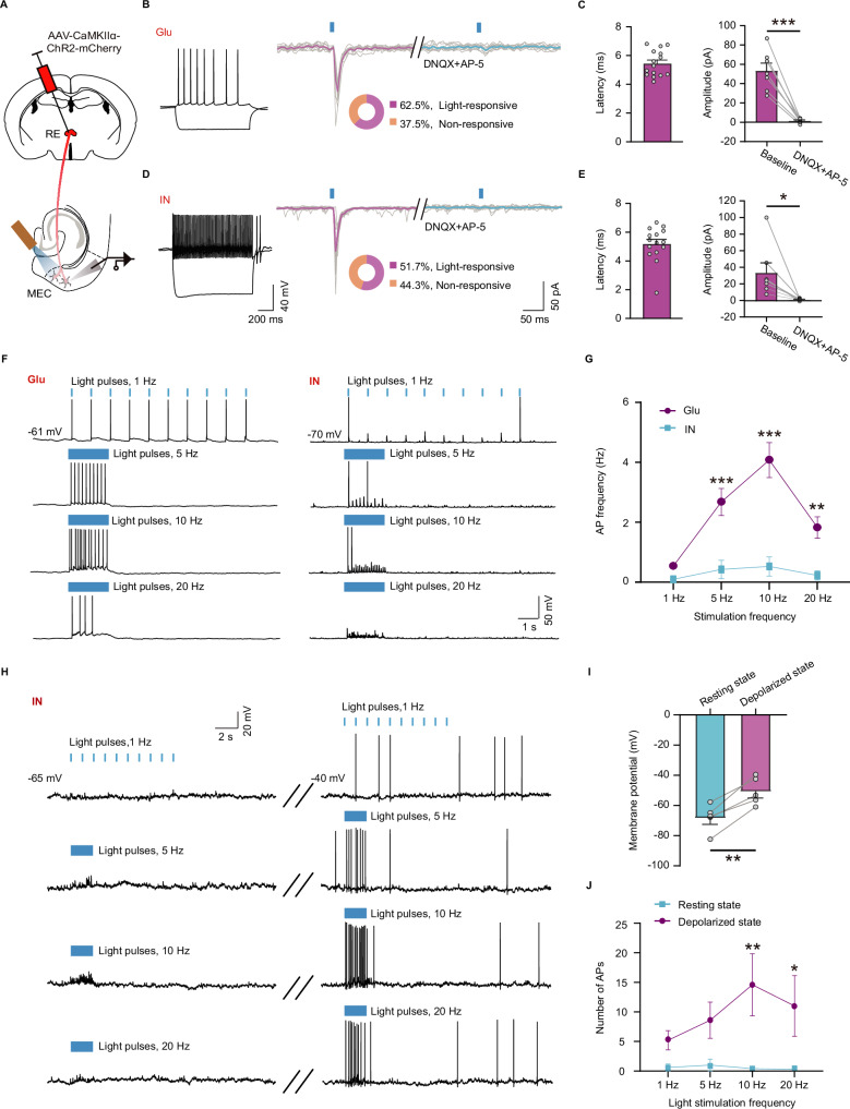
RE input does not drive all MEC cells identically. Excitatory cells respond more readily to lower-frequency stimulation, while interneurons are less readily recruited at their resting membrane potential. Depolarizing the interneurons enhances their response. The recipient cell’s existing state changes the effect of an arriving signal.
Acetylcholine also depolarizes these interneurons. The researchers propose that differences in cellular state between sleep and wakefulness may help shape distinct rhythms. This explanation derives from slice response properties and adds a cellular clue to understanding why isolated theta appears during sleep.
Source: Xiao Q et al. · Nature communications · CC BY-NC-ND
Original figure and English caption ↗
Brief peaks persist within lower overall activity
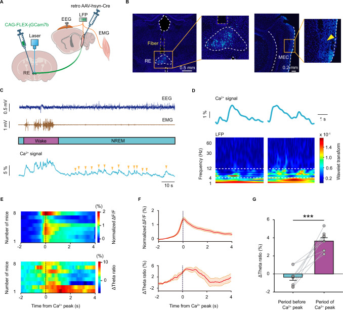
The overall calcium signal of MEC-projecting RE cells falls upon entry into NREM sleep, yet discrete activity peaks remain. MEC theta ratio rises around these peaks. Lower average activity does not make the input silent: brief events retain an association with local rhythmic changes.
Source: Xiao Q et al. · Nature communications · CC BY-NC-ND
Original figure and English caption ↗
Suppressing the input makes finding the goal less direct
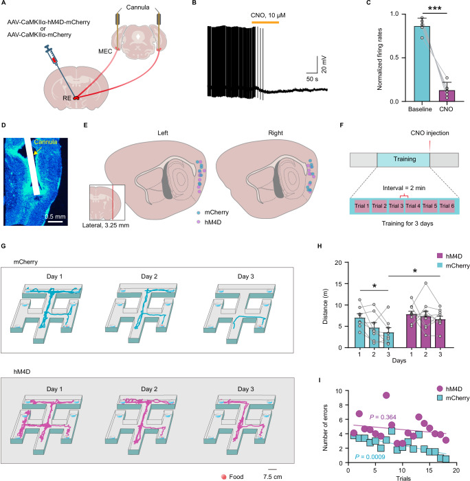
The researchers suppress RE-to-MEC input after training. Mice subsequently take longer paths to food and perform worse in wrong-arm entries. Intervention after learning links the connection to processing that follows experience.
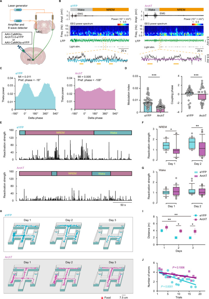
A more precise closed-loop experiment suppresses the input when isolated theta is detected during NREM sleep. Theta activity and its coupling to delta weaken, sleep ensemble reactivation decreases, and subsequent maze performance worsens. Wake reactivation shows no significant difference.
Source: Xiao Q et al. · Nature communications · CC BY-NC-ND
Original figure and English caption ↗
Memory processing also involves brief rhythmic events
Closed-loop intervention does not significantly alter measured sleep–wake architecture or EEG power spectra. Changes in task performance therefore accompany weakening of particular within-sleep rhythms and reactivation, rather than an obvious loss of sleep overall. Brief theta events connect input, ensemble activity and spatial-task performance.
These findings, obtained mainly in male mice, give a concrete picture of memory processing during sleep: brief rhythms embedded in slower activity can help recruit recent experience again. How these events cooperate with hippocampal sharp-wave ripples and sleep spindles remains a further question about the broader consolidation process.
Source: Xiao Q et al. · Nature communications · CC BY-NC-ND
Original figure and English caption ↗