What new windows does AAV11 open onto connected cells?
AAV11 offers new options for retrograde projection labeling and astrocyte transduction in mice, with pathway-dependent advantages and informative labeling differences in a disease model.
To understand communication between brain regions, researchers first need to identify the cells sending signals to a given destination. Viral vectors can travel retrogradely from projection terminals to cell bodies, revealing these sources. Yet the same vector performs differently across connections, potentially missing some populations. A study published in Nature Communications on June 26, 2023 examines the less-used natural serotype AAV11 in adult male mice. It retrogradely labels several projection populations with properties complementary to AAV2-retro and supports enhanced astrocyte transduction in selected regions with an astrocyte-related promoter. These findings open windows onto connectivity, activity and neuron–glia relationships.
AAV11 performance is connection-dependent: it labels more strongly in several pathways but less strongly than AAV2-retro in others. With suitable expression control, it supports projection-population calcium recording and astrocyte labeling, expanding the circuit toolkit.
Contents · 7 sections
Labels travel from the destination toward the source
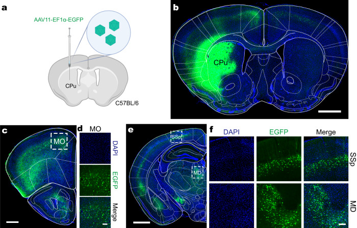
In caudate-putamen, AAV11 produced local fluorescence and labeled upstream cells projecting to the region. Signals in multiple cortical and thalamic areas demonstrate its utility for identifying input sources, selecting cells by their projection destination.
In a primary visual cortex test, the researchers did not observe labeled cell bodies in known downstream regions when checking anterograde transsynaptic spread. Alongside the interpretation of uptake at terminals and retrograde transport, these results support its use as a retrograde tracer under the tested conditions.
Source: Han Z et al. · Nature communications · CC BY
Original figure and English caption ↗
Two vectors reveal different strengths at one destination
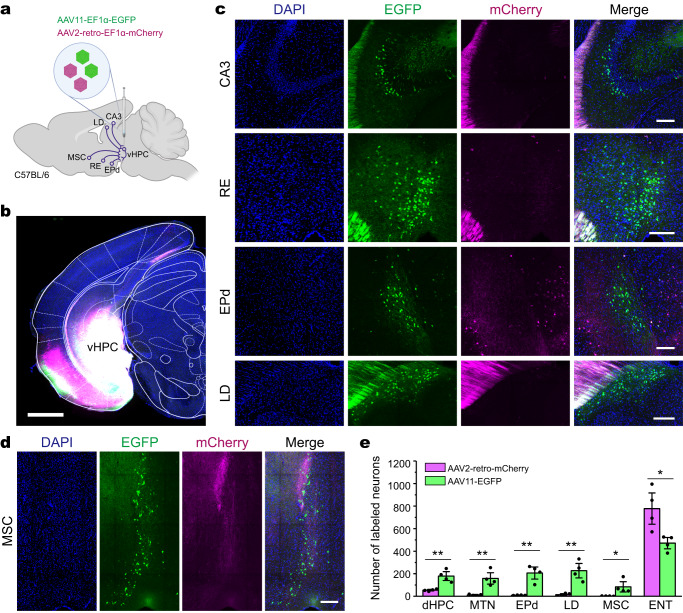
For projections to ventral hippocampus, AAV11 labeled more cells in dorsal hippocampus, midline thalamus, dorsal endopiriform nucleus, lateral dorsal thalamus and medial septal complex. AAV2-retro labeled entorhinal input more effectively. The advantage did not run uniformly in one direction.
Separate-vector tests, batch comparisons and fluorophore exchange provided additional support. Different vectors reveal different populations more readily, so a labeling map reflects both connections and transduction properties. Complementary tools can broaden what becomes visible.
Source: Han Z et al. · Nature communications · CC BY
Original figure and English caption ↗
Changing the destination changes the advantage
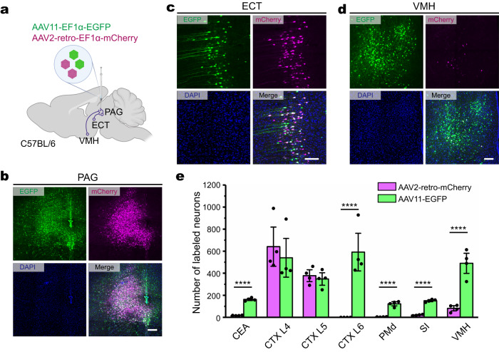
For projections to periaqueductal gray, AAV11 labeled more cells in central amygdala, cortical layer 6, dorsal premammillary nucleus, substantia innominata and ventromedial hypothalamus. Cortical layers 4 and 5 did not show significant differences.
This connection dependence matters because fewer labels with one vector do not automatically establish a weaker pathway. Entry, transport and expression can influence the result. AAV11 adds a route to observing populations that may be less readily revealed by another vector.
Source: Han Z et al. · Nature communications · CC BY
Original figure and English caption ↗
Adding cell type to projection identity
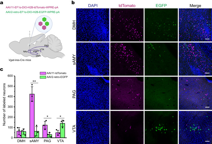
Using Vgat-Cre mice, the study examined GABAergic cells projecting to nucleus accumbens. AAV11 labeled more in striatum-like amygdalar regions and periaqueductal gray, whereas AAV2-retro labeled more in VTA. Dorsomedial hypothalamus did not significantly differ.
Projection destination and genetic identity jointly define the observed population. Again, an advantage in one source cannot be assumed in others. Different projection populations may benefit from different vectors to build a broader network picture.
Source: Han Z et al. · Nature communications · CC BY
Original figure and English caption ↗
Seeing when the identified cells respond
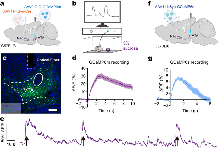
The researchers next observed VTA cells projecting to accumbens. Calcium signals increased when mice licked sucrose solution. Two expression strategies recorded related responses, demonstrating that AAV11 can support activity measurements in a projection-defined population.
This connects a structural map with behavioral timing: researchers can observe when a projection-defined population changes its activity. These population calcium responses provide a basis for further study of cellular identity, function and behavioral relationships.
Source: Han Z et al. · Nature communications · CC BY
Original figure and English caption ↗
The window also opens onto astrocytes
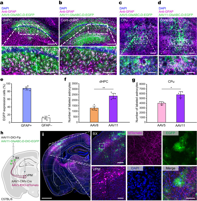
With the GfaABC1D promoter, AAV11 expression occurred predominantly in astrocytes, with a smaller non-GFAP-colabeled fraction. It labeled more astrocytes than AAV8 in dorsal hippocampus and more than AAV5 in caudate-putamen and hippocampal comparisons.
Combined with AAV1, the study labeled projection neurons and astrocytes in the ventral posteromedial thalamus–barrel cortex system. This coordinated labeling offers possibilities for examining neuron–astrocyte relationships; functional studies are needed to clarify how the labeled cells interact.
Source: Han Z et al. · Nature communications · CC BY
Original figure and English caption ↗
Fewer labels provide a clue to altered connections
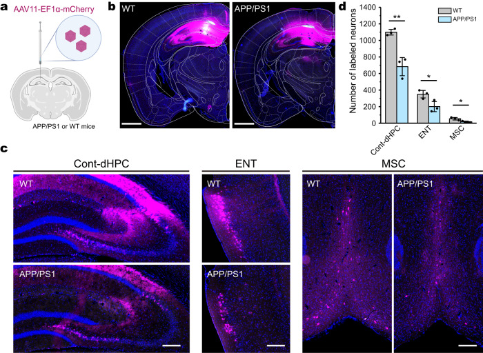
Tracing inputs to dorsal hippocampus in 10-month-old APP/PS1 and wild-type mice revealed fewer labeled cells in contralateral dorsal hippocampus, entorhinal cortex and medial septal complex in the disease model. These findings identify a disease-associated labeling difference for further study.
Fewer labels may reflect reduced connectivity, altered retrograde transport or different immune responses to the vector. Distinguishing these possibilities remains necessary. AAV11 expands visibility of selected projections and astrocytes while encouraging closer examination of the biology behind a fluorescent map.
Source: Han Z et al. · Nature communications · CC BY
Original figure and English caption ↗