Lighting one site while listening across brain regions
A male-mouse study integrates distributed microwires and optical fibers into a lightweight electrode covering 18 recording sites, with tests of field potentials, single units and multisite changes during basal-forebrain activation.
Brain activity is distributed across many locations. Examining how an input at one site accompanies changes elsewhere requires a shared timeline. A study published in Frontiers in Neuroscience on June 13, 2023 develops an electrode for synchronous recording and optical stimulation: microwires reach different depths, optical fibers deliver light, and sites connect through one module. Tests in freely moving male mice examine field potentials, single-unit signals using another bundled-wire version, and distributed responses during basal-forebrain cholinergic activation.
The integrated electrode supports distributed field-potential recordings, while a bundled-wire version separates single units. Multiple cortical sites show weaker theta components during basal-forebrain cholinergic activation. Tested field-potential and single-unit recording durations span weeks and days, respectively.
Contents · 7 sections
Distributed brain sites require a distributed design
Regular probe layouts suit some compact regions, while functional networks extend across hemispheres and depths. The study integrates microwire and fiber positions into one module to reduce interactions among separate components during placement. The goal is to bring distributed signals to the recording system synchronously.
The prototype weighs about 0.28 grams and targets 16 default-mode-network-related sites plus bilateral basal forebrain, totaling 18 locations. Another version uses bundled wires for single units in basal forebrain and selected cortex. The two versions serve different recording tasks.
Anatomical location gives recordings their context
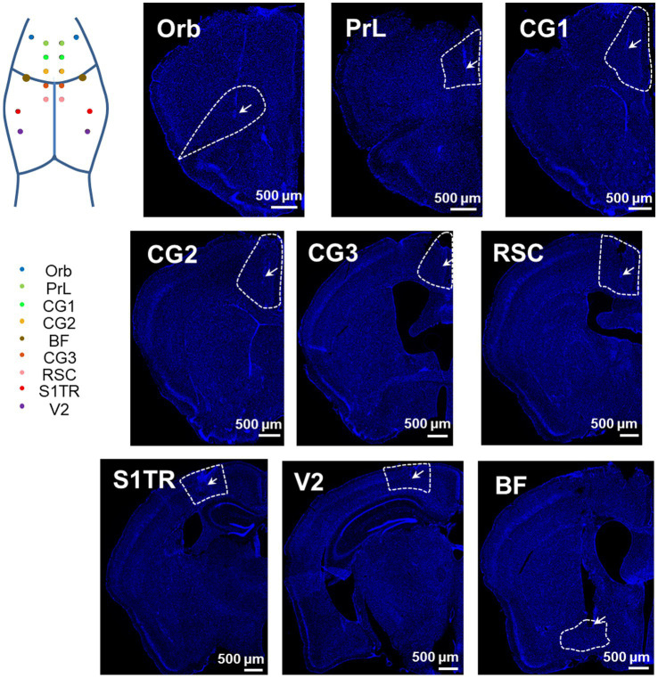
Tissue sections locate tested microwire tips and fibers near intended regions. Prefrontal, cingulate, retrosplenial, sensory-related and basal-forebrain images associate channels with tissue locations, providing anatomical context for their labels.
The sites span cortex and subcortical regions. Their localization supports the prototype’s tested placement, while waveforms, spectra and animal state reveal the content of the signals.
Source: Zhang Q et al. · Frontiers in neuroscience · CC BY
Original figure and English caption ↗
Recording slow rhythms and hippocampal ripples
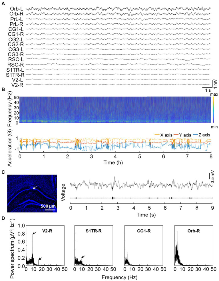
Simultaneous field-potential traces display activity across regions within one time window. Longer records pair spectra with head acceleration to show signals during activity and rest in freely moving mice. Field potentials and related electrophysiological signals remain recordable for at least four weeks in the evaluation.
The team records high-frequency ripple-like activity in hippocampal CA1 and lower-frequency responses associated with visual flicker. These examples illustrate the signal range while retaining the regional and contextual specificity of different rhythms.
Source: Zhang Q et al. · Frontiers in neuroscience · CC BY
Original figure and English caption ↗
A different wire configuration resolves individual units
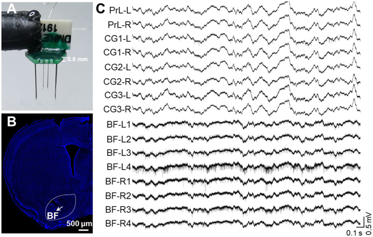
Field potentials describe local population activity, while single-unit recordings attempt to separate cellular spikes. The bundled-wire version samples basal forebrain and selected default-mode-network-related regions, revealing spikes superimposed on slower activity across multiple channels.
Most tested channels detect single-unit activity, with some unit signals continuously detectable for at least five days. This differs from weeks of field-potential recording. Capturing population rhythms and separating units are different performance tests.
Source: Zhang Q et al. · Frontiers in neuroscience · CC BY
Original figure and English caption ↗
Separated units differ between rest and exploration
Spike shapes, clustering and firing features separate basal-forebrain units. Some are classified electrophysiologically as putative PV-positive cells and fire more during rest than free exploration. This classification follows waveform features rather than individual genetic confirmation.
The example connects device testing with behavioral state: recordings yield unit changes associated with particular settings. Increased resting-period firing supplies an association rather than establishing that the cells cause rest.
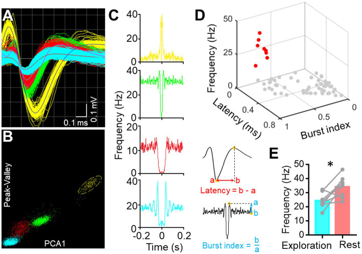
Source: Zhang Q et al. · Frontiers in neuroscience · CC BY
Original figure and English caption ↗
Illuminating basal forebrain while observing distributed responses
The study expresses ChR2 in basal-forebrain cholinergic cells, with most fluorescent cells colabeled for ChAT. Multiple cortical recording sites show weaker theta components during activation, placing a controlled input and remote field-potential changes on a shared timeline.
Frequency responses at different sites can be observed together rather than assembled from separate animals or time periods. This example tests integration of optical input with multisite electrical recordings and provides data for examining regional state changes.
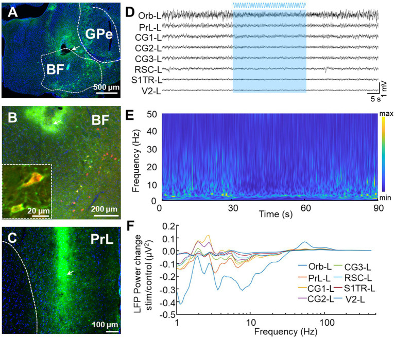
Source: Zhang Q et al. · Frontiers in neuroscience · CC BY
Original figure and English caption ↗
Bringing dispersed locations into one temporal question
The testing uses adult male mice, with extended-recording evaluation principally involving three animals. Localization, field-potential and unit examples demonstrate prototype capabilities, while long-term tracking remains sensitive to material–tissue interactions, position shifts and unit-identification stability.
Its contribution is recording distributed activity together with an optical input. Network studies can move from what occurs at each site to how changes appear at the same time, while connection direction, cellular identity and behavioral roles require corresponding experimental tests.