What do cerebellar climbing fibers signal when mice stand to explore?
Population calcium recordings from cerebellar climbing fibers in freely behaving male mice show stronger responses around tiptoe standing and rearing than around grooming, turning or ordinary locomotion.
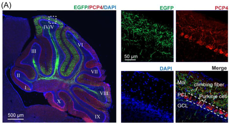
A mouse explores an open field and suddenly lifts its forepaws to stand upright. What signal accompanies that brief action in the cerebellum? A study published in Biology on June 13, 2022 examines climbing fibers, which originate in inferior olive and provide input to Purkinje cells. Combining projection tracing with axonal calcium recording reveals population responses closely associated with standing exploration in cerebellar vermis lobule IV/V of freely behaving male mice. The work both identifies a behavioral association and establishes a way to follow this input during free behavior.
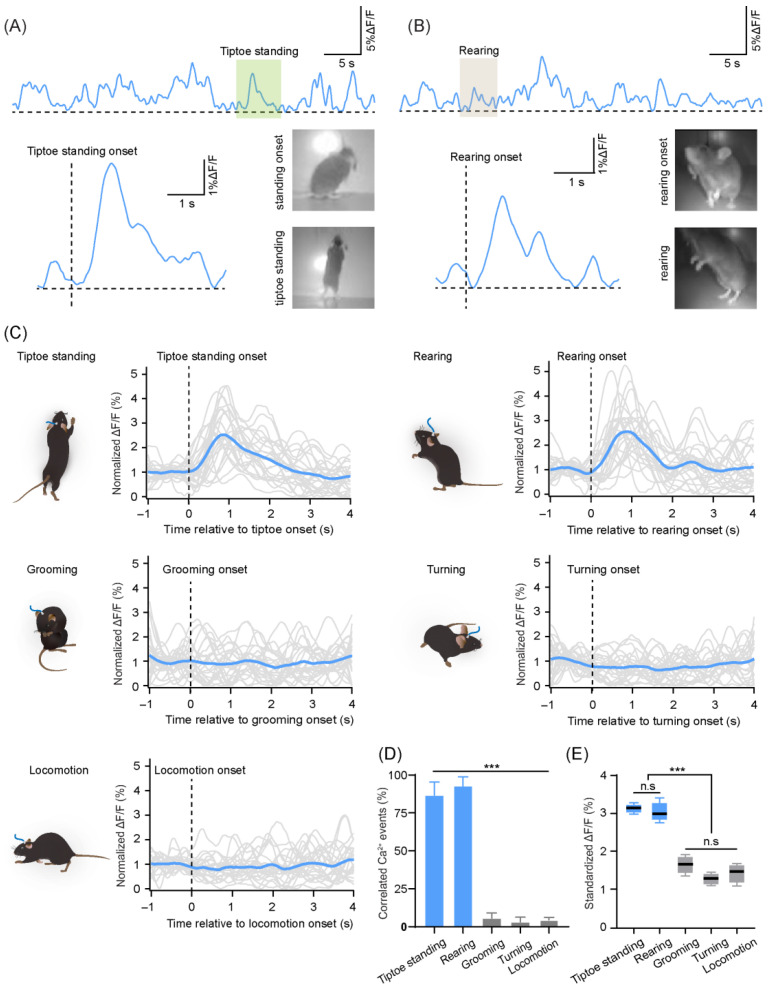
Climbing-fiber calcium responses in lobule IV/V more often accompany tiptoe standing and rearing, whose peak amplitudes differ nonsignificantly. Response amplitude and frequency also show no significant differences across recording days1,2and5.
Contents · 6 sections
From recipient cells to the input they receive
Climbing fibers form an important input within cerebellar coordination and learning circuits. Purkinje-cell complex spikes have often provided an indirect view of the olivocerebellar system. Looking at terminals instead focuses on the input itself and its relationship with natural actions.
Climbing-fiber signals were historically described as relatively simple all-or-none teaching events, while later work proposed richer information in bursting and response magnitude. This study does not directly test a learning rule; it establishes observation of this input during free behavior.
Free behavior is itself part of the scientific question. Standing, balancing and changing posture integrate motor and vestibular-related information, and head-fixed actions do not cover the full range of natural behavior. Allowing animals to choose when to stand adds a different observational setting for climbing-fiber research.
Tracing first establishes where the input arrives
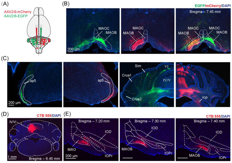
Bilateral tracing shows predominantly contralateral projections with zonal organization in cerebellar cortex. Lobule IV/V of the vermis contains relatively dense labeling in the examined distribution and becomes the recording region. Retrograde tracing links the lobule with corresponding inferior-olive regions.
The recording location therefore has an anatomical basis. Climbing fibers reach particular lobules across regions, and different projection distributions can support different functional observations even within the same input system.
Source: Tang J et al. · Biology · CC BY
Original figure and English caption ↗
Fibers in the molecular layer are distinguished from Purkinje somata
Histology places labeled fibers in the molecular layer, with separate markers identifying Purkinje cells. Purkinje somata do not colabel with the fiber tracer, supporting an upstream climbing-fiber origin for the observation.
This establishes identity for later recordings. Population fluorescence integrates a region, so interpretation depends on the fibers’ origin and whether expression includes nearby recipient cells. Anatomy and colabeling together define the observation more closely.
Source: Tang J et al. · Biology · CC BY
Original figure and English caption ↗
Signals can be followed across days of free movement
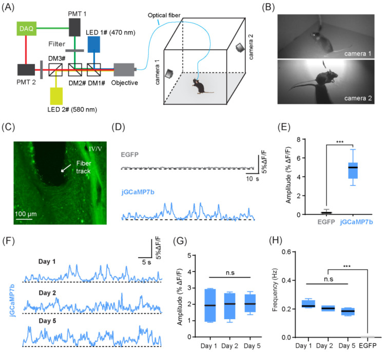
Calcium-indicator animals show larger fluorescence transients than control-fluorescence animals. Another channel in the same animals also lacks changes of comparable magnitude. These comparisons support calcium-related activity beyond fluctuations shared with movement.
Across four mice, mean amplitude and transient frequency differ nonsignificantly on days1,2and5. Recordings can span multiple observation days, although they do not track whether each individual fiber remains identical. Stability concerns the measured population metrics.
Source: Tang J et al. · Biology · CC BY
Original figure and English caption ↗
Standing exploration stands out among limb movements
Open-field actions are classified as tiptoe standing, rearing, grooming, turning and locomotion. The first two frequently begin alongside climbing-fiber calcium transients, while the other actions much less often accompany comparable events. Limb movement alone does not produce the same population response.
Peak amplitudes are higher for tiptoe standing and rearing than for the other actions, with no significant difference between those two motifs. The findings associate the sampled lobule with exploratory movements, while posture, movement form and exploration remain intertwined rather than identifying an abstract curiosity signal.
Source: Tang J et al. · Biology · CC BY
Original figure and English caption ↗
Visible input dynamics open further functional questions
Stronger population fluorescence could involve more coactive fibers, but individual fibers and their synchrony are not independently resolved here. Calcium–action association likewise does not establish that the fibers initiate or drive exploration.
The work uses adult male mice, with open-field behavioral analyses focused on four animals and recordings centered on lobule IV/V. It establishes observation of input during natural movement; other lobules, learning tasks and disease models require their own tests.