Can a tiny focusing sphere help neural modulation use less light?
Improving light use starts with its spatial distribution. Microspheres concentrate optical fields and enhance responses in cells, brain slices and mice, providing a proof of concept for reducing incident-light requirements.
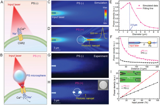
Light used to modulate neurons spreads and is absorbed by tissue. Must stronger cellular responses always require stronger illumination? A study published in Advanced Science on February 20, 2022 explored a different approach: tiny transparent spheres focus part of the optical energy nearby. The localized field, called a photonic nanojet, enhanced currents in light-sensitive cells, neural responses in brain slices and motor-cortex-associated behavioral changes in mice. The central idea is to redistribute light rather than simply deliver more of it.
Microsphere focusing enhances optogenetic responses under specific conditions. Evidence spans optical fields, cellular currents, brain-slice activity and mouse behavior, while long-term compatibility, off-target effects and clinical applications remain to be established.
Contents · 6 sections
A sphere reshapes the local optical field
The sphere acts as a microlens. Without it, incident light spreads along its path; with it, a more concentrated region appears on the shadow side. Simulations and optical-field imaging both support this local focusing effect.
Enhancement depends on sphere size, position and distance from the light source, with modest differences between simulations and measurements. Local concentration therefore does not imply equal enhancement throughout a tissue.
A concentrated optical field begins the evidence chain. Whether it increases cellular current, neuronal firing and behavior requires observations at successive levels. The study links these readouts to examine how optical changes translate into biological responses.
Source: Guo J et al. · Advanced science (Weinheim, Baden-Wurttemberg, Germany) · CC BY
Original figure and English caption ↗
Cellular current can increase under matching illumination
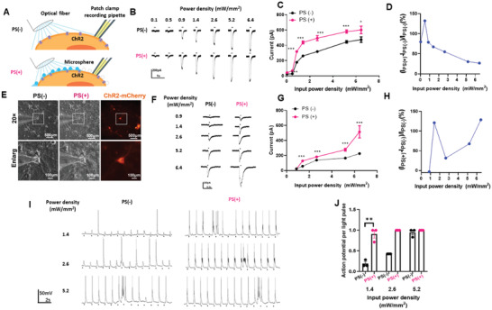
Light-sensitive cultured cells showed larger inward currents with spheres, followed by similar findings in primary hippocampal neurons. The first system addressed channel responses, while the second involved neurons, providing complementary evidence.
The gain varied with illumination rather than increasing indefinitely. At selected lower-light conditions, neurons generated action potentials more successfully with spheres; at stronger illumination, success rates converged. Responses approaching saturation leave less room for additional gain.
Source: Guo J et al. · Advanced science (Weinheim, Baden-Wurttemberg, Germany) · CC BY
Original figure and English caption ↗
Responses also increase in brain slices
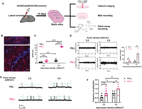
Brain tissue adds optical complexity and network interactions. Slice experiments therefore combined calcium signals, electrode recordings and membrane-potential measurements. Several readouts showed stronger light responses with spheres, extending the evidence into tissue.
Readouts differed in their conditions: calcium responses remained limited in many cells at the lower illumination, while enhancement was clearer at the higher condition. Electrical recordings showed more spikes across the tested conditions. Complementary measures thus retained distinct response patterns.
Source: Guo J et al. · Advanced science (Weinheim, Baden-Wurttemberg, Germany) · CC BY
Original figure and English caption ↗
A motor response emerges at lower illumination
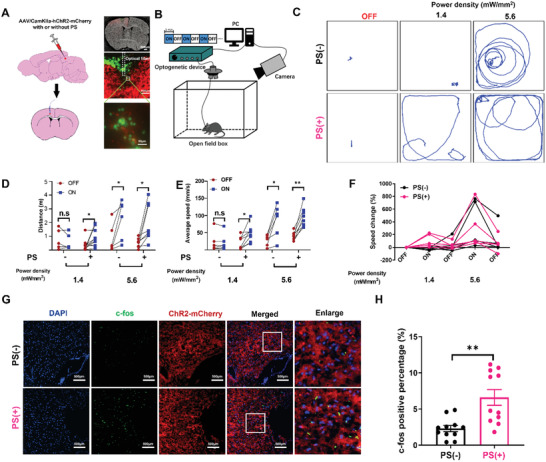
At the lower illumination, mice without spheres did not show an appreciable light-on motor change, whereas sphere-treated mice increased distance and speed. At stronger illumination, both groups showed increased movement, relating focusing effects to incident intensity.
An activity-related marker was also higher in the sphere group at lower illumination, supporting the behavioral observation. This was a motor-cortex response study rather than a motor-disorder treatment trial or proof of a uniform reduction across brain regions and light-sensitive channels.
Source: Guo J et al. · Advanced science (Weinheim, Baden-Wurttemberg, Germany) · CC BY
Original figure and English caption ↗
Focusing light raises material and spatial questions
Reducing incident-light requirements motivates the approach, while reduced off-target effects require dedicated comparisons in neighboring cells and tissue. Long-term compatibility, distribution and stability of foreign spheres cannot be inferred from short-term response enhancement.
The work still relies on invasive light delivery rather than external illumination of deep brain regions. Local focusing addresses how light is used once nearby; getting light into tissue remains a distinct problem.
Efficiency can improve by reshaping the optical path
From optical fields to currents and from slices to behavior, this proof of concept shows that responses depend on light distribution as well as cellular expression. Combining optical structures with neural readouts opens a research direction, while durable applications need further material, spatial-selectivity and biological evidence.