Two hippocampal inputs cooperate in lasting potentiation
A dual-opsin strategy distinguishes CA3 and entorhinal inputs in male-mouse hippocampal slices, revealing potentiation after coactivation and enabling study of neuropeptide-related circuit plasticity.
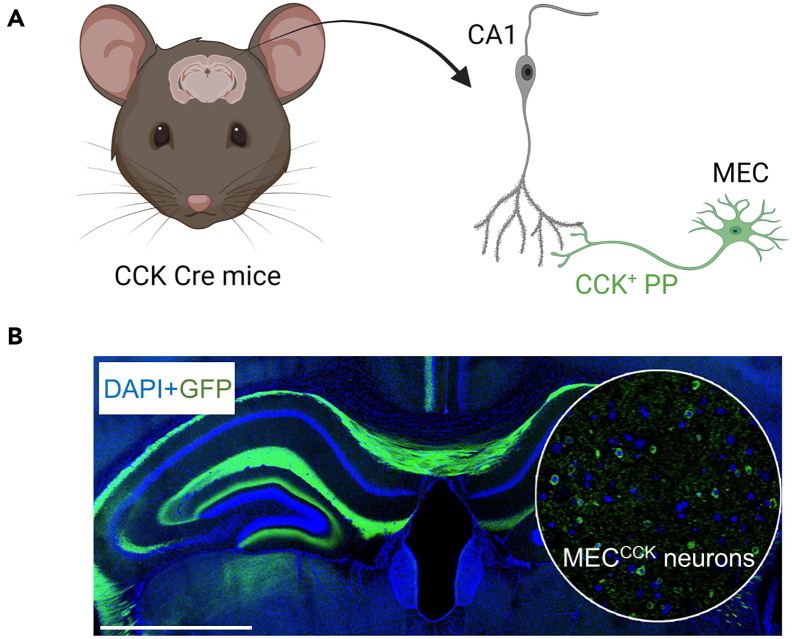
The strength of one neural pathway can depend on another input. How can such cooperation be distinguished within a converging hippocampal network? A STAR Protocols paper published on February 1, 2024 presents a dual-opsin strategy separating CA3 input from CCK-related entorhinal input, then examining lasting changes in CA3-to-CA1 responses after coactivation. Building on the team’s 2023 study, it makes two identifiable pathways available for investigating neuropeptides and synaptic plasticity.
Coactivating CA3-to-CA1 and CCK-positive entorhinal inputs increases field excitatory postsynaptic responses in the CA3-to-CA1 pathway. The paper also reports no significant potentiation without the second input, referring to control data in the preceding study.
Contents · 6 sections
Plasticity can depend on a second input
Long-term potentiation describes synaptic responses remaining stronger after activity. Beyond changes within an activated pathway, heterosynaptic potentiation asks whether another converging input helps shape the lasting change in that connection.
Here, two inputs converge on CA1: Schaffer collaterals from CA3 and perforant-path input from medial entorhinal cortex, including CCK-expressing cells. Anatomical labeling shows this CCK-related input reaching hippocampus, providing a substrate for studying cooperation.
The focus is the change retained after coactivity. Overlapping inputs can enlarge an immediate response, whereas long-term potentiation concerns a subsequent change in the tested pathway. Distinguishing input identity from later response separates coincidence from a lasting alteration in connectivity.
Distinct opsins distinguish input origins
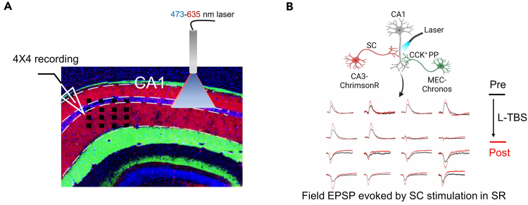
Activating converging inputs together makes their individual contributions difficult to assign. The strategy gives them distinct optical identities: ChrimsonR in CA3 input and Chronos in CCK-related entorhinal input, allowing different sources to be examined and combined in the same tissue.
Expression images and light-evoked field potentials link anatomical labeling to measurable functional responses. The value of the two markers lies in distinguishing circuit origins, beyond simply adding two colors to tissue.
Coactivation strengthens the CA3-to-CA1 response
The central result comes from dorsal hippocampal slices of male CCK-Cre mice. After coactivation, the slope of the light-evoked field excitatory postsynaptic potential tracks the CA3-to-CA1 response. Its increase reflects lasting plasticity in the measured pathway.
The field signal pools synaptic responses in the recorded region rather than resolving every synapse. It enables before-and-after comparison of the tested pathway while distinguishing whether the second input participates.
Source: Huang F et al. · STAR protocols · CC BY-NC-ND
Original figure and English caption ↗
A neuropeptide-linked input enters the plasticity question
CCK is a widely distributed neuropeptide. Selecting CCK-related entorhinal input connects pathway cooperation with neuromodulation: can a second input provide conditions that support potentiation of a classical excitatory pathway?
The expected outcomes report no significant potentiation without CCK-positive perforant-path participation, referring to the 2023 study for those control data. This protocol focuses on the capability to distinguish input contributions; it does not itself present separate measurements of peptide release or receptor interventions.
Light-evoked responses can decline during sustained activity
A defined circuit label does not guarantee equally stable responses throughout activation. The authors observe attenuation of field potentials and population spikes during induction, potentially affecting action-potential generation and depolarization.
Local inhibitory networks can also influence whether potentiation develops. The paper identifies response dynamics and GABA-related regulation as limitations. Coactivation starts the question, while recruitment of the surrounding slice network also shapes the outcome.
From a response curve to interactions between circuits
The work demonstrates a plasticity capability in male-mouse slices rather than directly measuring improved spatial learning in living animals. Separating input origin, coactivation and later response makes interactions between converging pathways easier to examine.
The hippocampus receives many inputs, and strengthening one connection can reflect influences from others. Distinguishing pathway origins helps identify such cooperation within pooled responses: which inputs enter a network, and which help shape the lasting change it retains.