Turning neuropeptide messages into visible light
A panel of genetically encoded sensors converts neuropeptide binding into fluorescence, revealing signals from local islet fluctuations to reward learning and stress, and clarifying when and where these messages appear.
Labeling a cell as somatostatin- or CRF-expressing does not reveal when it releases that peptide. Observing where messages appear, how long they persist and how they change with behavior requires another kind of readout. A 2023 Science study develops GRAB neuropeptide sensors that convert receptor recognition of peptides into green fluorescence. Across cultured neurons, pancreatic islets, brain slices and live mice, the sensors reveal spatial and temporal patterns of previously elusive peptide signals.
The study characterizes sensors for SST, CRF, CCK, NPY, NTS and VIP, and uses SST and CRF sensors to reveal local islet signals, reward-associated activity and stress-related peptide dynamics. These fluorescence observations report peptide-related changes, distinct from cellular firing or causal behavioral effects.
Contents · 8 sections
Translating receptor movements into fluorescence
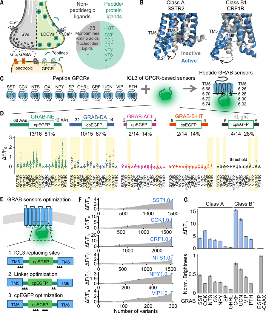
Most neuropeptide receptors are G-protein-coupled receptors that change shape on ligand binding. GRAB sensors link this movement to brightening of an embedded fluorescent module. The study transfers a module from existing sensors to different peptide receptors, providing a shared starting point.
The approach works across two major receptor-scaffold classes and yields several sensor candidates after optimization. Detailed characterization of SST, CRF, CCK, NPY, NTS and VIP sensors then moves from producing fluorescence to reliably reporting peptide signals.
Source: Huan Wang et al. · Science ·
Original figure and English caption ↗
Sensitive observation requires understanding sensor properties
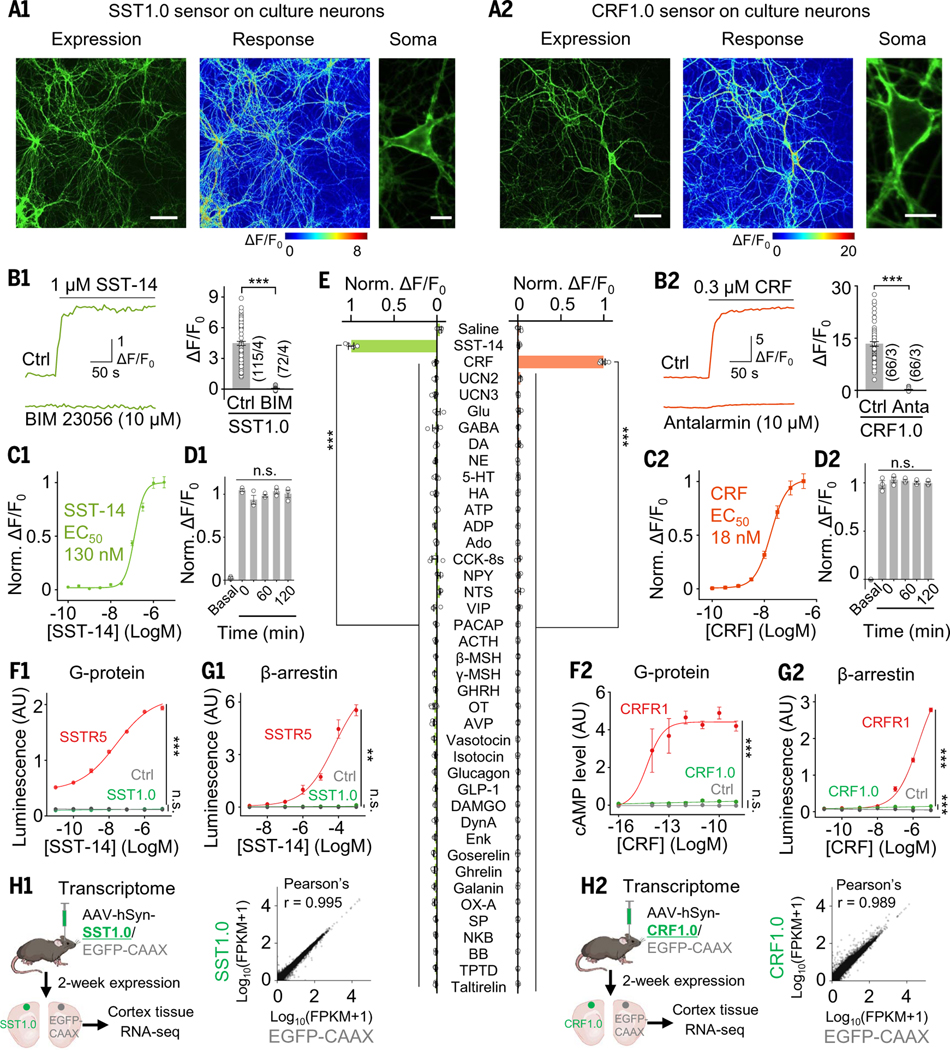
The six sensors localize to cell membranes and neuronal processes and respond clearly to their corresponding peptides. Signals can rise on a subsecond timescale and generally decay more slowly. Their response kinetics shape how fluorescence follows local peptide changes.
Selectivity reflects the parent receptor: the CRF sensor also responds to related peptide UCN1, whereas UCN2 and UCN3 do not elicit the same response. Most sensors have weak downstream coupling, while the CRF sensor retains residual coupling. Tested transcriptomic features and several behavioral measures show no obvious changes.
Source: Huan Wang et al. · Science ·
Original figure and English caption ↗
Peptide signals do not rise uniformly across an islet
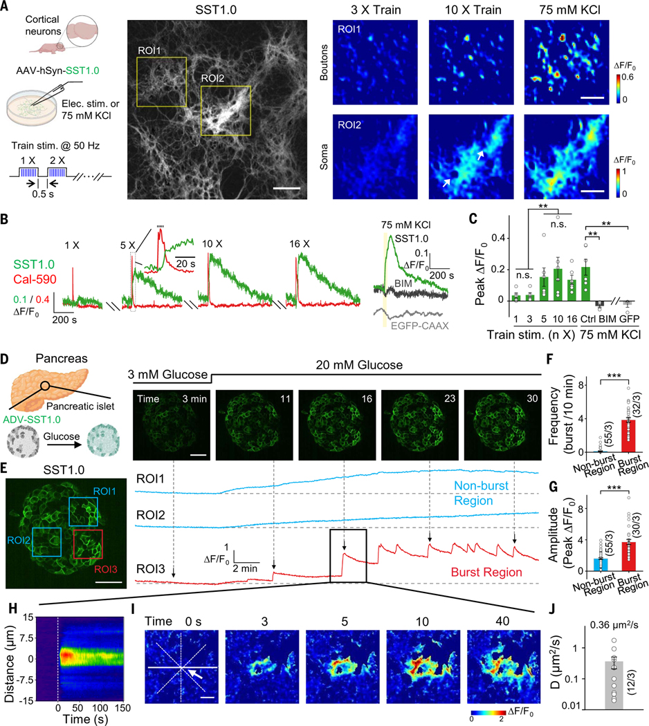
Stimulating cultured neurons increases SST-sensor signals in association with intracellular calcium changes. In isolated mouse pancreatic islets, increased glucose also raises SST-related fluorescence, but different locations follow different rhythms.
Some regions change gradually, while others show stronger, more frequent pulses. During individual events, signals begin at local hotspots and expand to neighboring locations; nearer responses are faster and stronger. This reveals spatial detail in local paracrine signaling within islets.
Source: Huan Wang et al. · Science ·
Original figure and English caption ↗
Another signal appears before reward arrives
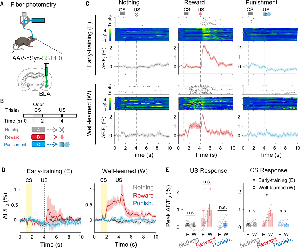
In mouse basolateral amygdala, reward delivery early in training accompanies a transient SST-signal rise, unlike punishment or no-outcome trials. After odor–reward learning, recordings also show a signal during the odor period that persists until reward arrives.
Responses at reward delivery differ across training stages, linking peptide dynamics with learning stage. No-outcome and punishment trials lack similarly prominent signals in both stages. Recordings spanning cue and outcome place responses at reward delivery alongside changes occurring beforehand.
Source: Huan Wang et al. · Science ·
Original figure and English caption ↗
Local spread of CRF signals in brain slices
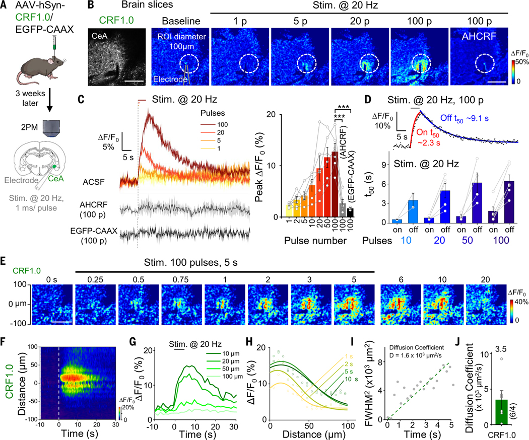
In central-amygdala slices, electrical stimulation increases CRF-sensor signals, with stronger responses as tested pulse numbers rise. Blocking peptide binding, using a CRF-insensitive sensor or reducing local CRF-gene expression weakens signals, supporting peptide-dependent detection.
Time-resolved images show signals spreading during stimulation. This range may reflect peptide diffusion as well as additional local release. The images show their combined distribution, describing the local spatial range of the signal.
Source: Huan Wang et al. · Science ·
Original figure and English caption ↗
Hypothalamic peptide signals accompany stress
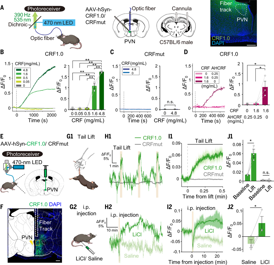
In paraventricular hypothalamus, the responsive sensor reports administered CRF whereas a control mutant does not show the same change. Tail lifting evokes a time-locked signal rise, while an abdominal-malaise stimulus produces a more persistent increase than saline.
Different stimuli leave different temporal profiles. The sensor adds a view of stress-related local peptide dynamics over time alongside behavioral measures.
Source: Huan Wang et al. · Science ·
Original figure and English caption ↗
Observing the same peptide locally in cortex
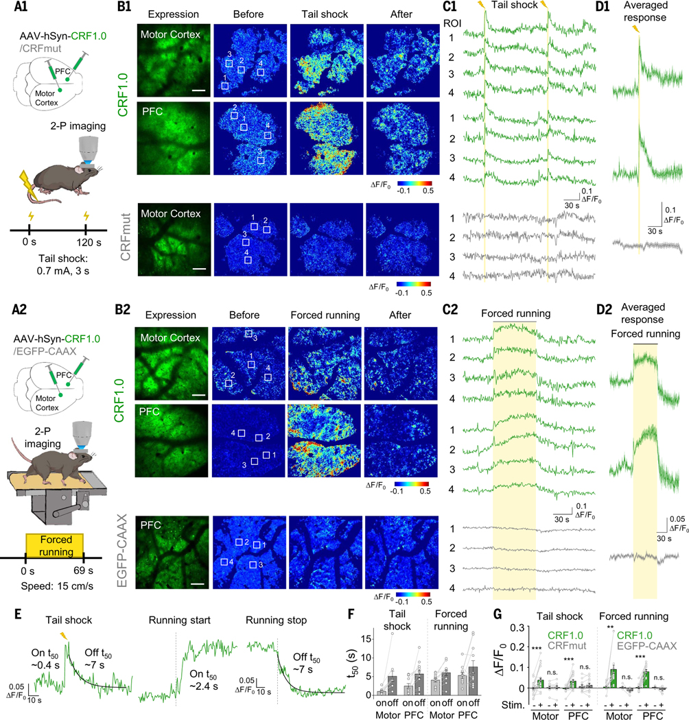
Imaging in motor and prefrontal cortex reveals transient, reversible CRF-related fluorescence increases during tail stimulation. Signals rise and plateau during forced running, then decline after it stops. Peptide-insensitive control signals do not show matching increases.
Imaging distinguishes locations within the field rather than supplying only one regional average. Both stressful conditions produce increases with different temporal profiles, allowing peptide dynamics to be examined in relation to specific events.
Source: Huan Wang et al. · Science ·
Original figure and English caption ↗
From cell identity to the timing and location of messages
The toolkit separates which cells express peptides from when peptide signals appear. Cultures, isolated islets, slices and mouse behavioral recordings show local hotspots and different rhythms during reward and stress.
Fluorescence retains the sensor’s affinity and response kinetics and does not directly report downstream activation of native receptors. Its contribution is making the occurrence of peptide signals observable alongside cellular activity and behavior.