Two colors reveal norepinephrine and cellular responses
Next-generation GRABNE sensors improve norepinephrine responses and combine with red calcium signals to follow sleep, sensation and movement: broad transmitter fluctuations coexist with distinct cellular rhythms.
After a sound, where does brain norepinephrine change, and how do recipient cells respond? These are related but distinct questions. A 2024 Neuron study introduces next-generation GRABNE sensors and combines green transmitter signals with red calcium signals in mice. From sleep transitions and whisker sensation to spontaneous movement, it reveals broad norepinephrine changes alongside cellular responses with distinct spatial and temporal features.
GRABNE2m and GRABNE2h improve tested norepinephrine responses. Dual-color observation reveals broad transmitter changes alongside localized neuronal calcium hotspots or delayed astrocytic calcium responses.
Contents · 7 sections
Making small transmitter changes easier to see
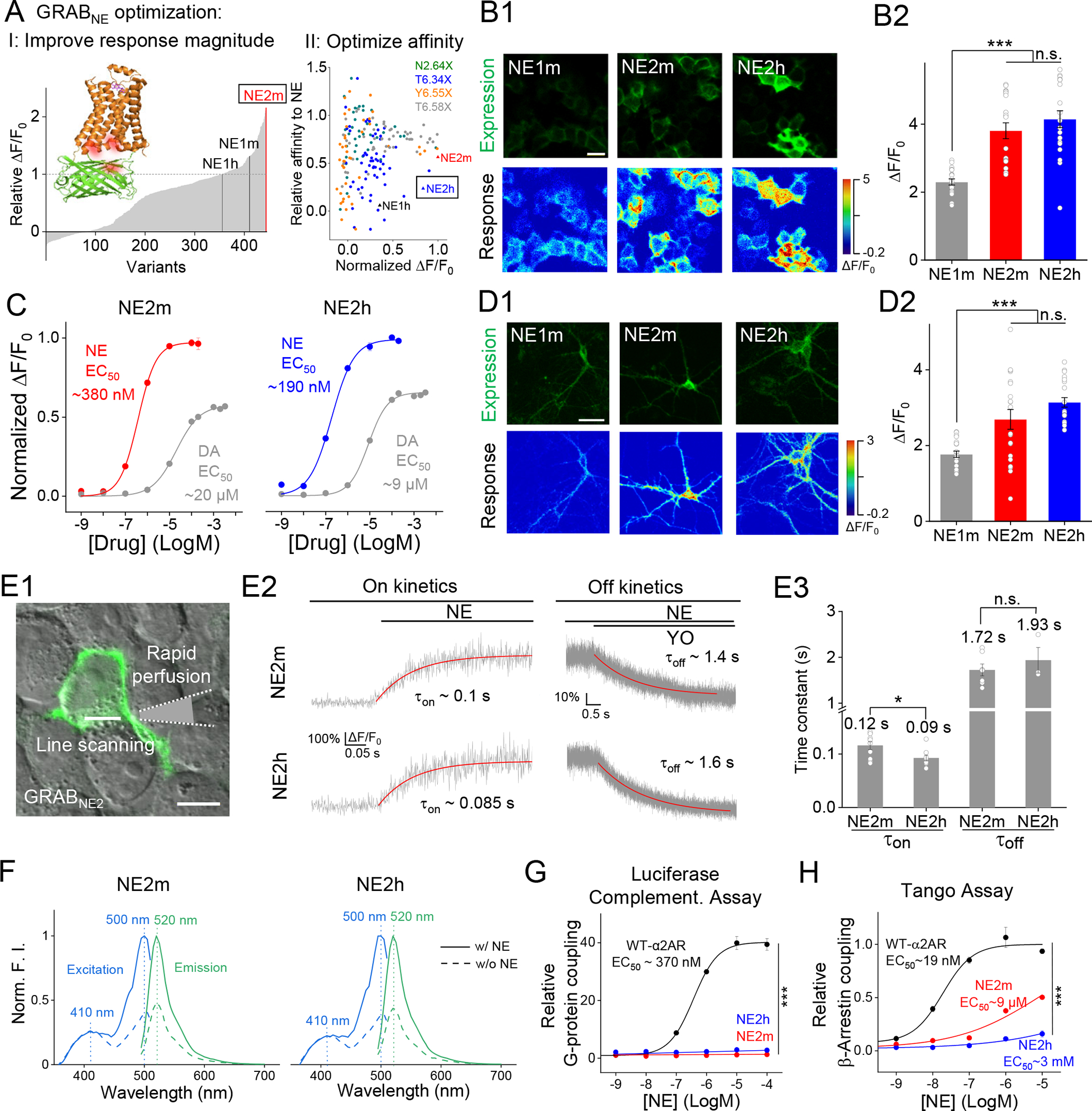
GRAB sensors turn the conformational change accompanying receptor–transmitter binding into fluorescence. The new NE2m and NE2h variants show larger tested responses in cultured cells and neurons. NE2h has higher apparent affinity and responds to lower norepinephrine concentrations.
Sensitivity does not make every property faster. Both new sensors respond rapidly, yet their kinetics are slightly slower than first-generation NE1m. Weak downstream G-protein and beta-arrestin coupling helps separate signal observation from cellular responses driven by the receptor itself.
Source: Jiesi Feng et al. · Neuron · TDM
Original figure and English caption ↗
Distinguishing closely related molecules
Norepinephrine and dopamine are structurally similar. Cellular dose responses show greater sensitivity to norepinephrine, although dopamine can still elicit responses at higher concentrations. The sensors also respond to epinephrine, making selectivity dependent on the particular comparison.
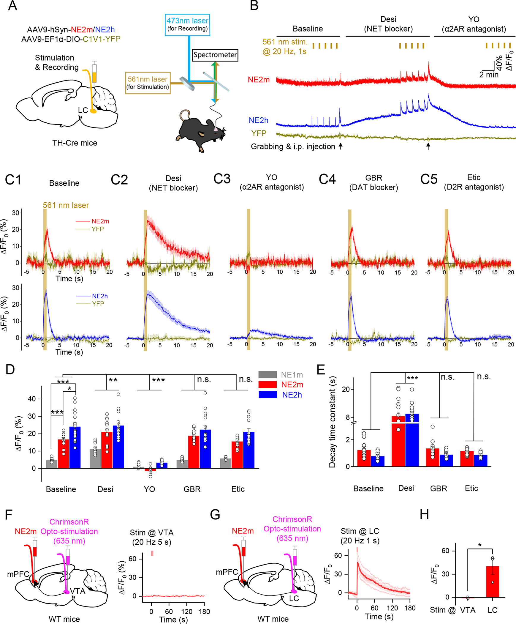
Evoking locus-coeruleus activity in mice produces stronger signals in both new sensors without a matching control-fluorescence change. In separate prefrontal recordings, locus-coeruleus stimulation produces a clear response, whereas the tested ventral-tegmental stimulation causes no detectable increase, supporting discrimination of norepinephrine dynamics under these conditions.
Source: Jiesi Feng et al. · Neuron · TDM
Original figure and English caption ↗
Behavioral events reveal different response magnitudes
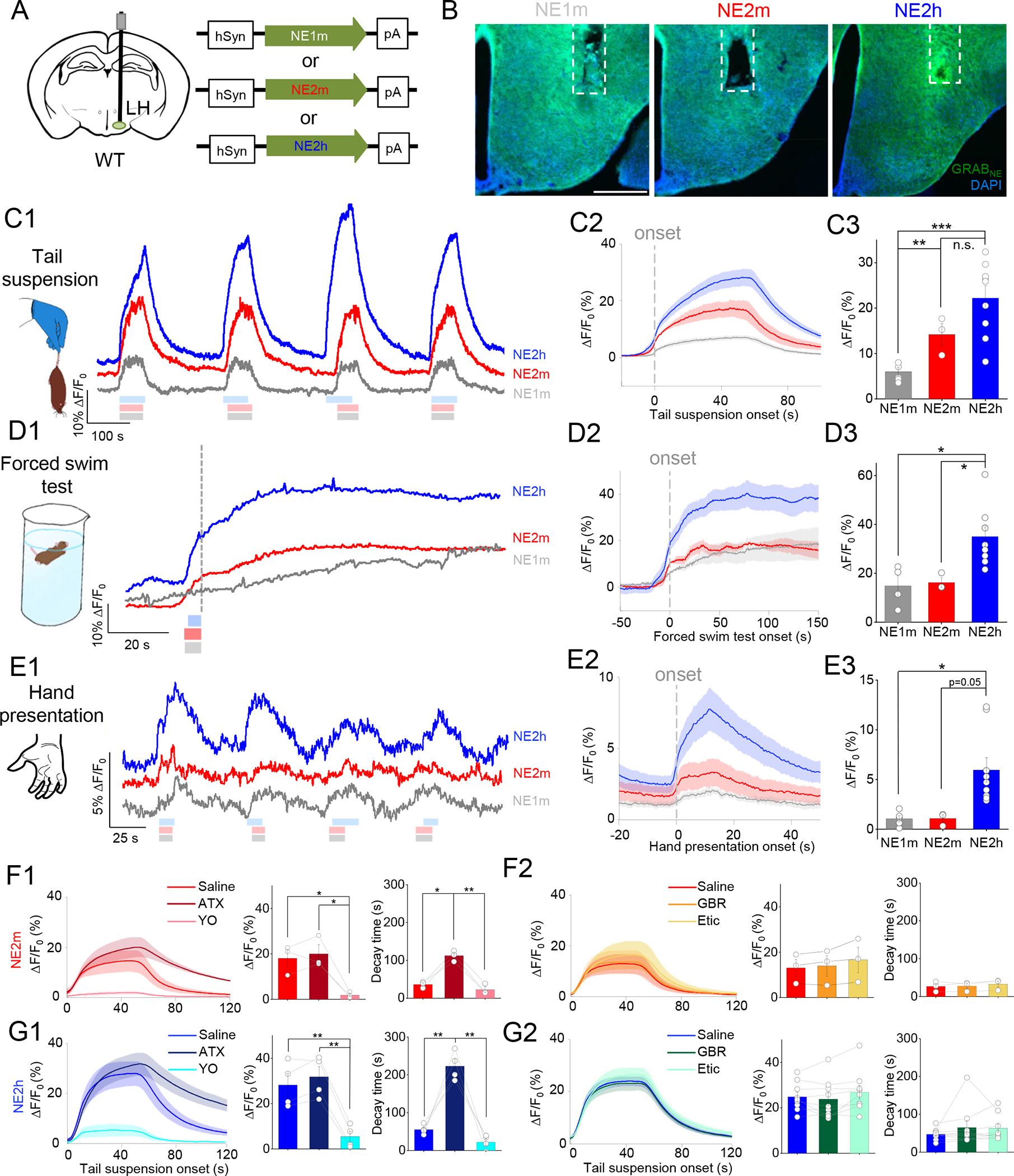
In lateral hypothalamus, tested events including tail suspension, swimming and hand presentation accompany sensor-signal increases. Improvements vary by context: both new sensors have larger responses during tail suspension, whereas NE2h is stronger during swimming and NE2m does not significantly differ from the first generation.
After norepinephrine-transporter blockade, the suspension-related signal decays more slowly without a significant peak change. Height and duration provide different information, showing how transmitter clearance affects the observed dynamics.
Source: Jiesi Feng et al. · Neuron · TDM
Original figure and English caption ↗
Waking and falling asleep follow different dynamics
Simultaneous prefrontal and preoptic recordings show closely synchronized norepinephrine signals across sleep–wake cycles. Signals are relatively high during waking and non-REM sleep and low during REM. Increases on waking are faster than decreases on entering sleep.
Dual-color mice also show oscillatory norepinephrine during non-REM sleep while cellular calcium activity remains relatively low. Compared with viral-expression recordings, tested transmitter-signal amplitudes and kinetics are similar, with lower within-group variation.
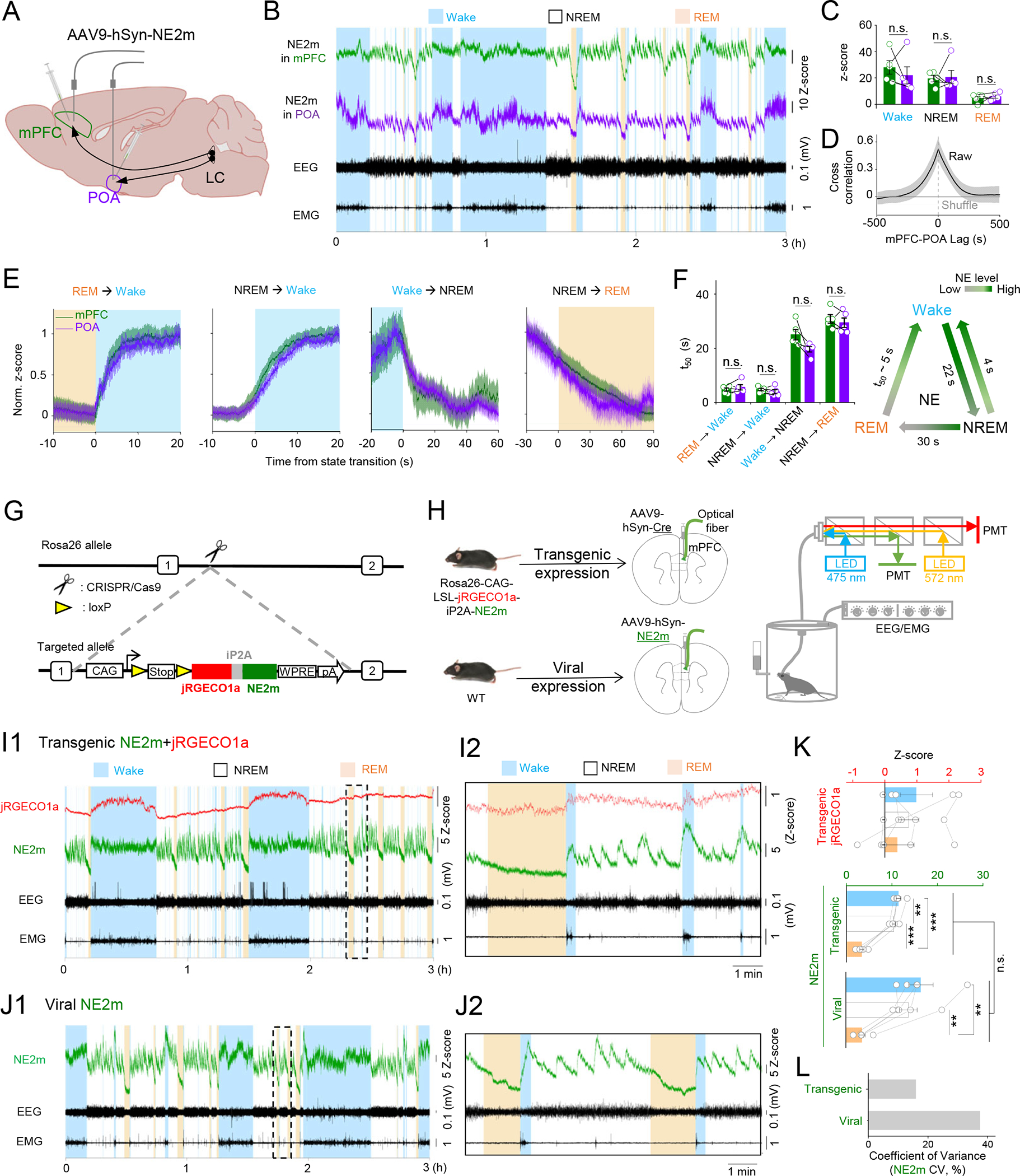
Source: Jiesi Feng et al. · Neuron · TDM
Original figure and English caption ↗
Broad transmitter changes meet localized cellular responses
After unilateral whisker stimulation, sensors on excitatory neurons show broad norepinephrine changes across imaged dorsal cortex, whereas calcium responses concentrate contralaterally. Astrocyte-associated transmitter signals are also broad, with smaller, delayed calcium responses.
The distinction concerns cells bearing the sensors and their calcium responses, rather than norepinephrine release from those cells. Different senses also produce different results: tested visual stimuli cause no detectable transmitter or calcium-signal change in astrocytic recordings.
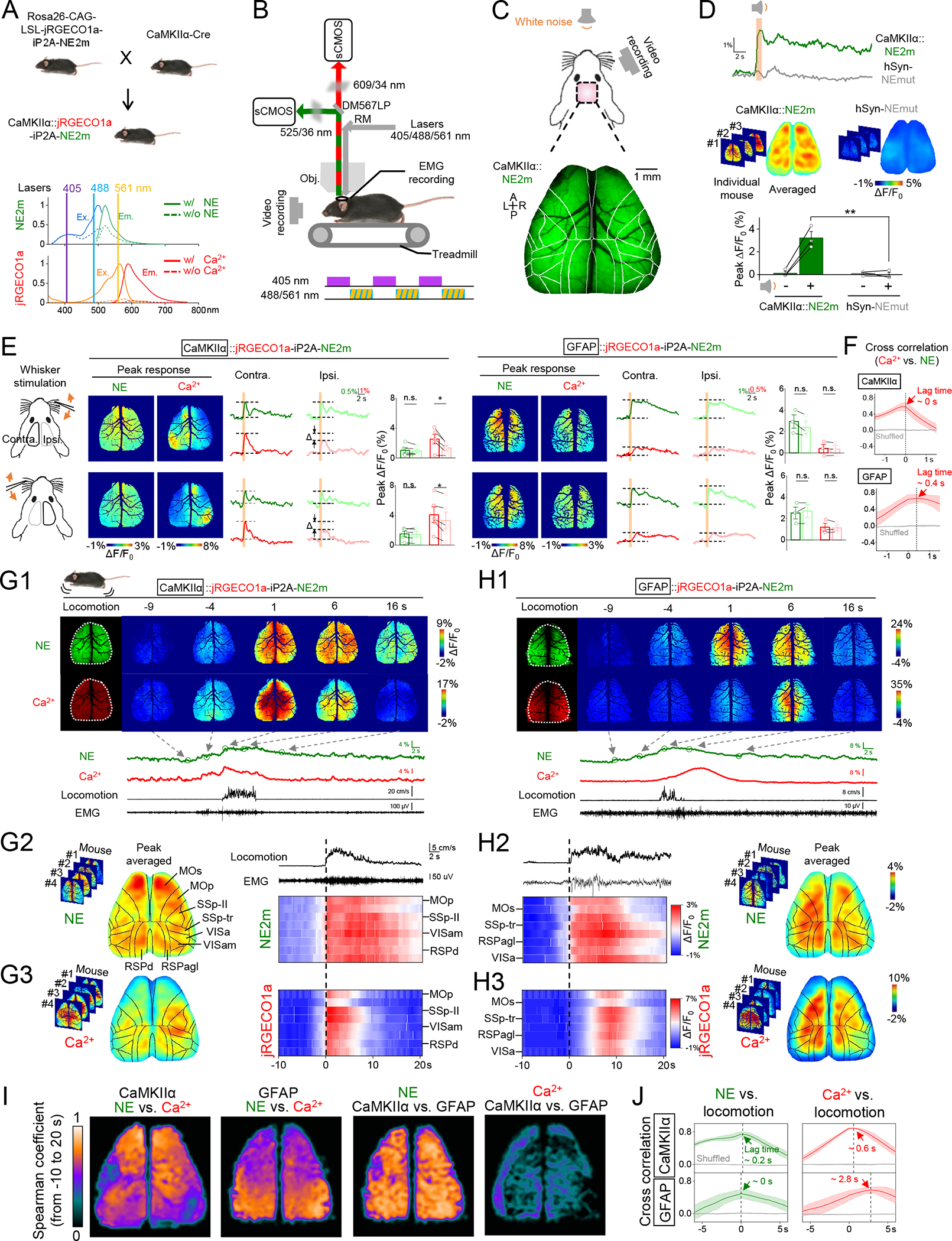
Source: Jiesi Feng et al. · Neuron · TDM
Original figure and English caption ↗
Two cell types follow different rhythms at movement onset
At spontaneous movement onset, both cell-associated recordings show broad norepinephrine signals. Excitatory-neuronal calcium changes form sensorimotor hotspots, whereas astrocytic calcium changes are broader and follow movement onset by about three seconds.
The two colors reveal more than synchrony. Similar large-scale transmitter changes accompany cellular responses with different distributions and speeds, helping describe how neuromodulatory signals relate to ongoing brain activity.
Following incoming signals and cellular responses
The study validates sensors in cultures and adult mice of both sexes, extending transmitter observation across cell types and cortical scales. Fluorescence retains the sensors’ sensitivity and temporal properties, reporting transmitter-related changes rather than counts of neuronal action potentials.
The central insight is that broad norepinephrine fluctuations and specific cellular responses can be observed together while interpreted separately. Where transmitter arrives, where cells respond and how far apart those events occur become questions that can be compared within one setting.