Turning prostaglandin into a live optical signal: how do cortical dynamics differ in inflammation and seizure?
Using the genetically encoded sensor GRAB-PGE2-1.0 in awake mice, researchers reveal distinct spatiotemporal prostaglandin E2 patterns during systemic inflammation and seizure.
Fever, pain, neuroinflammation, and epileptic seizures all involve a pivotal lipid messenger: prostaglandin E2 (PGE2). As a major product of the cyclooxygenase (COX) pathway and an indirect target of nonsteroidal anti-inflammatory drugs, when and where does PGE2 surge in the living brain, and does it spread from blood vessels or neurons? Because PGE2 is hydrophobic and present at low basal concentrations, tissue homogenate mass spectrometry lacks live temporal resolution, while microdialysis typically requires 20- to 30-minute sampling intervals. In a study published online in Neuron on March 13, 2026, Yulong Li and colleagues at Peking University developed the first-generation G-protein-coupled receptor activation-based PGE2 sensor, GRABPGE2-1.0 (PGE2-1.0), turning endogenous PGE2 dynamics in the living mouse brain into a high-resolution optical readout.
Engineered from the human EP2 receptor, PGE2-1.0 offers nanomolar affinity, subsecond on-kinetics, high selectivity, and negligible downstream Gs or beta-arrestin coupling; combining fiber photometry and dual-color wide-field imaging, the study shows that systemic inflammation drives a slow, microvascular-associated cortical PGE2 rise, whereas seizures trigger rapid anterior cortical PGE2 waves immediately following neuronal calcium bursts.
Contents · 5 sections
Screening nearly 3,000 variants to build a nanomolar sensor with minimal signaling perturbation
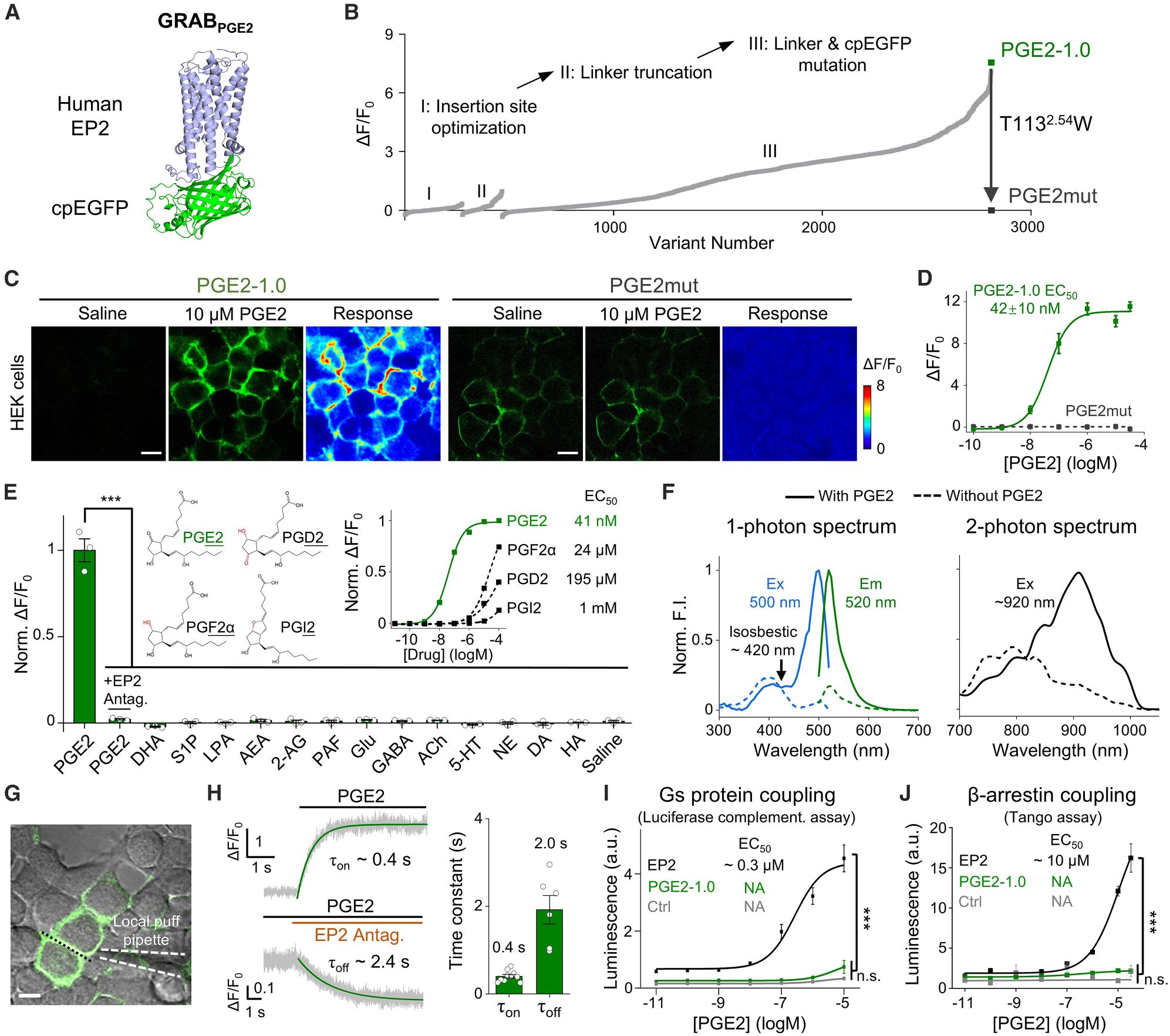
To make endogenous PGE2 optically visible, the team screened the four human PGE2 receptors (EP1-EP4) and inserted circularly permuted enhanced green fluorescent protein (cpEGFP) into the third intracellular loop (ICL3) of the human EP2 receptor. Across three rounds of systematic optimization covering insertion sites, linker truncations, and cpEGFP/linker mutations among nearly 3,000 variants, they identified the top-performing sensor PGE2-1.0, and introduced a single binding-pocket point mutation (T113^2.54W) to create the ligand-insensitive control sensor PGE2mut.
In HEK293T cells, PGE2-1.0 localized cleanly to the plasma membrane and produced a ~1,100% (~11-fold) peak fluorescence increase (dF/F0) to 10 uM PGE2 with an EC50 of 42 +/- 10 nM, whereas PGE2mut showed no response. The signal was abolished by the selective EP2 antagonist PF-04418948, and the sensor displayed orders-of-magnitude lower affinity for related prostanoids (PGF2alpha, PGD2, and PGI2) and no response to other tested lipids or neurotransmitters. Local puffing revealed rapid kinetics (tau_on ~0.4 s, tau_off ~2.4 s), while split-luciferase and Tango assays confirmed negligible Gs protein or beta-arrestin coupling.
Source: Lei Wang et al. · Neuron · © 2026 Elsevier Inc.
Original figure and English caption ↗
From cultured neurons to acute hippocampal slices: capturing endogenous release evoked by cytokines and hyperexcitability
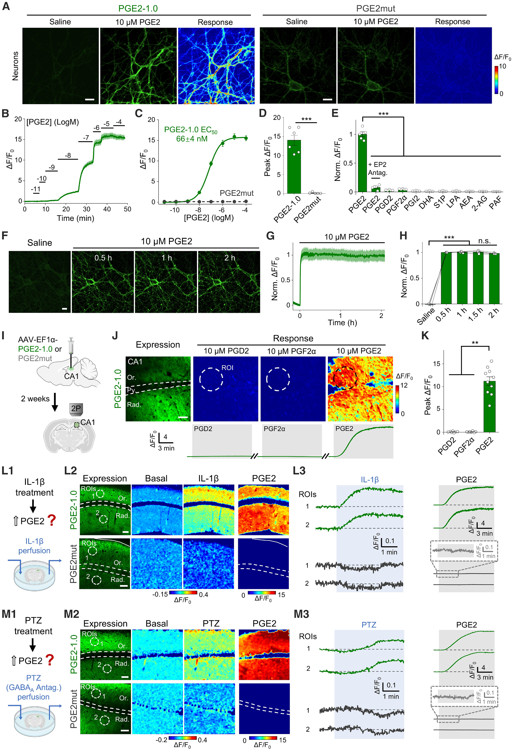
Moving into neural preparations, the team tested PGE2-1.0 in primary cultured rat cortical neurons. Both somata and neurites displayed robust membrane fluorescence, yielding a ~1,400% peak fluorescence increase to exogenous PGE2 (EC50 ~66 nM). Continuous exposure to 10 uM PGE2 for up to 2 hours produced a stable fluorescence plateau without noticeable internalization or desensitization, supporting long-duration physiological and pathological recordings.
In acute mouse hippocampal slices expressing the sensor in CA1 via AAV, two-photon microscopy confirmed high selectivity over PGD2 and PGF2alpha. Perfusing either the proinflammatory cytokine IL-1beta or the GABAA receptor antagonist pentylenetetrazol (PTZ) evoked sustained fluorescence increases across stratum oriens and stratum radiatum in PGE2-1.0 slices, while PGE2mut control slices remained at baseline, demonstrating reliable detection of endogenous PGE2 release in intact neural tissue.
Source: Lei Wang et al. · Neuron · © 2026 Elsevier Inc.
Original figure and English caption ↗
From the preoptic hypothalamus to cortex-wide windows: tracking slow inflammatory waves and rapid seizure surges
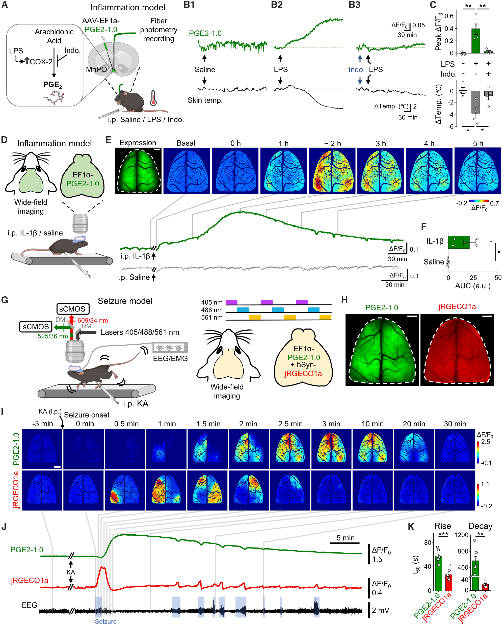
The median preoptic nucleus (MnPO) within the hypothalamic preoptic area is a central hub for inflammatory thermoregulation. Using fiber photometry in the mouse MnPO, the authors found that intraperitoneal lipopolysaccharide (LPS) injection triggered a progressive rise in PGE2-1.0 fluorescence accompanied by a drop in skin temperature under these experimental conditions; pretreatment with the COX inhibitor indomethacin suppressed both the optical surge and the temperature shift.
To visualize cortex-wide dynamics, the team combined dorsal cranial windows with mesoscopic wide-field imaging. Intraperitoneal IL-1beta triggered a slow cortical PGE2 wave that peaked around 2 hours and gradually subsided. By contrast, in a kainic acid (KA)-induced seizure model combining dual-color imaging of green PGE2-1.0 and red calcium indicator jRGECO1a with EEG recordings, each generalized ictal calcium burst was followed within tens of seconds by a sharp cortical PGE2 surge (rise t50 ~58 s, decay t50 ~560 s)—longer-lasting than the calcium transient yet dozens of times faster than the inflammatory response.
Source: Lei Wang et al. · Neuron · © 2026 Elsevier Inc.
Original figure and English caption ↗
Microvascular periphery versus anterior cortex: contrasting spatial maps point to distinct cellular origins
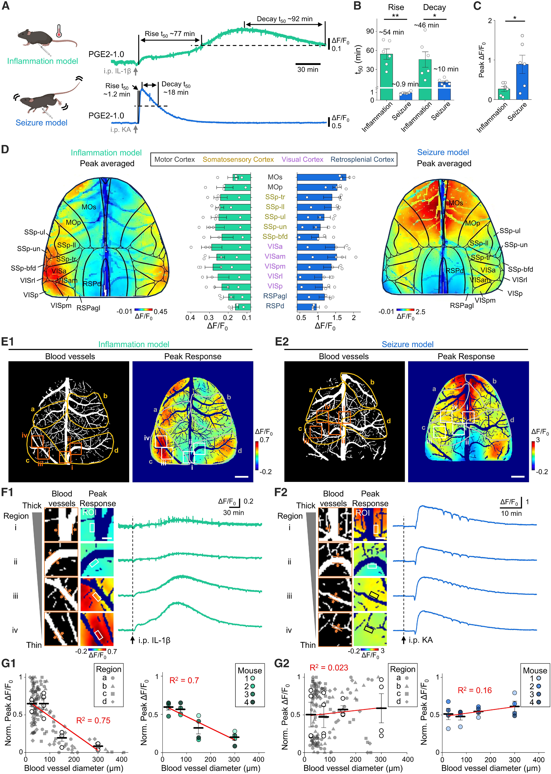
Beyond the stark temporal contrast between slow inflammatory waves (rise t50 ~54 min, decay t50 ~46 min) and fast ictal surges (rise t50 ~0.9 min, decay t50 ~10 min), mesoscopic mapping uncovered fundamentally different spatial topologies. During IL-1beta-induced systemic inflammation, PGE2 increases concentrated near the lateral and posterior cortical periphery and correlated negatively with adjacent blood vessel diameter (ROI R2 = 0.75, mouse-averaged R2 = 0.7), peaking around thin microvessels and vascular bifurcations.
During KA-induced seizures, however, peak PGE2 responses clustered in anterior cortical regions such as secondary and primary motor cortex (MOs/MOp) and showed virtually no correlation with blood vessel diameter (ROI R2 = 0.023, mouse-averaged R2 = 0.16). This topological divergence supports the view that systemic inflammation recruits PGE2 predominantly from microvascular endothelial cells, whereas seizure-evoked PGE2 surges are driven locally by hyperactive neural parenchyma.
Source: Lei Wang et al. · Neuron · © 2026 Elsevier Inc.
Original figure and English caption ↗
Providing a high-resolution tool for neuroimmune research while defining optical and mechanistic boundaries
By rendering prostaglandin E2 dynamics visible across spatial and temporal scales, PGE2-1.0 fills a critical gap in lipid-signaling biosensors and demonstrates how the same messenger obeys distinct spatiotemporal rules across disease states. Notably, the rapid seizure-evoked PGE2 surge occurs within seconds—far faster than de novo transcriptional induction of COX-2—leaving open whether it arises from constitutive microglial COX-1 or basal activity-regulated neuronal COX-2.
The authors also note that, like other fluorescent protein-based sensors, PGE2-1.0 can be affected by hemodynamic optical artifacts during intense physiological perturbations: brief transient fluorescence dips coinciding with ictal discharges likely reflect seizure-induced vasodilation and cerebral blood volume increases that alter light absorption and scattering. Pairing PGE2-1.0 with the ligand-insensitive control PGE2mut and multi-wavelength correction will help dissect prostanoid signaling across diverse physiological and pathological contexts.