Reading deep-brain temperature through two bands of light
A male-mouse study uses upconversion luminescence to read local temperature during feeding, anesthesia and external thermal changes, and separates signals at two depths along one probe.
The brain can gradually warm as a mouse eats. Where that change occurs and how large it is cannot be answered easily from surface temperature alone. A study published online in Advanced Science on September 15, 2023 combines temperature-sensitive luminescent material with optical fibers to read thermal changes in deep regions of freely moving male mice. Rather than neuronal firing, it records local temperature associated with activity, metabolism and environment. Two sensing locations along one probe allow measurements at different depths during the same experience.
Local luminescence ratios report thermal changes: hypothalamic temperature rises during feeding, hippocampal readings fall during anesthesia, and external thermal changes influence cortex. Dual-site recording demonstrates spatially resolved sensing rather than a whole-brain temperature map.
Contents · 7 sections
Temperature information in emitted light
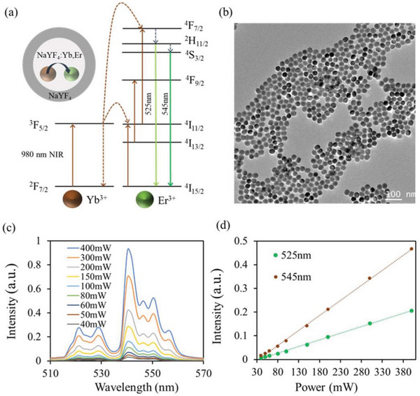
Upconversion nanoparticles convert near-infrared light into visible emissions. The relative strengths of two emission bands vary with temperature, allowing their ratio to form a thermal readout. A ratio provides a reference beyond the brightness of one band and helps reduce effects of overall intensity fluctuations.
The fiber carries the emissions back, avoiding reliance on visible light traversing deep tissue to an external camera. A localized sensing window confines the measurement, so the readout concerns its surroundings rather than averaging every region along the fiber.
Source: Zhou B et al. · Advanced science (Weinheim, Baden-Wurttemberg, Germany) · CC BY
Original figure and English caption ↗
Stability and timing depend on the test conditions
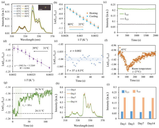
During heating and cooling in vitro, emission ratios follow similar relationships, showing reversibility. Readings also track temperature near the physiological range. Fluctuation-based estimates under constant conditions correspond to about 0.3 degrees Celsius, a performance observation tied to those conditions.
Response timing differs across tests. Local heating produces a clear rise within about ten seconds, whereas a larger environmental-temperature transition takes roughly a hundred seconds. Heat transfer around the probe influences its time course. Repeated checks over days show overall optical stability alongside some drift.
Source: Zhou B et al. · Advanced science (Weinheim, Baden-Wurttemberg, Germany) · CC BY
Original figure and English caption ↗
Hypothalamic temperature rises gradually during feeding
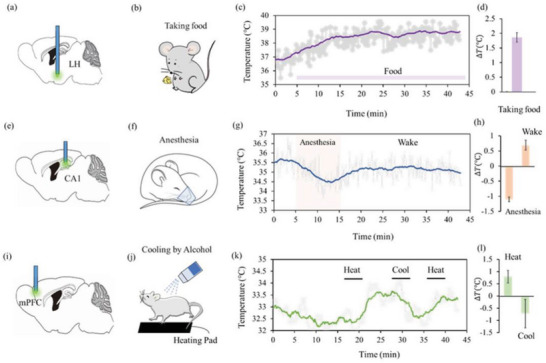
The team records first in lateral hypothalamus, a region involved in feeding, metabolism and sleep. During eating, temperature gradually rises from approximately 36.8 to 38.8 degrees Celsius over minutes. The record aligns a visible behavior with a slower thermal process inside the brain.
This demonstrates local deep-brain thermometry. Several physiological processes may accompany warming; the trace alone cannot assign contributions to cellular heat production, blood flow or body temperature, nor translate temperature rise into the activity of a particular neuronal group.
Source: Zhou B et al. · Advanced science (Weinheim, Baden-Wurttemberg, Germany) · CC BY
Original figure and English caption ↗
Cooling during anesthesia and partial rewarming
Hippocampal CA1 provides a different trajectory. During the tested anesthesia period, readings fall from around 35.5 to 34.5 degrees Celsius. During subsequent recovery they rise by about half a degree, without fully returning to the initial level within the displayed interval.
Feeding and anesthesia show opposite temperature directions, but also involve different sites and contexts. The advance is continuous access to local thermal dynamics, rather than ranking regional activity or treating temperature as a direct scale of wakefulness.
External thermal changes reach cortical readings
Medial prefrontal readings rise under the tested warm condition, fall with cooling and rise again upon rewarming. Displayed changes are below approximately one degree. The brain is not thermally isolated; bodily heat exchange can influence its local environment.
In a same-mouse cortical comparison with a commercial thermocouple, readings do not differ significantly. This provides an in-vivo reference under the tested conditions, without establishing the same error performance across every activity and brain region.
From one local reading to two depths
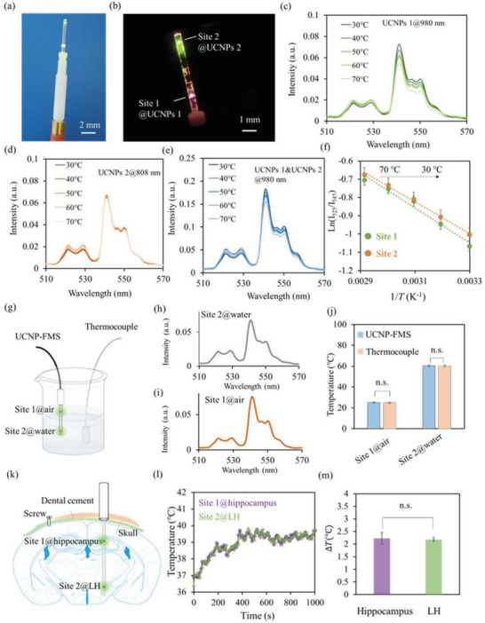
Two sensing positions along one fiber use distinct material responses to separate readings. In vitro, one site experiences a hotter environment while the other remains near room temperature; both agree with thermocouple references rather than collapsing into one average.
The in-vivo demonstration follows hippocampus and lateral hypothalamus together. Both warm during feeding, with no significant difference in the measured rise. Two depths can therefore be observed during the same behavioral episode rather than in separate experiences.
Source: Zhou B et al. · Advanced science (Weinheim, Baden-Wurttemberg, Germany) · CC BY
Original figure and English caption ↗
Local thermal dynamics add a distinct observational scale
These results come from male mice aged eight to nine weeks, with about three animals per demonstrated behavioral condition. The paper reports no significant change in the assessed inflammation-related tissue markers weeks later, a limited observation rather than a universal long-term implant response. The sensing window does not define the diameter of the entire probe.
The work turns deep local temperature from a difficult background variable into a continuously recorded, spatially comparable signal. It complements spikes and transmitters with another dimension of observation. Separating local activity from bodily and environmental contributions will require complementary physiology.