Can light bring a colonic contraction forward?
In isolated mouse colon, activating Kit-labeled interstitial cells of Cajal brings forward low-frequency, high-amplitude contractions and increases their frequency without significantly increasing amplitude. ANO1-related blockade removes light-evoked contractions.
Colonic muscle need not wait for a fresh neural instruction before every contraction. Interstitial cells of Cajal, ICC, help generate electrical rhythms and pass signals to smooth muscle. How slow waves relate to individual contractions is a key question in intestinal motility. Can this pacemaker be accelerated from outside? A study published in Journal of Neurogastroenterology and Motility on July 30, 2023 expresses light-sensitive ChR2 in Kit-labeled mouse cells and examines isolated colonic muscle strips. Light brings forward normally intermittent large contractions and makes them more frequent, chiefly changing timing rather than increasing the strength of every contraction.
In isolated colonic strips, single light activation elicits premature low-frequency, high-amplitude contractions, while periodic activation increases their frequency without significantly increasing amplitude. ANO1-related pharmacological blockade suppresses spontaneous phasic and light-evoked contractions.
Contents · 6 sections
An intestinal network passes electrical rhythms to muscle
ICC occupy different layers of the intestinal wall, cooperate with smooth muscle and other interstitial cells, and receive enteric-neural influences. Their slow waves are rhythmic membrane-potential changes transmitted to neighboring muscle, providing an electrical basis for phasic contractions. Several partners contribute to intestinal movement: smooth muscle generates force, ICC help organize electrical rhythms, and nerves and other interstitial cells influence their coordination. The study focuses on this pacemaker component to examine which contractions its activity can alter.
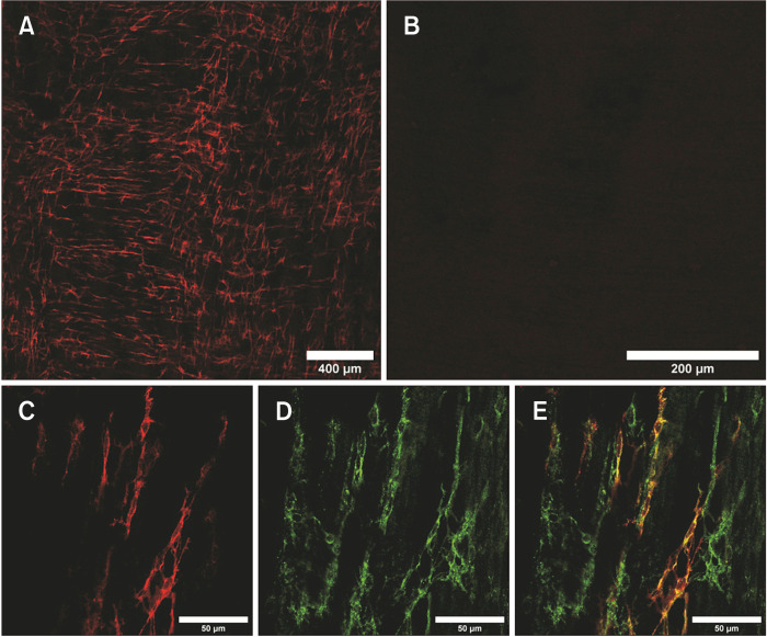
The study targets Kit-labeled cells and expresses ChR2 in a subset. Red fluorescence in colonic muscularis overlaps substantially with Kit labeling without covering every Kit-positive cell. Tissue images connect the light-responsive cellular network with subsequent muscle-strip observations.
Source: Zhao S et al. · Journal of neurogastroenterology and motility · CC BY-NC
Original figure and English caption ↗
A light pulse brings the next large contraction forward
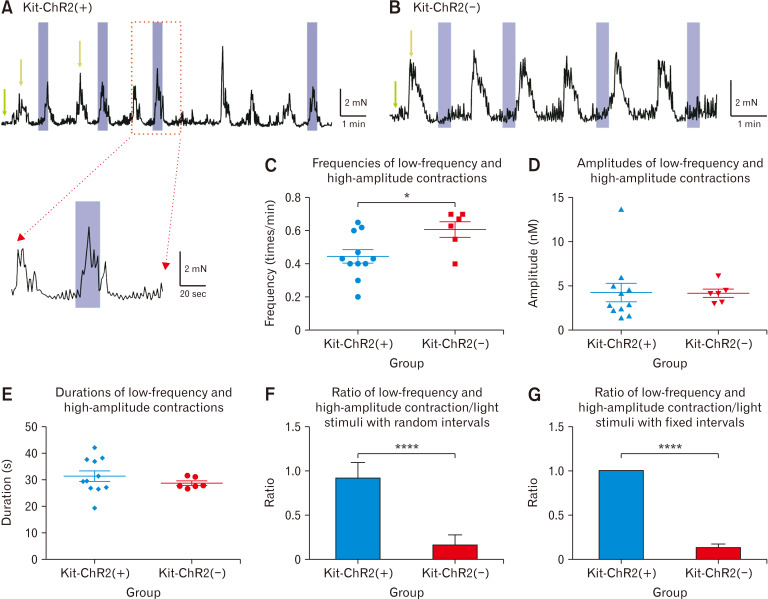
Strips naturally show frequent small contractions and intermittent low-frequency, high-amplitude contractions. The expression group has a lower baseline frequency of large contractions, with similar amplitude and duration to controls. Light-associated premature large contractions occur mainly in that group, without the same change in frequent small contractions.
The study also examines light soon after a spontaneous large contraction, when the next one would not usually occur. The expression group still shows more new large contractions. This temporal relationship clarifies the advance in timing and distinguishes the two contraction patterns.
Source: Zhao S et al. · Journal of neurogastroenterology and motility · CC BY-NC
Original figure and English caption ↗
Repeated pacing increases number while amplitude stays similar
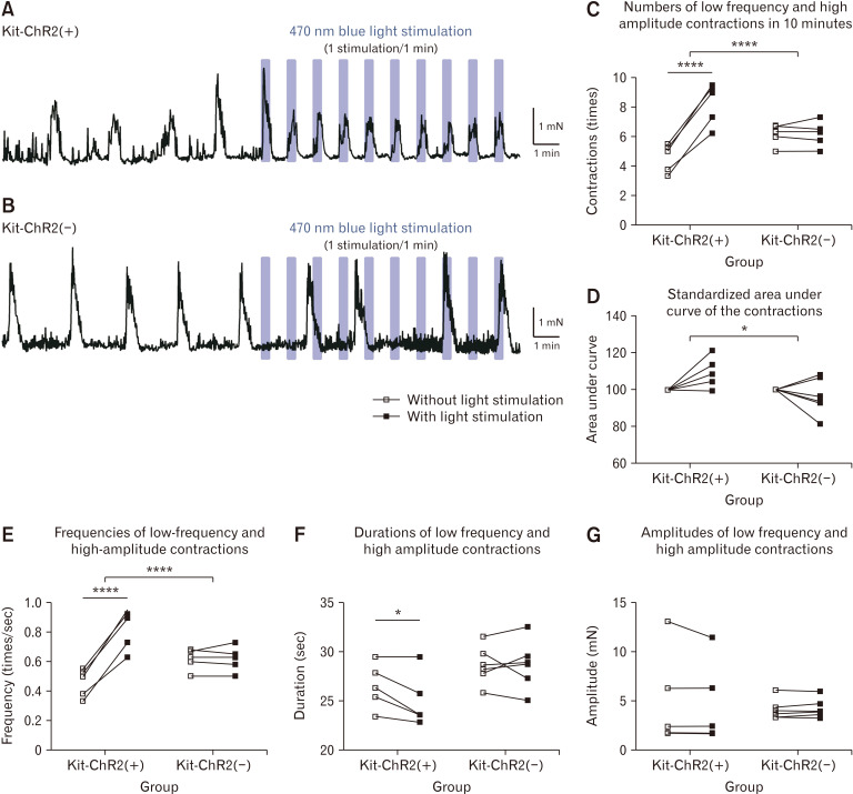
Periodic activation increases the number of large contractions in the expression group, with repeated contractions following light timing and a higher frequency. Control strips show no significant number or frequency change. Previously sparse contractions become more closely spaced.
Single-contraction amplitude does not significantly increase, nor does integrated force over the recording significantly increase within the expression group. More events do not automatically imply greater accumulated force, separating rhythm modulation from strengthening contraction. Force records contain both frequent small activity and intermittent large contractions. Making one pattern more frequent leaves the overall trace shaped by several activities, explaining why frequency and force offer different observations.
Source: Zhao S et al. · Journal of neurogastroenterology and motility · CC BY-NC
Original figure and English caption ↗
ANO1-related blockade removes the light effect
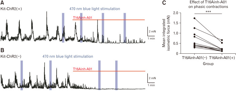
ANO1 is a calcium-activated chloride channel closely associated with ICC activity. Pharmacological blockade suppresses spontaneous phasic contractions in both groups and lowers mean recorded force. Premature large contractions previously evoked by light in the expression group no longer appear.
Light-driven contraction changes depend on the tissue’s existing activity system. The blockade links evoked contractions with ANO1-related processes, focusing mechanistic interpretation on coordination between ICC activity and muscle contraction.
Source: Zhao S et al. · Journal of neurogastroenterology and motility · CC BY-NC
Original figure and English caption ↗
How might light-responsive cells relay signals to muscle?
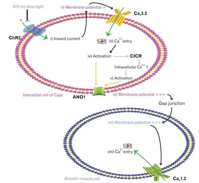
The authors’ model begins with ChR2-associated inward current: ICC depolarization may recruit calcium channels, with calcium changes interacting with ANO1 to amplify depolarization. Electrical changes pass through intercellular connections to smooth muscle and influence its calcium activity and contraction.
This model links electrical signaling with force. The study mainly measures muscle-strip force rather than directly following membrane potential, calcium and slow waves at every step. Contributions from different ICC layers remain unresolved; the model offers an explanation whose complete relay requires further testing.
Source: Zhao S et al. · Journal of neurogastroenterology and motility · CC BY-NC
Original figure and English caption ↗
Changing rhythm leaves transport as a separate question
The findings come from isolated colonic strips of otherwise normal mice, without in-vivo measurements of content transit. More frequent contractions and coordinated propulsion through the colon are different levels of motility, with the latter involving enteric nerves and cooperation across muscle regions.
The contribution is linking intestinal pacemaker-cell activity with the timing of individual contractions. A contraction brought forward and a more closely spaced rhythm provide observable phenomena for investigating how ICC contribute to colonic motility.