Cell-bearing fiber probes track chemical signals inside animals
FOPECs combine fluorescent recognition by engineered cells with fiber-optic readout, without requiring prior sensor expression in the host animal. The study demonstrates brain noradrenaline, dopamine and ATP dynamics and drug-related responses in rabbit vasculature.
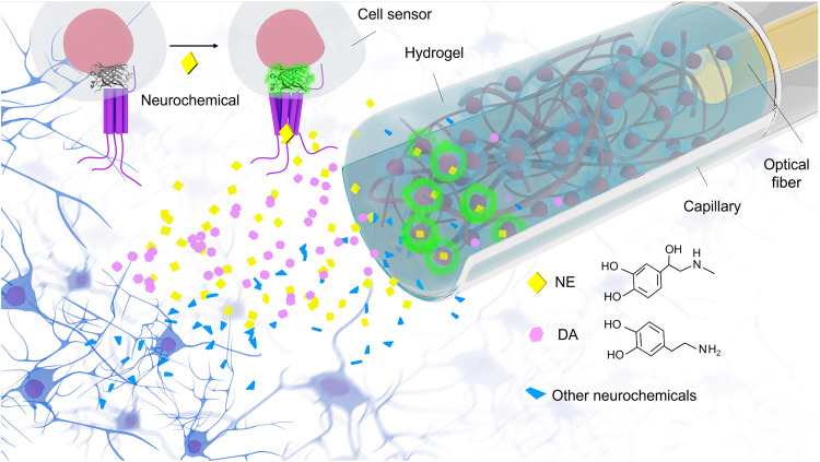
Could cells that recognize chemical molecules become the sensing element of an implanted probe? A study published in Science Advances on June 2, 2023 introduces FOPECs: engineered cells provide chemical recognition, and an optical fiber collects fluorescence changes. Preparing the sensing unit outside the animal removes the wait for host-cell sensor expression. The study demonstrates several neurochemical measurements in brain and flowing blood.
These cell-based fiber probes distinguish structurally similar noradrenaline and dopamine and detect chemical responses in several animal tests. Stress-associated signals remain recordable in freely moving mice on day 7 but decline in amplitude, highlighting the importance of the cellular probe’s working lifetime.
Contents · 7 sections
Cells sense within the probe and the fiber carries the readout
Following neurochemical dynamics requires identifying the molecule as well as detecting its change. FOPECs place engineered cells expressing GRAB fluorescent sensors at the sensing end. Nearby small molecules reach these cells and interact with surface sensors, producing green fluorescence changes that the fiber conveys outward.
A red reference signal does not respond appreciably to the target molecule. The two channels help relate target responses to shared fluctuations. Host brain cells need not express the sensor beforehand because recognition is supplied by cells carried in the probe.
Source: Zhou B et al. · Science advances · CC BY-NC
Original figure and English caption ↗
Closely related monoamines produce different responses
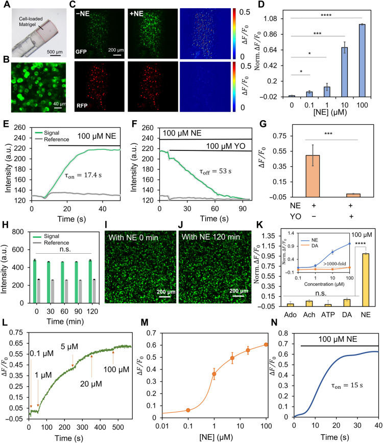
In vitro, an NE-sensitive probe shows a marked green increase in response to noradrenaline (NE), with little reference-red change. Responses vary with NE concentration and decline with receptor antagonism, supporting the intended recognition process.
Comparisons with dopamine and other molecules show substantial selectivity of the NE probe. The study also characterizes dopamine- and ATP-sensitive versions. Recognition components on the cells determine the chemical target, while fiber-based readout provides a common approach.
Source: Zhou B et al. · Science advances · CC BY-NC
Original figure and English caption ↗
Diffusion sets a timescale for sensing
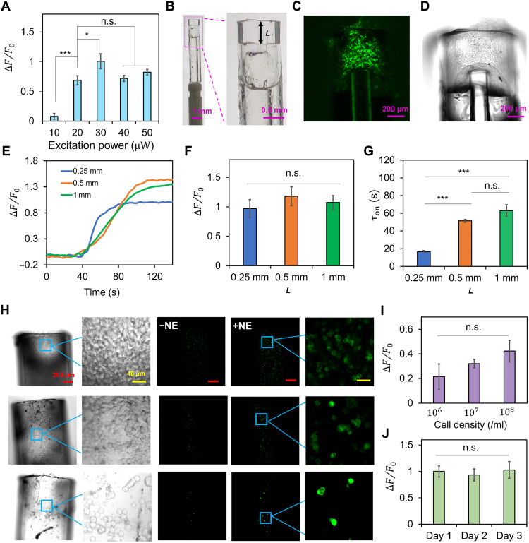
Molecular access to the sensing region influences timing. In vitro tests with a shorter sensing region show NE rise times of about 15–17 seconds; the decline after antagonist addition takes about 53 seconds. Changes in sensing-region size alter both signal strength and response time.
A larger sensing region can yield more fluorescence while requiring longer diffusion. This trade-off suits following slower chemical dynamics associated with stress or drugs. Signal strength and response speed are coupled within the same cellular probe.
Source: Zhou B et al. · Science advances · CC BY-NC
Original figure and English caption ↗
Responses repeat but weaken across days
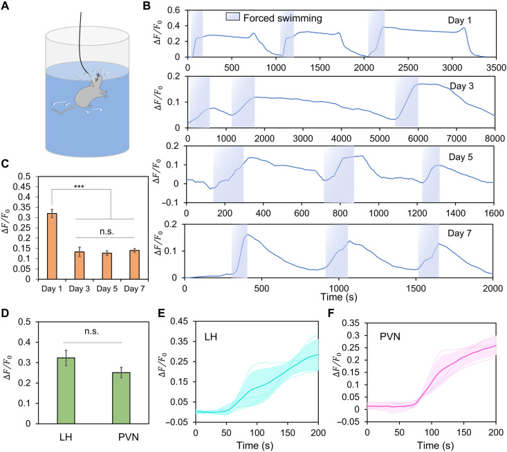
During swimming stress in freely moving mice, NE-related signals in the lateral hypothalamus rise and decline after removal from water. Responses recur across repeated episodes and remain detectable with similar directional dynamics on days 3, 5 and 7.
Later responses are smaller than on day 1. Cell survival within the probe also declines over time, making working lifetime part of its practical performance. Stress-response amplitudes do not differ significantly between the lateral hypothalamus and paraventricular nucleus in the reported comparison.
Source: Zhou B et al. · Science advances · CC BY-NC
Original figure and English caption ↗
From dopamine increases to seizure-associated ATP changes
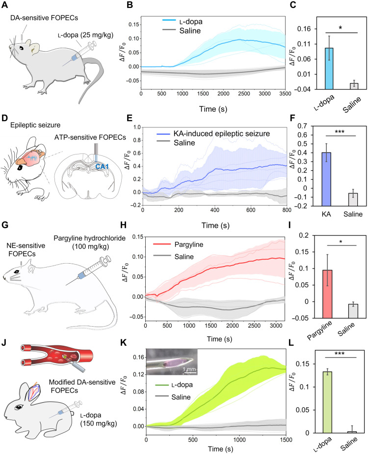
After levodopa, dopamine-probe signals in mouse hippocampal CA1 gradually rise and peak at about 40 minutes, unlike saline controls. This demonstrates tracking of a drug-related chemical process, with a time course that can be compared with other measurement approaches.
ATP-sensitive probes detect a sustained increase in a mouse seizure model, with EEG supporting the occurrence of seizures. The signal accompanies the pathological process and reports extracellular chemistry without by itself identifying the cells contributing ATP.
Source: Zhou B et al. · Science advances · CC BY-NC
Original figure and English caption ↗
Responses can also be detected in flowing blood
In rat hypothalamus, the study records an NE increase after drug treatment. Dopamine-sensitive probes in the rabbit ear vein also respond after levodopa. This peripheral blood measurement extends the demonstrated setting from brain tissue to a vascular environment.
Tests in chicken brain examine responses to externally administered NE. The animals demonstrate portability through different tasks: behavioral and pathological dynamics in mice, drug responses in rats and rabbits, and sensing validation under a defined condition in chickens.
Moving preparation outside the host creates new lifetime questions
FOPECs separate chemical recognition from host-cell expression, expanding options for animals and sites where prior sensor expression is difficult. Their performance also depends on cellular survival, molecular diffusion and the interface between a rigid probe and soft tissue.
One-week tissue observations under the study’s immune-management conditions find no significant inflammatory difference, while declining signals already highlight cellular maintenance. The work demonstrates a portable chemical readout approach whose longer-term recording capacity depends on further progress in materials and cellular stability.