From brain to heart: viewing multiple layers in a seizure model
A STAR Protocols article brings locus-coeruleus-associated cells, cardiac molecular measures and ECG observation into one framework for studying brain–body changes accompanying seizures.
A seizure begins in the brain, but associated physiological changes can extend to breathing and the heart. A STAR Protocols article published on June 30, 2023 builds on earlier locus-coeruleus–heart research, bringing neural activity, cardiac molecular measures and ECG observation into one framework. Using DBA/1 mouse models, it describes complementary views of a brain–body question and acknowledges limitations including movement interference and incomplete temporal coverage.
Neural, cardiac molecular and ECG measures provide evidence at different levels. The protocol supports multidimensional observation without establishing a unique direct brain–heart pathway or human SUDEP treatment efficacy.
Contents · 6 sections
Including the heart broadens the question
Understanding SUDEP requires attention to multiple physiological systems. This protocol considers seizure-induced respiratory arrest alongside cardiac changes, placing neural regulation and peripheral readouts within the same model.
Differences across several measures do not automatically establish a complete causal chain. Neural signals, receptor-related expression and cardiac rhythm answer distinct questions. The framework enables comparisons while leaving the timing and pathways between them open to testing.
Cell labeling localizes a neural change
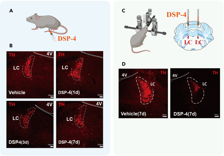
The locus coeruleus, or LC, is an important noradrenergic region. TH-associated tissue images show its location and labeling changes after intervention. TH relates to catecholamine synthesis and helps identify relevant cells, while labeling changes need interpretation alongside functional measures.
The images show reduced TH-positive labeling after intervention. This supports a change in the model’s cellular state, without directly measuring all neuronal activity or brain-wide transmitter levels. Identity, expression and function are related but distinct levels.
Source: Xu Q et al. · STAR protocols · CC BY
Original figure and English caption ↗
TH abundance and activity can move differently
The protocol summarizes earlier findings of increased TH abundance but reduced enzyme activity in cardiac tissue and related blood samples after respiratory arrest. Separating these measures avoids equating more detected protein with necessarily greater synthetic function.
The article also considers cardiac β1-adrenoceptors and their phosphorylation-related state. Receptor abundance and modification are different measures and do not individually describe total cardiac function. They provide mechanistic clues without fully resolving origins and temporal order.
Electrical signals place cardiac changes in time
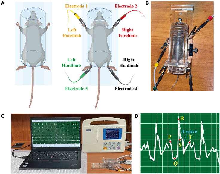
ECG adds dynamic information unavailable from tissue or molecular images alone. Figure 5 presents mouse ECG observation and a waveform example, enabling assessment of rhythm and electrical patterns around seizure-related stages.
Vigorous movement can interfere with recordings, particularly early in a seizure. The article also notes restricted temporal coverage in the acoustic model. A clear segment cannot substitute for complete observation of the entire event.
ECG primarily reflects electrical activity, while effective pumping also involves mechanical function. The article identifies more complete cardiac monitoring, including ejection fraction, as a future direction. Some functional dimensions therefore remain unmeasured even when neural, molecular and ECG information are available.
Source: Xu Q et al. · STAR protocols · CC BY
Original figure and English caption ↗
Different waveforms illustrate different electrical states
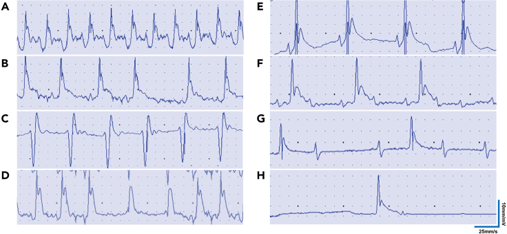
Figure 8 gathers author-labeled examples of normal rhythm, conduction abnormalities and ventricular rhythm disturbances. It makes cardiac electrical changes more specific than a simple faster-or-slower heart-rate measure.
The examples do not define a fixed sequence in one mouse or report the prevalence of every pattern across all animals. They concern model observations, not a human diagnostic atlas or evidence of established treatment efficacy.
Source: Xu Q et al. · STAR protocols · CC BY
Original figure and English caption ↗
Neural, molecular and ECG evidence retain distinct scopes
The protocol also summarizes an increase in LC population calcium activity during the clonic phase. This links neural and cardiac observations within one question, without assigning individual-cell firing or establishing direct LC control over every cardiac change.
Building on the original Lian study, the protocol contributes a research and interpretation framework. It includes DBA/1 mice of either sex, with findings bounded by model and observation conditions. More complete cardiac function measures, temporal coverage and pathway validation remain useful next questions; human SUDEP intervention efficacy requires clinical evidence.