Eye movements follow the beat: temporal interference stimulation tracks mouse superior-colliculus responses
From electric-field models to recordings in living animals, a study tracks superior-colliculus calcium signals and eye movements across beat frequencies, while finding different response ranges for behavior and local cellular signals.

When two nearby sound frequencies meet, their combination produces a waxing and waning beat. Temporal interference stimulation uses a related superposition principle: high-frequency electric fields form a slower envelope in tissue to explore modulation of deep brain regions. A study published on April 11, 2025 in Neuroscience Bulletin focuses on the mouse superior colliculus, a midbrain structure involved in eye movements. Activity and eye-movement recordings show rhythms changing with stimulation beat frequency. Changing the frequency difference changes the rhythm of eye movement, linking the field’s timing, neural responses and visible action.
Temporal interference stimulation evokes superior-colliculus-associated calcium responses and eye movements in mice, with their rhythms following beat frequency; field concentration and region-exclusive responses remain distinct questions.
Contents · 6 sections
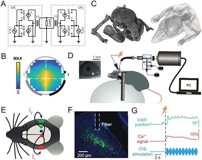
Beat frequency offers a different stimulation principle
Conventional transcranial electric fields spread through tissue, making it difficult to concentrate effects in small deep regions. Temporal interference combines two high-frequency fields into a slowly varying envelope. Their frequency difference sets its rhythm, and the study asks how that slower variation relates to neural responses.
Because the superior colliculus participates in visual orienting and eye-movement control, the eyes provide a visible observation window. The study tracks both eye movements and population calcium signals in the superior colliculus, connecting external stimulation, internal activity and visible movement beyond field calculations alone.
Source: Wang M et al. · Neuroscience bulletin · CC BY
Original figure and English caption ↗
Field concentration can shift without moving electrodes
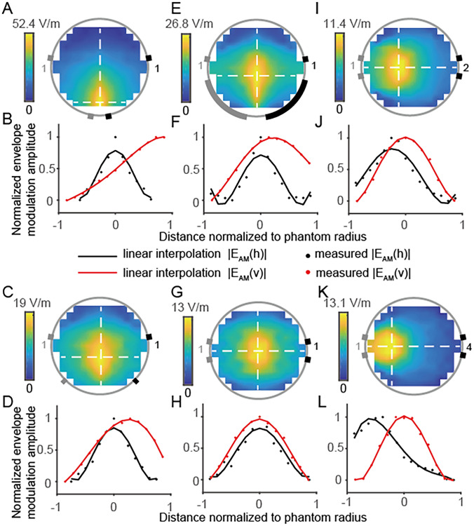
A tissue phantom reveals how envelope fields are distributed under different conditions. Changing electrode positions changes the distribution; changing the relative currents also shifts envelope concentration. Spatial adjustment therefore need not rely solely on moving external electrodes.
Changing electrode size has a smaller effect on distribution than placement or current ratio under the tested conditions. The result distinguishes factors that substantially change spatial distribution from those with weaker effects in this setup, providing a physical basis for subsequent mouse-head modeling.
Source: Wang M et al. · Neuroscience bulletin · CC BY
Original figure and English caption ↗
Temporal-interference fields are more concentrated
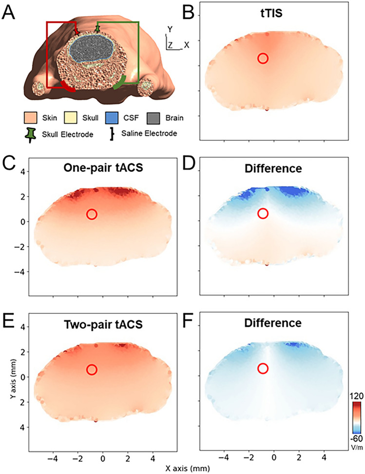
Finite-element modeling includes head-tissue structure and conductivity differences. Comparing temporal interference with conventional alternating-current stimulation under the same electrode configuration shows a more concentrated distribution near the target while retaining target-region field strength. Conventional alternating stimulation applied only from the skull top concentrates more superficially.
The advantage is initially a modeled one: current passage through tissues and envelope strength can be compared directly. Whether cells respond and whether neighboring tissue also responds requires in vivo observation. Modeling supplies a spatial prediction that the subsequent experiment relates to activity and movement.
Source: Wang M et al. · Neuroscience bulletin · CC BY
Original figure and English caption ↗
Responses do not appear without a beat
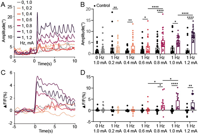
When the two high-frequency currents have equal frequencies, the experiment detects no clear eye-movement or superior-colliculus calcium response. A nonzero beat evokes both. Under the tested conditions, high-frequency current alone does not produce the same effect, linking the slower envelope to the responses.
Eye-movement amplitude and local calcium signals change with stimulation intensity, but the ranges producing clear responses differ. Eye movements emerge from many cells and pathways, whereas fiber recordings sample part of the superior colliculus. Their different sampling scopes help interpret this discrepancy.
Source: Wang M et al. · Neuroscience bulletin · CC BY
Original figure and English caption ↗
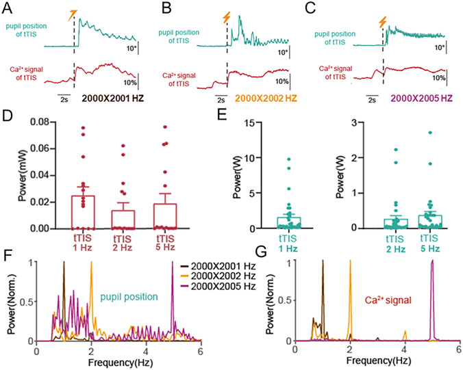
Changing the beat changes eye-movement rhythm
Across the tested low beat frequencies, the dominant rhythm in the eye-movement spectrum shifts and superior-colliculus calcium signals show corresponding rhythmic changes. Slow field fluctuations are thereby linked to the temporal structure of internal activity and movement, demonstrating rhythmic modulation by temporal interference.
This goes beyond eliciting a single movement: changing the temporal features of stimulation changes those of the response. Observing both signals describes this correspondence and raises a specific question about the contributions of different superior-colliculus cells and other eye-movement regions.
Source: Wang M et al. · Neuroscience bulletin · CC BY
Original figure and English caption ↗
A shared rhythm links fields, neural activity and eye movements
The study uses six adult male mice, with superior-colliculus calcium recordings from three. Electrode fixation involves skull damage, and neural recordings use implanted fibers; these preparation conditions matter when interpreting the field–response relationship. Modeled distribution advantages are examined in vivo within this specific setup.
The work connects physical envelopes, deep activity and eye-movement rhythm. Broader neural recordings are needed to distinguish target and neighboring responses and explain why movement and local recordings do not always respond over the same range, translating a more concentrated field into clearer knowledge of where it acts.